

DNA Damage Induction by Laser Microirradiation
激光微辐射诱导DNA损伤


发布: 2016年12月05日第6卷第23期 DOI: 10.21769/BioProtoc.2039 浏览次数: 21925
评审: HongLok LungMarco Di GioiaAnonymous reviewer(s)
参见作者原研究论文
The authors used this protocol in:

Feb 2016
Advertisement


Abstract
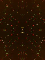
Genome instability can lead to cell death, senescence and cancerous transformation. Specific repair pathways have evolved to prevent accumulation of DNA lesions. Studying these highly dynamic and specific repair pathways requires precise spatial and temporal resolution, which can be achieved through a combination of laser microirradiaiton and live cell microscopy. DNA lesions are introduced at pre-determined sub-nuclear sites and repair can be analyzed in real time in living cells when using fluorescently tagged repair proteins (Mortusewicz et al., 2008). Alternatively, laser microirradiation can be combined with immunofluorescence analysis to study recruitment of endogenous proteins to laser-induced DNA damage tracks that can be visualized by positive controls like, e.g., γH2AX that mark sites of DNA breaks.
Keywords: Microirradiation (微辐照)Background
The genomic integrity of mammalian cells is constantly challenged by DNA damage introduced through external and internal sources. Amongst the most common DNA lesions are oxidized bases, double strand breaks, single strand breaks, inter- and intra-strand crosslinks and UV adducts. Various DNA damage signalling and repair pathways have evolved to deal with these lesions. For DNA repair to be fast, precise and efficient, numerous proteins involved in sensing, signalling and repairing specific DNA lesions have to be coordinated in space and time. Furthermore, DNA is organized into higher order chromatin structures and thus for DNA lesions to be accessible to DNA repair enzymes, chromatin has to be remodeled. Laser microirradiation in combination with advanced live cell microscopy allows studying these highly dynamic processes in the context of living cells (Mortusewicz et al., 2008). The protocol described here uses a 405 nm laser that should be readily available at most confocal or spinning disk microscopes to induce DNA damage in living cells and should therefore be cost effective and feasible in most standard cell biology laboratories. Using different sensitization methods (e.g., Hoechst versus BrdU sensitization) and laser energies, the ratio between double strand breaks, single strand breaks, oxidative lesions and UV damage can be modified to the experimental needs.
Materials and Reagents
- Live cell microscopy compatible Petri dish or chambers (e.g., Ibidi, catalog number: 35 mm µ-Grid )
- Adherent cell lines, e.g., U2OS
- If no CO2 control is available at your microscope, use CO2 independent medium without phenol red (e.g., Leibovitz's L-15 medium or CO2 independent medium, Thermo Fisher Scientific, GibcoTM, catalog number: 18045088 ) supplemented with 10% FBS (Thermo Fisher Scientific, GibcoTM, catalog number: 10082139 ) and antibiotics (Thermo Fisher Scientific, GibcoTM, catalog number: 15140-122 ). Alternatively, HEPES (Sigma-Aldrich, catalog number: H4034 ) can be added to your medium of chose.
- 5-bromo-2’-deoxycytidine (e.g., Santa Cruz Biotechnology, catalog number: 1022-79-3 )
- Hoechst 33342 (Thermo Fisher Scientific, Molecular ProbesTM, catalog number: H1399 )
- 4% formaldehyde in PBS (Santa Cruz Biotechnology, catalog number: 30525-89-4 )
- Triton X-100 (Sigma-Aldrich, catalog number: 234729 )
Note: This product has been discontinued. - Mouse-anti-γH2AX (EMD Millipore, catalog number: 05-636 )
- Donkey anti-Mouse IgG (H+L) secondary antibody, Alexa Fluor® 488 (Thermo Fisher Scientific, InvitrogenTM, catalog number: A-21202 )
- Bovine serum albumin (BSA) (Sigma-Aldrich, catalog number: A4503-500g )
- Tween 20 (Sigma-Aldrich, catalog number: P1379-500ML )
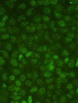
- Optional: plasmids encoding for fluorescently tagged proteins of interest, e.g., GFP-Timeless (Figure 1). Fluorescently tagged proteins known to be involved in different DNA repair pathways, like PARP-1, XRCC1 or 53BP1, can be used as controls to set up optimal conditions.
- Optional: transfection reagent of choice, e.g., self-made PEI solution or commercial distributor
Equipment
- Inverted confocal or spinning disk microscope equipped with a 405 nm laser and an environmental chamber or insert to control temperature and CO2/humidity for long-term live cell experiments, e.g., Zeiss LSM710 or LSM780 confocal laser scanning microscope equipped with a UV-transmitting Plan-Apochromat 63x/1.40 Oil DIC M27 or Plan-Apochromat 40x/1.30 Oil DIC M27 objective, respectively
- Cell incubator
Software
- Microscope software, e.g., ZEN from Zeiss (Zeiss)
- Microsoft Excel (Microsoft)
- Image J (https://imagej.nih.gov/ij/)
Procedure
文章信息
版权信息
© 2016 The Authors; exclusive licensee Bio-protocol LLC.
如何引用
Tampere, M. and Mortusewicz, O. (2016). DNA Damage Induction by Laser Microirradiation. Bio-protocol 6(23): e2039. DOI: 10.21769/BioProtoc.2039.
分类
癌症生物学 > 基因组不稳定性及突变 > 细胞生物学试验 > DNA结构和改变
细胞生物学 > 细胞成像 > 活细胞成像
您对这篇实验方法有问题吗?
在此处发布您的问题,我们将邀请本文作者来回答。同时,我们会将您的问题发布到Bio-protocol Exchange,以便寻求社区成员的帮助。