

A 3D Culture System of Human Immortalized Myometrial Cells
人永生子宫肌层细胞的三维培养系统


发布: 2016年10月20日第6卷第20期 DOI: 10.21769/BioProtoc.1970 浏览次数: 9218
评审: Andrea IntroiniJalaj GuptaAnonymous reviewer(s)
参见作者原研究论文
The authors used this protocol in:
Jun 2012
Advertisement


Abstract
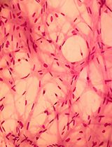
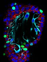
Myometrium forms the middle layer of the uterus and is mainly composed of the smooth muscle cells. The cells in vitro are usually grown in a single layer (2-dimensional; 2D) format, whereas in vivo cells are structured in an extracellular matrix scaffolding that allows the cells to communicate and respond to environmental cues. We have developed human myometrium and leiomyoma 3-dimensional (3D) culture, wherein the cells retain their molecular characteristics and respond to environmental cues (Malik and Catherino, 2012; Malik et al., 2014).
Keywords: Myometrium (子宫肌层)Background
In the last decade a certain shift is observed as more laboratories move from using the artificial 2D format of cell culture into 3D cell culture model system, where the cells are grown in a matrix that allows them to attach and attain a more physiologic configuration. This model system provides the cells with a more natural state of differentiation and the cultured cells develop an in vivo tissue-like environment. This is a detailed protocol for myometrium 3D cell culture growth in a collagen-I matrix, modified from Malik and Catherino (2012).
Materials and Reagents
- 8 chamber glass slides (8 well culture slides) (Corning, Falcon®, catalog number: 354108 )
- 5 ml, 10 ml and 25 ml serological pipettes, sterile, individually wrapped (Thermo Fisher Scientific)
- 15 ml and 50 ml conical sterile polypropylene centrifuge tubes (Thermo Fisher Scientific)
- Microscope slides (Thermo Fisher Scientific, Fisher Scientific, catalog number: 12-5446 )
- Coverslips (22 x 60 mm) (Thermo Fisher Scientific, Fisher Scientific, catalog number: 12-544G )
- 6-well culture plate
- 12-well cell culture plate
- T75 flask
- Aerosol barrier pipette tips (10 µl to 1,000 µl) (Thermo Fisher Scientific)
- Unfiltered 1 ml pipette tips (Thermo Fisher Scientific, Fisher Scientific, catalog number: 13-611-101 )
- 0.45 µm millex syringe filter unit (EMD Millipore, catalog number: SLHA02510 )
- Millex vented 0.22 µm syringe filter unit (EMD Millipore, catalog number: SLGSV255F )
- Kimberly-ClarkTM Kimwipes (Thermo Fisher Scientific, Fisher Scientific, catalog number: 06-666A )
- Myometrial cells
- Rat tail collagen-I
*Cultrex® 3-D culture matrix rat collagen I: 4 mg/ml (Trevigen, Cultrex®, catalog number: 3447-020-01 )
Collagen type I: 4.48 mg/ml (EMD Millipore, catalog number: 08-115 )
Collagen I, high concentration: 8-11 mg/ml (Corning, catalog number: 354249 )
*Note: Rat tail collagen-I from Trevigen is the most commonly used matrix in the lab but depending on the concentration of the gel to be made, we routinely use collagen-I from other vendors as listed. Follow the manufacturer’s conditions on storage as improper storage can lead to increased viscosity of the collagen and difficult to handle. - 10x PBS (filtered through 0.2 µm filter) (Thermo Fisher Scientific, GibcoTM, catalog number: 70011044 )
- Sodium hydroxide (NaOH, 6 N) (VWR, catalog number: JT5672-2 )
- Double distilled water (DD water; Filtered through 0.2 µm filter)
- Trypsin-EDTA (0.05%) (Thermo Fisher Scientific, GibcoTM, catalog number: 25300054 )
- Dulbecco’s modified Eagle’s medium/F12 (DMEM/F12, with phenol red) (Thermo Fisher Scientific, GibcoTM, catalog number: 11320-033 )
- Fetal bovine serum (FBS) (Defined) (GE Healthcare, HycloneTM, catalog number: SH30070.03 )
- Glutamax (Thermo Fisher Scientific, GibcoTM, catalog number: 35050061 )
- Penicillin-streptomycin (Thermo Fisher Scientific, GibcoTM, catalog number: 15140122 )
- Fungizone (Thermo Fisher Scientific, GibcoTM, catalog number: 15290018 )
- Paraformaldehyde (Sigma-Aldrich, catalog number: P6148 )
Or 16% paraformaldehyde solution (Electron Microscopy Sciences, catalog number: 15710 ) - Glycine (Sigma-Aldrich, catalog number: G5417-100G )
- Triton X-100 (Sigma-Aldrich, catalog number: 93443 )
- Normal goat serum (NGS) (Abcam, catalog number: ab156046 ) (Dilution in 1x PBS)
- Secondary antibody: Alexa 488 (follow manufacturers dilution instructions) (Thermo Fisher Scientific, InvitrogenTM, catalog number: A11008 )
- Primary antibody to smooth muscle alpha actin (0.2 mg/ml) (Abcam, catalog number: ab5694 )
- Prolong® Gold antifade mountant with DAPI (Thermo Fisher Scientific, Molecular ProbesTM, catalog number: P36931 )
- DPBS (no magnesium or calcium) (Thermo Fisher Scientific, GibcoTM, catalog number: 14190144 )
- Bovine serum albumin (BSA) (35% solution in DPBS) (Sigma-Aldrich, catalog number: A7979 ), Dilution in 1x PBS
- 10% growth media (see Recipes)
- 5% growth media (see Recipes)
- 1 N NaOH (see Recipes)
- 3 mg/ml collagen gels (see Recipes)
- 0.15 M glycine/PBS (see Recipes)
- 4% paraformaldehyde/PBS (see Recipes)
- Blocking buffer in PBS (see Recipes)
- Primary ab dilution buffer in PBS (see Recipes)
Notes:
a.Keep #1-3 at -20 °C if possible or 4 °C overnight (I have a small -20 °C freezer right next to my culture hood).
b.Keep #15-18 on ice before the start of experiment.
Equipment
- Microscope (Nikon, model: Eclipse TS100 )
- Centrifuge (Eppendorf, model: 5804R )
- Automated cell counter (Bio-Rad Laboratories, model: TC 20TM )
- Hemocytometer (if automated cell counter not available)
- Metal 50ml holder (e.g., Blue anodized aluminum [Thomas Scientific, catalog number: 1225W71 ])
Note: In -20 °C if possible or 4 °C overnight. - 95% air/5% CO2 incubator at 37 °C and 95% relative humidity
- Shaker at 4 °C and one at room temperature
- Vacuum aspirator
- Hot plate/stirrer (Fisher Scientific)
- Pyrex beaker (100 ml) (Fisher Scientific)
- Flat tip forceps (Thermo Fisher Scientific, Fisher Scientific, catalog number: 16-100-111 )
- Microscope: confocal laser microscope: (e.g., Carl Zeiss, model: ZEISS LSM 800 )
Procedure
文章信息
版权信息
© 2016 The Authors; exclusive licensee Bio-protocol LLC.
如何引用
Malik, M., Britten, J. and Catherino, W. H. (2016). A 3D Culture System of Human Immortalized Myometrial Cells. Bio-protocol 6(20): e1970. DOI: 10.21769/BioProtoc.1970.
分类
细胞生物学 > 细胞分离和培养 > 3D细胞培养
您对这篇实验方法有问题吗?
在此处发布您的问题,我们将邀请本文作者来回答。同时,我们会将您的问题发布到Bio-protocol Exchange,以便寻求社区成员的帮助。