

Ear Inflammation and Whole-mount Ear Staining
耳部炎症和耳部整体染色

发布: 2016年10月20日第6卷第20期 DOI: 10.21769/BioProtoc.1967 浏览次数: 11351
评审: Ivan ZanoniGriselda Zuccarino-CataniaBenoit Stijlemans
参见作者原研究论文
The authors used this protocol in:
Apr 2015
Advertisement


Abstract
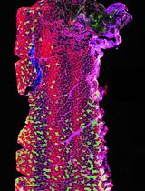
The recruitment of circulating neutrophils from the bloodstream to the site of inflammation represents one of the earliest events during an innate immune response. During this response, neutrophils tether and roll along the vessel walls before transmigrating across the endothelium into the interstitial space to exert their functions. Here, we describe a protocol for the staining of intravascular and tissue-localized neutrophils following contact sensitization of the skin with croton oil. Visualization of the neutrophilic distribution in skin provides for a better interpretation of the local immune response.
Keywords: Neutrophils (中性白细胞)Background
Characterisation of neutrophil distribution within the skin following inflammation represents an important avenue for the understanding of their specialized functions. Even though the recruitment of neutrophils to the inflamed skin has been widely characterized by flow cytometry (Hampton et al., 2015; Stock et al., 2014), such technique provides limited insight on the intravascular versus interstitial localization of neutrophils. Recirculating and tissue-localized neutrophils exhibit different phenotypes and functions, which necessitates their discrimination to identify key players of the local immune response. With the development of intravital microscopy (IVM), the direct visualization of fluorescently tagged immune cells in vivo is made possible. However, due to the high cost of IVM, the accessibility of this powerful imaging tool to researchers is limited. Here, we present an alternative protocol for the high resolution static imaging of neutrophils in skin by making use of cost-effective reagents and commonly available confocal laser-scanning microscopy.
Materials and Reagents
- Cotton swabs
- SterilinTM Petri dish (90 mm) (Thermo Fisher Scientific, Thermo ScientificTM, catalog number: 101IRR )
- 24-well cell culture plates (Thermo Fisher Scientific, Thermo ScientificTM, catalog number: 142475 )
- Glass slides (Biomedical Sciences Institutes, catalog number: BMH.880103 )
- FisherbrandTM cover glass (Thermo Fisher Scientific, Fisher Scientific, catalog number: 12-545-81 )
- Clear nail polish
- Mice
- Croton oil (Sigma-Aldrich, catalog number: C6719 )
- Veet hair removal cream
- Phosphate buffered saline (PBS) (Thermo Fisher Scientific, GibcoTM, catalog number: 10010023 )
- Acetone (Thermo Fisher Scientific, Fisher Scientific, catalog number: 67-64-1 )
- Bovine serum albumin (BSA) (Sigma-Aldrich, catalog number: 10735108001 )
- Sodium azide (Sigma-Aldrich, catalog number: S2002 )
- Antibodies
- PE anti-mouse Ly-6G antibody (clone 1A8) (BioLegend, catalog number: 127607 )
- PE rat IgG2a, κ Isotype control antibody (clone RTK2758) (BioLegend, catalog number: 400507 )
- APC anti-mouse CD31 (clone MEC 13.3) (BD, PharmingenTM, catalog number: 561814 )
- Mounting medium (ibidi, catalog number: 50001 )
- DAPI (Sigma-Aldrich, catalog number: D9564 )
- Stain and wash buffer (1% BSA in PBS) (see Recipes)
Equipment
- Pipette (Eppendorf, model: Eppendorf Research plus )
- Dissection scissors (Roboz Surgical Instrument, catalog number: RS-6702 )
- Forceps (Roboz Surgical Instrument, catalog number: RS-5240 ; RS-5135 )
- Shaker (Labnet)
- Zeiss LSM 510 Meta confocal microscope (Zeiss)
Software
- ImageJ software (National Institutes of Health) (https://imagej.nih.gov/ij/index.html)
Procedure
文章信息
版权信息
© 2016 The Authors; exclusive licensee Bio-protocol LLC.
如何引用
Loh, J. T., Gunawan, M. and Su, I. (2016). Ear Inflammation and Whole-mount Ear Staining. Bio-protocol 6(20): e1967. DOI: 10.21769/BioProtoc.1967.
分类
免疫学 > 免疫细胞成像 > 共聚焦显微镜技术
细胞生物学 > 细胞成像 > 共聚焦显微镜
您对这篇实验方法有问题吗?
在此处发布您的问题,我们将邀请本文作者来回答。同时,我们会将您的问题发布到Bio-protocol Exchange,以便寻求社区成员的帮助。