

Lipid Extraction from HeLa Cells, Quantification of Lipids, Formation of Large Unilamellar Vesicles (LUVs) by Extrusion and in vitro Protein-lipid Binding Assays, Analysis of the Incubation Product by Transmission Electron Microscopy (TEM) and by Flotation across a Discontinuous Sucrose Gradient
以HeLa细胞研究蛋白质和膜相互作用的一系列方法:脂质提取和气相色谱定量;挤压法制备大单室脂质体(LUV);蛋白质和脂质体外孵育;采用透射电子显微镜(TM)和浮式离心法在不连续蔗糖梯度范围分析蛋白质和脂质互作;


发布: 2016年10月20日第6卷第20期 DOI: 10.21769/BioProtoc.1963 浏览次数: 17232
评审: Ivan ZanoniAnonymous reviewer(s)
参见作者原研究论文
The authors used this protocol in:
Dec 2015
Advertisement


Abstract
Dissecting the interactions established between proteins and membranes in a given type of cells is not an easy task. Using a cell-free system of large unilamellar vesicles (LUVs) to analyze these interactions may help decipher these interactions and identify potential membrane deformations induced by the proteins incubated with these LUVs. This article describes the protocols for 1) extraction of total lipids from eukaryotic cells using the method developed by Bligh and Dyer (1959), 2) the quantification of glycerophospholipids by gas chromatography after methanolysis, followed by 3) the formation of LUVs by extrusion, 4) protein-lipid binding assay, 5) analysis of the incubation product by transmission electron microscopy (TEM) and by flotation across a discontinuous sucrose gradient and finally, 6) analysis of the proteins by immunoblot and revelation of the glycerophospholipids by iodin fumigation.
Keywords: Large Unilamellar Vesicles (LUVs) (大单层脂质体(爱))Background
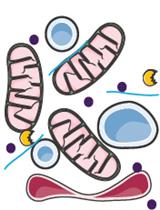
Cell-free systems consisting in giant unilamellar vesicles (GUVs; vesicles composed of a single bilayer of phospholipids and with a diameter greater than 1 μm) or liposomes incubated with recombinant proteins may help understand these interactions. Depending on their diameter and number of lamellae, liposomes are classified into small unilamellar vesicles (SUVs; vesicles constituted of a single bilayer of phospholipids and with a diameter comprised between 20 and 100 nm), large unilamellar vesicles (LUVs; vesicles constituted of a single bilayer of phospholipids and with a diameter comprised between 100 and 400 nm), large multilamellar vesicles (MLVs; vesicles constituted of multiple phospholipid bilayers and with a diameter comprised between 200 nm and 3 μm) and multivesicular vesicles (MVVs; large vesicles composed of a single bilayer of phospholipids and containing several smaller vesicles, each composed of a single bilayer of phospholipids).
When a dried mix of lipids is dispersed in an aqueous solvent, large multilamellar vesicles (LMVs) form spontaneously. Smaller liposomes (SUVs or LUVs) may then be formed by sonication or extrusion. Here we report the formation of LUVs by extrusion of LMVs formed from complex lipids extracted from HeLa cells and their use to investigate Toxoplasma proteins/membrane interactions by flotation across a sucrose gradient and by TEM.
Materials and Reagents
- PYREX® disposable glass conical centrifuge tubes without cap (capacity: 50 ml) (Sigma-Aldrich, catalog number: CLS9950250-72EA )
- Disposable screw thread culture tubes with marking spot (ø13 mm) (KIMAX test tubes with Teflon liner caps) (Thomas Scientific, catalog number: 9210J23 )
- Pasteur pipettes (non-plugged, L 5 ¾ in.) (Sigma-Aldrich, catalog number: S6018 )
- Polycarbonate membranes (Avanti Polar Lipids)
Note: The diameter of their pores is defined by the manipulator (Example: 100 nm, Avanti Polar Lipids, catalog number: 610005 ). - Grids for transmission electron microscopy (grid size 400 mesh x 62 μm pitch, copper) (Sigma-Aldrich, catalog number: G5026 )
- 0.80 ml open ultra-clear centrifuge tubes (size 5 x 41 mm) (Beckman Coulter; catalog number: 344090 ) and split adaptors (Beckman Coulter; catalog number: 356860 )
- Nitrocellulose membranes for protein transfer as for example: nitrocellulose membranes, 0.2 µm, 8 x 12 cm (Thermo Fisher Scientific, catalog number: 77012 )
- HeLa cells (ATTC, catalog number: CCL-2 )
- Dulbecco’s modified Eagle’s medium (DMEM) (Thermo Fisher Scientific, catalog number: 41966-029/052 )
- Fetal bovine serum (FBS) (Eurobio, catalog number: CVFSVF0001 )
- Penicillin/streptomycin (PAN-Biotech, catalog number: P06-07100 )
- L-glutamine (200 mM) (Thermo Fisher Scientific, catalog number: 25030-024 )
- Dulbecco’s phosphate-buffered saline (DPBS) (Thermo Fisher Scientific, GibcoTM, catalog number: 14190-094/069 )
- Chloroform (for HPLC, ≥ 99.8%, amylene stabilized) (Sigma-Aldrich, catalog number: 34854 )
- Methanol (for HPLC, ≥ 99.9%) (Sigma-Aldrich, catalog number: 34860 )
- Water sterile-filtered (BioReagent, suitable for cell culture) (Sigma-Aldrich, catalog number: W3500 )
- C21:0 fatty acid (Heneicosanoic acid) (Matreya, catalog number: 1241 )
- Hexane (anhydrous, 95%) (Sigma-Aldrich, catalog number: 296090 )
- Sulfuric acid (99.999%) (Sigma-Aldrich, catalog number: 339741 )
- HEPES free acid [N-(2-hydroxyethyl)piperazine-N’-(2-ethanesulfonic acid); 4-(2-hydroxyethyl)-1-piperazineethanesulfonic acid)] (Sigma-Aldrich, catalog number: H3375 )
- Sodium chloride (NaCl) (ACS reagent, ≥ 99.0%) (Sigma-Aldrich, catalog number: S9888 )
- Liquid N2
- Uranyl acetate (Electron Microscopy Sciences, catalog number: 22400 )
- Sucrose (≥ 99.5%) [α-D-Glucopyranosyl, β-D-fructofuranoside, D(+)-Saccharose] (Sigma-Aldrich, catalog number: S8501 )
- Iodine (ACS reagent, ≥ 99.8%, solid) (Sigma-Aldrich, catalog number: 207772 )
- Complete DMEM medium (see Recipes)
- Protein-lipid binding buffer (see Recipes)
Equipment
- Flasks (150 cm2) (Dominique Dutscher, catalog number: 190150 )
- 37 °C/5% CO2 cell culture incubator (Dominique Dutscher, catalog number: 911378M )
- Refrigerated bench centrifuge (Dominique Dutscher, catalog number: 472456 )
- Fume hood (standard equipment of any lab)
- Vortex (Dominique Dutscher, catalog number: 079030 )
- Bottle of nitrogen gas
- Freezer (-20 °C) (Dominique Dutscher, catalog number: 099288B )
- 10 μl positive displacement pipet such as microman Gilson model M10 (Gilson, catalog number: F148501 )
- Oven (for dry heat: 100 °C) (Dominique Dutscher, catalog number: 780405 )
- BPX70 gas chromatography column (30 m x 0.25 mm ID BPX70 0.25 μm) (Trajan Scientific, catalog number: 0 54622 )
- Gas chromatography apparatus (Shimadzu, ref GC-2010 Plus High-end GC)
- Water bath (37 °C) (Dominique Dutscher, catalog number: 910648 )
- Extruder set with holder/heating block (Avanti Polar Lipids, catalog number: 610000 )
- 2 x 250 μl Hamilton® GASTIGHT® syringes, 1800 series (1825N, volume 250 μl, needle size 22s ga [bevel tip], needles L51 mm [2 in.]) (Sigma-Aldrich, catalog number: 21394 )
- Fridge (4 °C) (Dominique Dutscher, catalog number: 670251B )
- Rotating agitator (Dominique Dutscher, catalog number: 062646 )
- Carbon evaporator (Q150T Turbo-Pumped sputter coater/carbon coater) (Quorum Technologies, model: Q150T )
- Electron transmission microscope such as JEOL-1400 plus (MET 120 kV) (JEOL, model: 1400 Plus )
- Ultracentrifuge type TL100 labtop equipped with an MLS-50 Swinging-Bucket rotor (Beckman Coulter, catalog number: 367280 )
- 12% Mini-PROTEAN® TGXTM precast protein gels (10-well, 30 µl) (Bio-Rad Laboratories, catalog number: 4561043 )
- Power supplier (Dominique Dutscher, catalog number: 049192 )
- SDS-PAGE apparatus (SDS-PAGE), as for example Mini-PROTEAN® Tetra vertical electrophoresis cell for mini precast gels, 2-gels (Bio-Rad Laboratories, catalog number: 1658005 )
- Protein transfer apparatus, as for example Trans-Blot® TurboTM transfer system (Bio-Rad Laboratories, catalog number: 1704150 )
- Thin layer chromatography plates: silica gel on TLC aluminium foils (silica gel matrix, L x W = 20 x 20 cm) (Sigma-Aldrich, catalog number: 60805 )
- Hermetic glass tank such as Aldrich® rectangular TLC developing tanks, complete (L x H x W = 27.0 x 26.5 x 7.0 cm) (Sigma-Aldrich, catalog number: Z126195 )
Procedure
文章信息
版权信息
© 2016 The Authors; exclusive licensee Bio-protocol LLC.
如何引用
Bittame, A., Lopez, J., Effantin, G., Blanchard, N., Cesbron-Delauw, M., Gagnon, J. and Mercier, C. (2016). Lipid Extraction from HeLa Cells, Quantification of Lipids, Formation of Large Unilamellar Vesicles (LUVs) by Extrusion and in vitro Protein-lipid Binding Assays, Analysis of the Incubation Product by Transmission Electron Microscopy (TEM) and by Flotation across a Discontinuous Sucrose Gradient. Bio-protocol 6(20): e1963. DOI: 10.21769/BioProtoc.1963.
分类
免疫学 > 免疫细胞功能 > 综合
生物化学 > 脂质 > 脂质分离
您对这篇实验方法有问题吗?
在此处发布您的问题,我们将邀请本文作者来回答。同时,我们会将您的问题发布到Bio-protocol Exchange,以便寻求社区成员的帮助。