

Imaging Thick Lymph Node Tissue Sections
淋巴结组织厚切片的成像技术


发布: 2016年09月20日第6卷第18期 DOI: 10.21769/BioProtoc.1938 浏览次数: 13644
评审: Ivan ZanoniFrancesca MingozziMeenal SinhaAnonymous reviewer(s)
参见作者原研究论文
The authors used this protocol in:
Sep 2015
Advertisement


Abstract
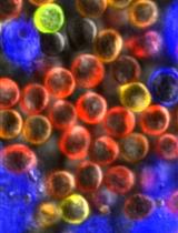
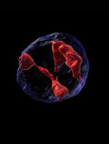
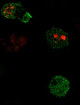
Our protocol describes a simple procedure for imaging thick lymph node sections by 2-photon microscopy. Lymph nodes are sectioned using a vibratome (vibrating microtome) to produce slices of tissue that can then be stained with fluorescently labeled antibodies. The thick tissue sections (150-200 μm depth) allow for the detection of cell clustering that is typically under-represented in thin sections (10-20 μm) used for conventional confocal microscopy. Application of 2-photon microscopy facilitates imaging through the thick volume of the vibratome sections. In combination with automated image processing software, a thick lymph node cross-section image also facilitates quantitation of cellular events within a relatively large area of the tissue, thus providing a clearer picture on the spatial distribution of cellular events of interest (e.g., T cell clustering). This method can also readily be applied to other tissues, such as the spleen or skin.
Keywords: 2-photon microscopy (双光子显微镜)Materials and Reagents
- Razor blades, single edge, No.9 (0.22 mm) (VWR International, catalog number: 55411-050 )
- Greiner CELLSTAR® 24-well flat-bottom plates (Sigma-Aldrich, catalog number: M8812 )
- 96-well round-bottom plates (Corning, catalog number: 3788 )
- Durapore tape (3M, catalog number: 1538-1 )
- SuperfrostTM Plus and ColorFrostTM glass slides (25 x 75 x 1.0 mm) (Thermo Fisher Scientific, Thermo ScientificTM, catalog number: 4951PLUS4 )
- Coverslips, No.1.5 (ProSciTech, catalog number: G425-2460 )
- Agarose (Sigma-Aldrich, catalog number: A6013-250G )
- Vetbond tissue adhesive (3M, catalog number: 1469SB )
- Phosphate-buffered saline (PBS) (Thermo Fisher Scientific, GibcoTM, catalog number: 10010023 )
- Protein block (Dako, catalog number: X0909 )
- Normal donkey serum (NDS) (Jackson ImmunoResearch, catalog number: 017-000-121 )
- Prolong gold antifade reagent (Thermo Fisher Scientific, Molecular ProbesTM, catalog number: P36930 )
- Antibodies [e.g., pacific blue anti-B220, RA3-6B2 (Biolegend, catalog number: 103230 ); goat anti-CD69 (R&D Systems, catalog number: AF2386 )]
- Sodium periodate (NaIO4) (Sigma-Aldrich, catalog number: 311448 )
- Sodium dihydrogen orthophosphate, monohydrate (NaH2PO4·H2O) (monobasic) (VWR, catalog number: 97062-412 )
- Di-sodium hydrogen orthophosphate, anhydrous (Na2HPO4) (dibasic) (Ajax Finechem, catalog number: 621-500G )
- L-lysine (Sigma-Aldrich, catalog number: L5501 )
- Gelatin from porcine skin (Sigma-Aldrich, catalog number: G1890-100G )
- Glycerol for molecular biology (≥ 99%) (Sigma-Aldrich, catalog number: G5516 )
- 16% paraformaldehyde aqueous solution (Electron Microscopy Sciences, catalog number: 30525-89-4 )
- Periodate-lysine-paraformaldehyde (PLP) fixative (see Recipes)
- P-buffer (see Recipes)
- L-lysine (see Recipes)
- 5% gelatin/glycerol mix (see Recipes)
Equipment
- Vibratome (Leica, model: VT1200S )
- Dumont No.5 forceps (Fine Science Tools, catalog number: 11251-10 )
- Scissors, straight, sharp (Roboz, catalog number: RS-6752 )
- Soft bristled paint brush (e.g., size 2/0)
- Microscope with tunable Coherent Chameleon Ti:Sa laser (ZEISS, model: LSM 710 NLO )
Note: This product has been discontinued (Replaceable item, e.g., ZEISS, model: LSM 800 ). - Scale for weighing 1 g
- Microwave
Software
- Imaris, Bitplane (http://www.bitplane.com/)
Procedure
文章信息
版权信息
© 2016 The Authors; exclusive licensee Bio-protocol LLC.
如何引用
Hor, J. L. and Mueller, S. N. (2016). Imaging Thick Lymph Node Tissue Sections. Bio-protocol 6(18): e1938. DOI: 10.21769/BioProtoc.1938.
分类
免疫学 > 免疫细胞成像 > 共聚焦显微镜技术
您对这篇实验方法有问题吗?
在此处发布您的问题,我们将邀请本文作者来回答。同时,我们会将您的问题发布到Bio-protocol Exchange,以便寻求社区成员的帮助。