

Quantification of the Adhesion Strength between Peroxisomes and Chloroplasts by Femtosecond Laser Technology
采用飞秒激光技术定量测定过氧化物酶体和叶绿体之间的粘附强度


发布: 2016年06月05日第6卷第11期 DOI: 10.21769/BioProtoc.1834 浏览次数: 10397
评审: Tie LiuJaroslav ĎurkovičDennis Nürnberg
参见作者原研究论文
The authors used this protocol in:

Feb 2015
Advertisement


Abstract
This is the detailed protocol to quantify adhesion strength between peroxisomes and chloroplasts in plant leaf palisade mesophyll cells described by Oikawa et al. (2015). The quantification was performed by utilizing local explosion induced by focusing femtosecond laser pulses into a mesophyll cell under a confocal microscope. When an impulsive force generated by an explosion is loaded on the interface between a peroxisome and a chloroplast, the peroxisome is frequently detached from the chloroplast. The probability of a peroxisome detaching from a chloroplast was estimated (left-top of Figure 1). Next, the magnitude of the impulsive force was quantified by an atomic force microscope (AFM) cantilever (right-top of Figure 1). On the basis of these results, the pressure to break adhesion between a peroxisome and a chloroplast was quantified as an index of the adhesion strength (bottom of Figure 1). In this protocol, these procedures are summarized. As the local explosion is induced not only in the medium of the mesophyll cells but also in aqueous medium generally, this method could be applied to various adhesions between organelles and between cells around 1 to 100 μm in diameter (e.g., adhesions between mitochondria and chloroplasts, between nucleus and cell membrane, and between two cells with weak physical interaction). Additionally, we have evaluated the interaction between peroxisomes and chloroplasts from the interaction length between two organelles. This protocol has been presented in Bio-protocol as “Measuring the interactions between peroxisomes and chloroplasts by in situ laser analysis” (Oikawa et al., 2015).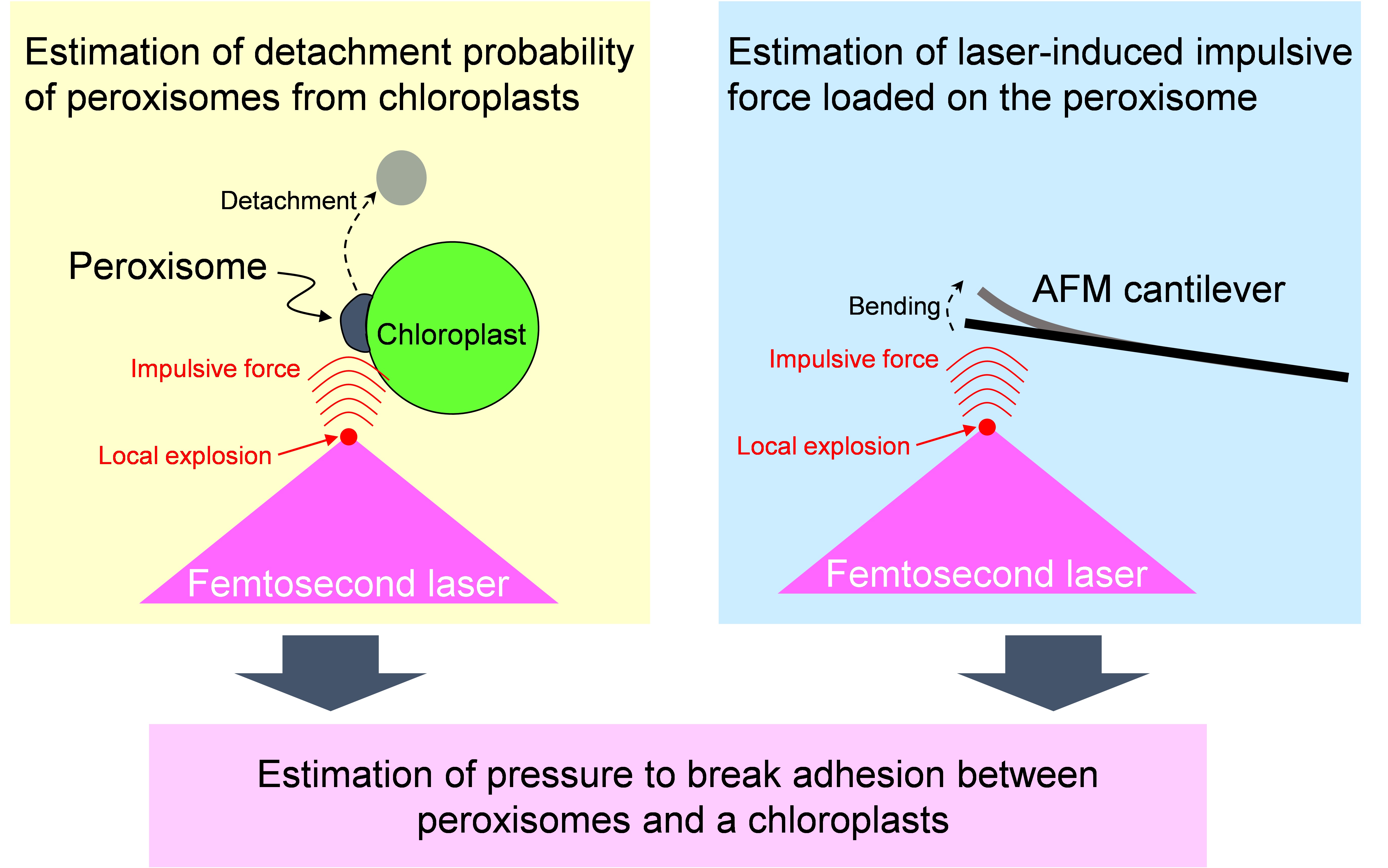
Figure 1. Flow chart estimating adhesion strength between a peroxisome and a chloroplast by utilizing femtosecond laser and atomic force microscope
Materials and Reagents
- Glass slide (26 x 76 No.1, thickness 0.8-1.0 mm) (Matsunami Glass Ind.)
Note: Any types of glass slide suitable for fluorescence observation can be used. We attached black tape on the glass slide to envelop the sample with the cover slip (see Figure 4D). - Cover slip (24 x 60 No.1, thickness 0.12-0.17 mm) (Matsunami Glass Ind.)
- 10 ml disposable syringe (Terumo Medical Corporation)
- Razor blade (43 x 23)
- Arabidopsis thaliana (ecotype Columbia) expressing peroxisome-targeted GFP (GFP-PTS1) (Mano et al., 2002)
- Agar powder (Funakoshi, catalog number: BA-10 )
- Distilled water
- 1/3x Murashige and Skoog salts (MS) medium (Wako Pure Chemical Industries, catalog number: 392-00591 )
Equipment
- Confocal microscope combined with amplified Ti:Sapphire femtosecond laser system (Figure 2)
- Ti:Sapphire femtosecond laser system (Cyber Laser Inc., model: IFRIT-SP-01 )
- Confocal microscope (Olympus Corporation of the America, model: FV300-IX71 )
- Objective (OLYMPUS CORPORATION, model: PlanN100x )
- Mechanical shutter (Gate time: 1/125 s) [SIGMAKOKI, model: 65GR (discontinued product); compatible product: SIGMAKOKI, model: SSH-25RA ]
- Collimator lenses
- λ/2 plate
- Polarizer
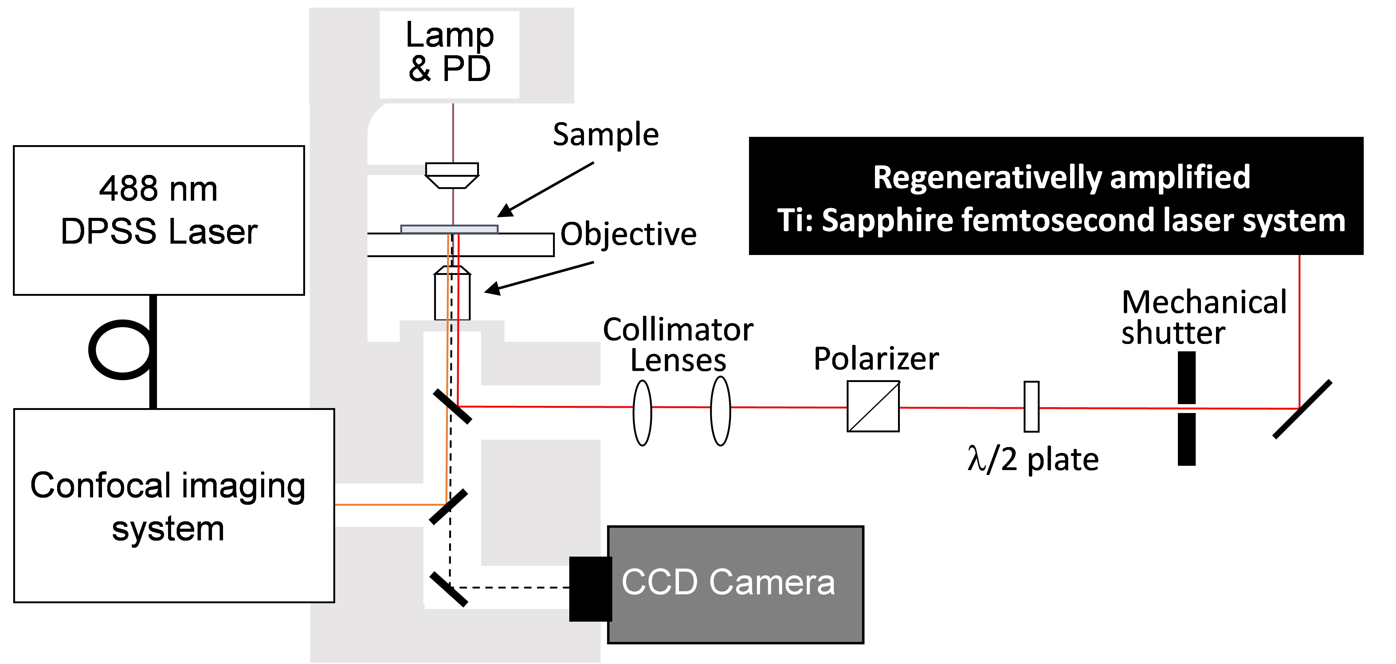
Figure 2. Experimental setup of confocal microscope and femtosecond laser system. Femtosecond laser pulses from an amplified Ti:Sapphire femtosecond laser system (Wavelength: 780 nm, Pulse duration: 230 fs, Pulse energy: <1 mJ/pulse, Repetition rate: 125 Hz) are introduced to a confocal microscope (Excitation wavelength: 488 nm, Detection wavelength: 515-550 nm). Mechanical shutter (Gate time: 1/125 s), collimator lenses, λ/2 plate and polarizer were placed in the beam line between the laser and the microscope. - Atomic force microscope (AFM) (Figure 3)
- AFM [Pacific Nanotechnology, model: Nano-R2 (discontinued product); compatible product: JPK Instruments, model: NanoWizard4 NanoScience ]
- Ti:Sapphire femtosecond laser system (see Equipment Section, 1a)
- Confocal microscope (see Equipment Section, 1b)
- Objective (see Equipment Section, 1c)
- A tipless AFM cantilever (NanoWorld AG, NANOSENSORSTM, model: TL-NCH )
- Motorized stage (SIGMAKOKI, model: BIOS-206T )
- Oscilloscope [TEKTRONIX, model: DP4104 (discontinued product); compatible product: TEKTRONIX, model: MDO4104c ]
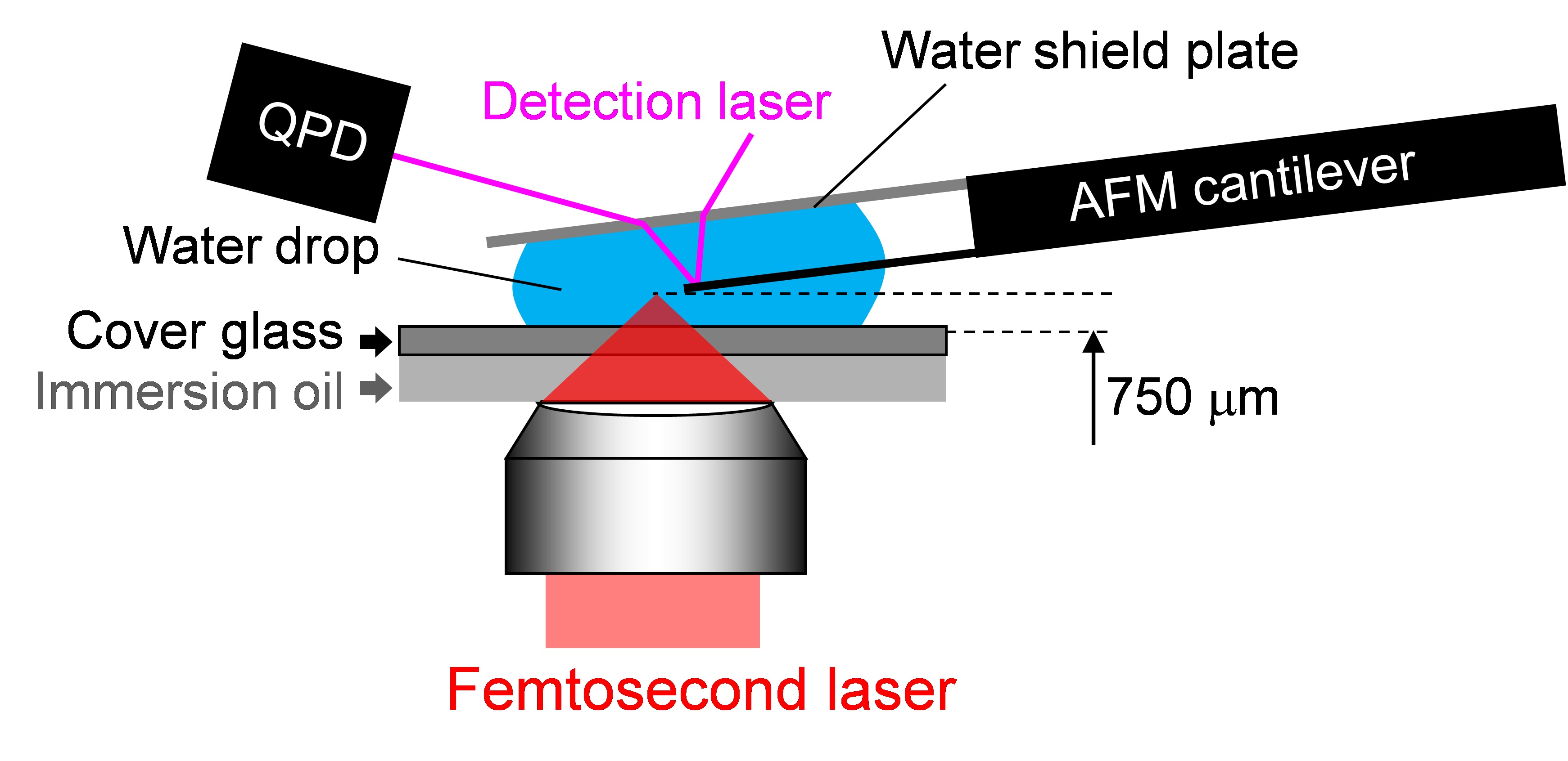
Figure 3. Experimental setup of AFM head on the microscope stage. The head of an AFM was mounted on the confocal microscope (Objective, PlanN100x) combined with the amplified Ti:Sapphire femtosecond laser system with the same setting as that mentioned in the former item. A tipless AFM cantilever is attached to the AFM head. The cantilever is placed between a water shield plate and cover glass, and immersed in distilled water or culture medium.
Note: The distance between the top of the cantilever and the laser focal point was tuned by a motorized stage equipped on the microscope. The normal function of the AFM system [feedback system between quadrant photo diode (QPD) and piezoelectric (PZT) motor] is deactivated. The signal of the QPD in the AFM head is directly monitored with an oscilloscope. The detailed settings are described in Hosokawa et al., 2011.
Software
- IGOR Pro 6.22J (WaveMetrics, https://www.wavemetrics.com)
- Visual Basic 6.0J (Microsoft, https://msdn.microsoft.com/en-us/vstudio/ms788229.aspx)
Note: Multivariate least-square fittings in procedures C and D were performed by a data analysis and graphing software, IGOR Pro 6.22J. For the least-square fitting in step C8, a user defined macro-program on the IGOR Pro was produced. The calculation program for step C9 was produced using a GUI programming language software, Visual Basic 6.0J.
Procedure
文章信息
版权信息
© 2016 The Authors; exclusive licensee Bio-protocol LLC.
如何引用
Hosokawa, Y., Iino, T., Oikawa, K., Mano, S., Yamada, K. and Nishimura, M. (2016). Quantification of the Adhesion Strength between Peroxisomes and Chloroplasts by Femtosecond Laser Technology. Bio-protocol 6(11): e1834. DOI: 10.21769/BioProtoc.1834.
分类
植物科学 > 植物细胞生物学 > 细胞成像
您对这篇实验方法有问题吗?
在此处发布您的问题,我们将邀请本文作者来回答。同时,我们会将您的问题发布到Bio-protocol Exchange,以便寻求社区成员的帮助。