

Mouse Mammary Intraductal (MIND) Method for Transplantation of Patient Derived Primary DCIS Cells and Cell Lines
小鼠乳腺管内(MIND)法移植病人来源的原代DCIS细胞和细胞系


发布: 2016年03月05日第6卷第5期 DOI: 10.21769/BioProtoc.1744 浏览次数: 10415
评审: Guillermo GomezKristina Y. AguileraVivien Jane Coulson-Thomas
参见作者原研究论文
The authors used this protocol in:

May 2009
Advertisement


Abstract
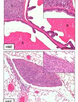
The MIND method involves intraductal injection of patient derived ductal carcinoma in situ (DCIS) cells and DCIS cell lines (MCF10DCIS.COM and SUM225) inside the mouse mammary ducts [Video 1 and Figure 1 in Behbod et al. (2009)]. This method mimics the normal environment of DCIS and facilitates study of the natural progression of human DCIS, i.e., their initial growth as carcinoma in situ within the ducts, followed by invasion into the stroma through the myoepithelial cell layer and basement membrane (Behbod et al., 2009; Valdez et al., 2011). In order to demonstrate that transplantation procedure is successful, the transplanted mammary glands may be excised as early as two weeks following intraductal injection of cells followed by Hematoxylin and Eosin (H&E) staining and/or immunofluorescence staining using human specific cytokeratin 5 and/or 19 [please see Figures 2-4 in Behbod et al. (2009)]. Additionally, the presence of trypan blue inside the mouse mammary ducts immediately following intraductal injection is the best indicator that the injection was successful (Video 1 starting at 4:33 sec).
Keywords: IntraductalMaterials and Reagents
- Hamilton syringe, 50 μl capacity, with a 30 gauge blunt-ended fixed 1/2-inch needle (Hamilton, catalog number: 80608 )
- 1 ml Tuberculin (TB) syringe (Benton Dickenson, catalog number: 309625 )
- Wound clips (Becton Dickinson, catalog number: 427631 )
- Animals: 8-12 week old female immunocompromised mice
Note: NSG, NOD scid gamma [NOD.Cg-Prkdcscid Il2rgtm1Wjl/SzJ (HE JACKSON LABORATORY, catalog number: 005557 )]. Alternatively, SCID/beige mice (Fox Chase SCID® Beige CB17.Cg-PrkdcscidLystbg-J/Crl) may be used for the injection of DCIS cell lines only. While a formal comparison has not been made, patient derived DCIS epithelial cells seem to grow more efficiently in the NSG mice. - Cells of interest [i.e., cell lines MCF10DCIS.COM and SUM225 (Behbod Lab, 2009 #329) or patient derived DCIS epithelial cells] in single cells
- PBS (pH 7.4, 1x), sterile (Life Technologies, catalog number: 10010023 )
Note: Currently, it is “Thermo Fisher Scientific, catalog number: 10010023”. - Trypan blue 0.4%, sterile (Sigma-Aldrich, catalog number: T8154 )
- 70% ethanol
- Sterile water
- Nembutal Sodium Solution (Pentobarbital sodium injection, USP) (Akorn, Oak Pharmaceuticals, catalog number: NDC 7647850120 )
- KetofenTM (Ketoprofen) (Patterson Veterinary Supply, catalog number: 10004029 )
Equipment
- Scissors Small scale suitable for mice
Sharp-Blunt (Fine Science Tool, catalog number: 14028-10 )
Castroviejo Spring Scissors (Fine Science Tool, catalog number: 15017-10 )
Balled Scissors (Fine Science Tool, catalog number: 14086-09 ) - Clippers (Fine Science Tool, catalog number: 1501710 )
- Rounded end flat handle tweezers (ROBOZ, model: RS5095 )
- Spring scissor (Fine Science Tool, catalog number: 1501710 )
- Balled scissor (Fine Science Tool, catalog number: 1408609 )
- Rounded end tweezers (Fine Science Tool, catalog number: 1105210 )
- Rat tooth tweezers (Fine Science Tool, catalog number: 1105310 )
- Clip applier (Becton Dickinson, catalog number: 427630 )
- Heat pad (Sunbeam Products, model: PN126985900 )
- Clip remover (Becton Dickinson, catalog number: 427638 )
Procedure
文章信息
版权信息
© 2016 The Authors; exclusive licensee Bio-protocol LLC.
如何引用
Kittrell, F., Valdez, K., Elsarraj, H., Hong, Y., Medina, D. and Behbod, F. (2016). Mouse Mammary Intraductal (MIND) Method for Transplantation of Patient Derived Primary DCIS Cells and Cell Lines. Bio-protocol 6(5): e1744. DOI: 10.21769/BioProtoc.1744.
分类
癌症生物学 > 侵袭和转移 > 动物模型 > 细胞侵袭
您对这篇实验方法有问题吗?
在此处发布您的问题,我们将邀请本文作者来回答。同时,我们会将您的问题发布到Bio-protocol Exchange,以便寻求社区成员的帮助。