

Craniotomy for Cortical Voltage-sensitive Dye Imaging in Mice
开颅手术后利用电压敏感性染料进行小鼠脑皮层成像

发布: 2016年02月05日第6卷第3期 DOI: 10.21769/BioProtoc.1722 浏览次数: 14698
评审: Soyun KimManuel SarmientoTifany Desprez
参见作者原研究论文
The authors used this protocol in:
Jun 2015
Advertisement


Abstract
Cortico-cortical interactions play crucial roles in various brain functions. Here, we present a detailed surgical procedure for cortical voltage-sensitive dye (VSD) imaging that allows monitoring of spatiotemporal dynamics in cortical activity in living mice. Cortical neurons in the upper layers (layer 1-3) are stained with a VSD, and an image sensor with a fast sampling rate (500 Hz) detects fluorescent changes in corrective activity. The procedure includes fixing a mouse brain to a stereotaxic apparatus, craniotomy on a large cortical area, VSD staining, and wide-field imaging of cortical activity. The entire procedure can be completed in 5 h (from the administration of anesthesia to the start of cortical VSD imaging).
Keywords: Voltage-sensitive dye imaging (电压敏感染料成像)Materials and Reagents
- Fine needle (Bonn Micro Probes) (Fine Science Tools, catalog number: 10030-13 )
- Cover glass (Matsunami Glass Ind, catalog number: C024241 )
- Wild-type mice (Japan SLC, model: C57BL / 6JJmsSlc )
- Isoflurane (e.g., Wako Pure Chemical Industries, catalog number: 099-06571 )
- Lidocaine solution (80 mg/ml) (e.g., Xylocaine Pump Spray 8%) (AstraZeneca) for local anesthesia
- Dental cements (Super-Bond C&B) (Sun medical) (GC's Global, UNIFAST II)
- Voltage-sensitive dye (OPTICAL IMAGING LTD, catalog number: RH1691 )
- NaCl
- KCl
- MgCl2.6H2O
- CaCl2.2H2O
- HEPES
- Distilled water
- RH1691
- Ringer’s solution (see Recipes)
- Voltage-sensitive dye (VSD) solution (see Recipes)
Equipment
- Anesthesia system for isoflurane (Shinano, catalog number: SN-487-0T )
- Electric clipper (e.g., Panasonic Corporation, catalog number: ER803P ) for cutting mouse hair
- Feedback-controlled heating pad (Bio Research Center, catalog number: BWT-100A )
Note: The pad monitors the mouse body temperature and maintains it at the desired temperature. - Head holder (stereotaxic apparatus) (NARISHIGE Group, model: SG-4N )
- Fine scissors (Fine Science Tools, catalog number: 91460-11 )
- Student Dumont #7 Forceps (Fine Science Tools, catalog number: 91197-00 )
- Dumont #5SF Forceps (Fine Science Tools, catalog number: 11252-00 )
- Cotton swab for absorbing the ringer’s solution
- Vacuum pump (e.g., AGC TECHNO GLASS CO., model: VPUMP-140 ) for removing the ringer's solution
- Surgical blade (e.g., Kai industries, catalog number: 310-A )
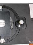
- Head-fixation plate (handmade, Figure 1A)
- Plate holder (custom-made, Figure 1B)
- Stereo Microscope (OLYMPUS, model: SZX7 )
- Green LED light (REVOX Inc., model: SLG-50S-G )
- Dental drill (SHOFU Inc., model: Tas-35LX )
- Dental round bur (SHOFU Inc., model: ELA Steel Bur HP-1 )
- Lens blower (e.g., HAKUBA, model: KMC-45 )
- Ultra-Fast CMOS Imaging System (Brainvision, model: MiCAM ULtima )
Note: For reference imaging (see procedure step 13), the cortex is illuminated with a blue LED light (center wavelength: 465 nm) through a 506-nm dichroic mirror; green fluorescence is corrected through a 536/40-nm filter.
For voltage-sensitive dye imaging, VSD fluorescence is excited with a red LED light (center wavelength: 625 nm). The excitation light is filtered with a 632/22-nm band-pass filter, reflected using a 655-nm dichroic mirror, and focused 375 μm below the cortical surface. Fluorescence is filtered with a 665-nm long pass filter. - Blue LED light (Brainvision, model: LEX2-B )
- Red LED light (REVOX Inc., model: SLG-50S-R )
- Stimulator (e.g., NIHON KOHDEN CORPORATION, model: SEN-5201 )
- Metal electrodes for hindpaw stimulation (handmade, Figure 1C)
- Vortex mixer (e.g., Scientific Industries Inc., model: VORTEX-GENIE 2 )
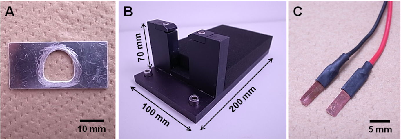
Figure 1. Head-fixation equipment. A. Handmade head-fixation plate; B. Custom-made plate holder; C. Handmade metal electrodes for hindpaw stimulation.
Software
- Acquisition software (Brainvision, model: UL-Acq)
- Analysis software (Brainvision, model: BV_Ana)
Procedure
文章信息
版权信息
© 2016 The Authors; exclusive licensee Bio-protocol LLC.
如何引用
Suzuki, T. and Murayama, M. (2016). Craniotomy for Cortical Voltage-sensitive Dye Imaging in Mice. Bio-protocol 6(3): e1722. DOI: 10.21769/BioProtoc.1722.
分类
神经科学 > 神经解剖学和神经环路 > 活细胞成像
您对这篇实验方法有问题吗?
在此处发布您的问题,我们将邀请本文作者来回答。同时,我们会将您的问题发布到Bio-protocol Exchange,以便寻求社区成员的帮助。