

Analysis of Murine Lung Tumors by Micro PET-CT Imaging
微型PET-CT成像分析鼠肺部肿瘤


(*contributed equally to this work) 发布: 2015年12月20日第5卷第24期 DOI: 10.21769/BioProtoc.1692 浏览次数: 15455
评审: HongLok LungAnonymous reviewer(s)
参见作者原研究论文
The authors used this protocol in:

Nov 2014
Advertisement


Abstract
Accurate live tumor imaging in mice is now possible by means of high-resolution positron emission tomography (micro-PET) and X-ray computed tomography (micro-CT). By providing a powerful tool to examine biological samples with complex structure in vivo, this technology generated a significant advance in the cancer research field, particularly regarding the ability to perform longitudinal studies in combination with a therapeutic intervention. Here, we describe methods to optimize visualization of murine lung tumors by micro-PET, micro-CT and combined micro-PET-CT.
Keywords: Cancer (癌症)Part I. Imaging by positron emission tomography (micro-PET)
The imaging of tumors at inner body locations in living animals is more challenging than the imaging of subcutaneous tumors. We have optimized the procedures outlined in the following protocol in order to study lung tumors in genetically modified mice and orthotopic models. Briefly, the mice are anesthetized prior to the administration of the [18F]-Fluorodeoxigluycose (18F-FDG) dose, and kept under anesthesia during the whole period of probe uptake and imaging, ensuring at all times that the mice are warm. The standardization of mouse handling and of anesthesia usage is essential to ensure data reproducibility and comparability.
Materials and Reagents
- 1-cc tuberculin syringes (B. Braun España, model: Omnifix-F )
- 30-G needles (B. Braun España, model: Sterican )
- Heating pads [e.g., Gaymar Mul-T-Pads (Gaymar industries) (http://www.gaymar.com/)]
- Heating pump to maintain temperature of heating pads [e.g., Gaymar TP600 (Gaymar industries) (http://www.gaymar.com/)]
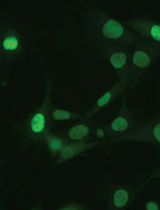
- Genetically modified mouse models bearing endogenous lung tumors (i.e., our own K-RasG12V inducible mouse model, Guerra et al., 2003), lung orthotopic implantation of primary tumors (Ambrogio et al., 2014), or tail-vein injected lung tumor cells (Ambrogio et al., 2014) (Figure 1)

Figure 1. Types of murine lung tumors detectable by PET-CT scan. PET-CT technology can be used to detect lung tumors of different origins: from the left, endogenous lung tumors (i.e., tumors induced by a K-RasG12V resident knock-in allele in a genetically engineered mouse model), tail vein-injected lung cancer cell lines or orthotopically implanted lung tumors (either murine or human lung adenocarcinomas). Scale bar: 200 μM. - Special mouse diets as necessary (Cussó et al., 2014)
- Inhalational anesthesia:
Sevoflurane [e.g., Sevoflo (Abbott Laboratories, catalog number: 05458-02 ) (http://www.abbottanimalhealth.com/veterinary-professionals/products/anesthesia/sevoflo.html)] - Oxygen obtained from an O2 concentrator
- [18F]FDG (0.01 to 0.1 μg/mCi), delivered daily from a local cyclotron (e.g., 40 mCi of [18F]FDG of 95% to 99% radiochemical purity in 1 ml of physiological saline solution buffered at pH 6.0, for ~10 micro-PET scans)
- Physiological saline: 0.9% (w/v) NaCl (B. Braun España)
- Lacryvisc Gel 10 G (3 mg/ml carbomere in benzalconium chloride) (Alcon) (http://www.alcon.com)
Equipment
- O2 concentrator (Eickemeyer Veterinary Equipment, model: Oxymat e3 )
- Infrared heating lamp Philips PAR38 IR 175W E27 (Royal Philips Electronics)
- Sevoflurane/oxygen-based anesthesia system fitted with an induction chamber and inhalation masks for mice McKinley, Type 2 (Everest tecnologia veterinaria)
- Dose calibrator (also known as activimeter) [e.g., VDC-505 dose calibrator (Veenstra Instruments) (http://www.dosecalibrator.com/)]
- micro-PET-CT imaging system [e.g., eXplore Vista PET-CT (GE Healthcare) (Figure 2); Argus PET-CT (SEDECAL) (http://www.sedecal.com/)]
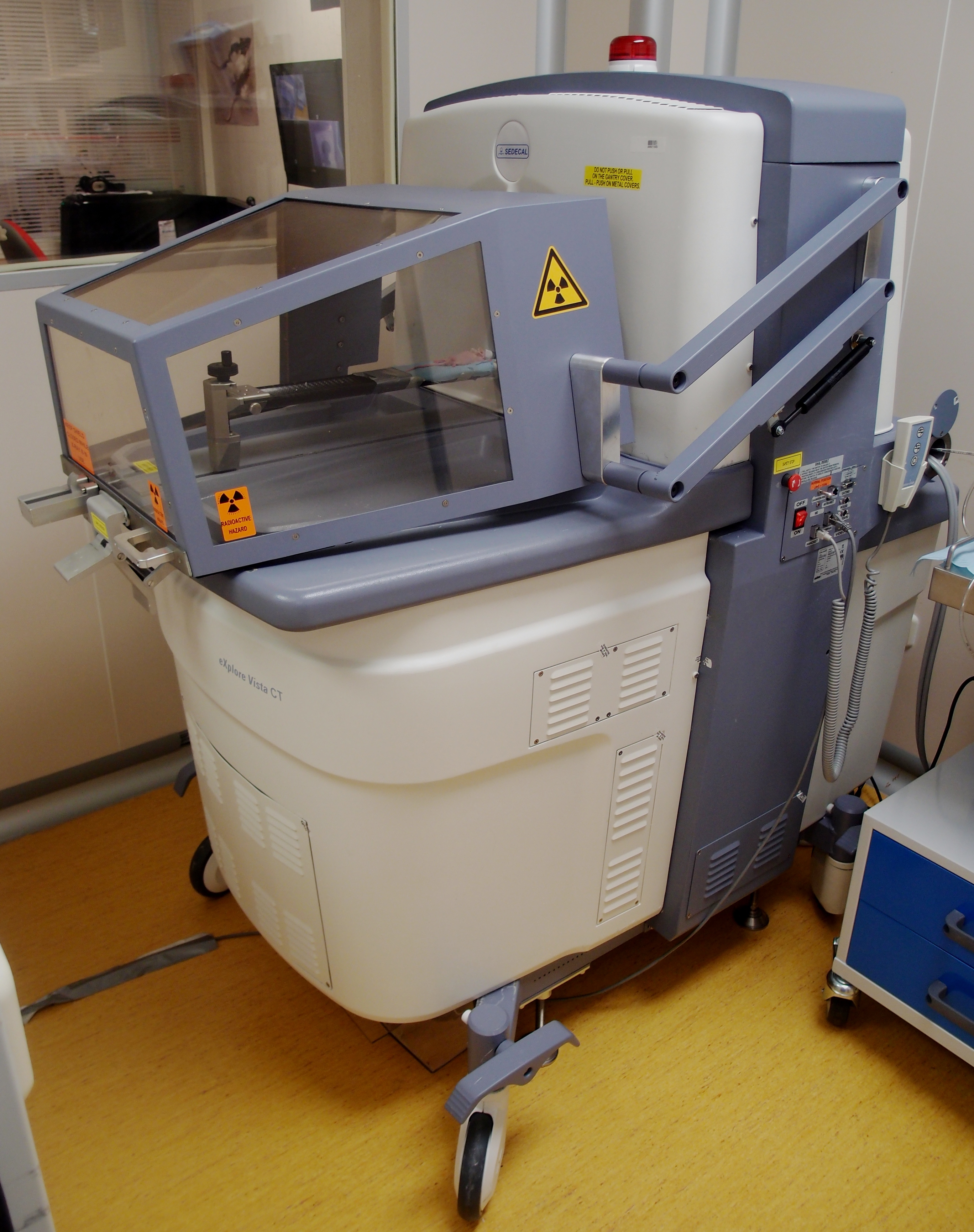
Figure 2. Micro-PET-CT machine (Argus SEDECAL) for mice - Workstation (e.g., Dell PowerEdge) for image acquisition, processing, and analysis meeting the following specifications:
- PE1950 Xeon 5120 1.86 GHz/4 MB 1066 FSB processor
- PE1950 PCIX Riser (2 slots)
- PE1950 Bezel Assembly
- 2 GB FB 667 MHz Memory (2 x 1 GB dual rank DIMMs)
- PE1950 Xeon 5120 1.86 GHz/4 MB 1066 FSB processor
- Alienware Dell Studio XPS Desktop 435 MT PC (for 3DOSEM image reconstruction) meeting the following specifications:
- Processor: Intel Core i7 Quad CPU 940 4 x 2.93 GHz
- Memory: 6144 MB (6 x 1,024) 1067 MHZ DDR3
- Graphics: ATI Radeon HD 3450 256 Mb GDDR2
- Processor: Intel Core i7 Quad CPU 940 4 x 2.93 GHz
Software
- eXplore Vista PET-CT MMWKS software (Desco et al., 2005) or AMIDE software (Loening and Gambhir, 2003) for image acquisition, processing, and analysis
Procedure
文章信息
版权信息
© 2015 The Authors; exclusive licensee Bio-protocol LLC.
如何引用
Ambrogio, C., Cámara, J. A., Nieto, P., Santamaría, D. and Mulero, F. (2015). Analysis of Murine Lung Tumors by Micro PET-CT Imaging. Bio-protocol 5(24): e1692. DOI: 10.21769/BioProtoc.1692.
分类
癌症生物学 > 通用技术 > 肿瘤形成
细胞生物学 > 细胞成像 > 活细胞成像
您对这篇实验方法有问题吗?
在此处发布您的问题,我们将邀请本文作者来回答。同时,我们会将您的问题发布到Bio-protocol Exchange,以便寻求社区成员的帮助。