

Insertional Mutagenesis of Chlamydomonas reinhardtii
莱茵衣藻的插入突变
发布: 2015年12月20日第5卷第24期 DOI: 10.21769/BioProtoc.1680 浏览次数: 9015
评审: Maria SinetovaIgor Cesarino Anonymous reviewer(s)
参见作者原研究论文
The authors used this protocol in:
Nov 2014
Advertisement


Abstract
The unicellular microalga Chlamydomonas reinhardtii (C. reinhardtii) has been used as a reference model for numerous fields of research. Principle research areas are eukaryotic flagellar structure and function, basal bodies (centrioles), cell-cell recognition, cell cycle control, chloroplast biogenesis, phototaxis, nonphotochemical quenching, and especially photosynthesis for C. reinhardtii can grow in the dark on an organic carbon (e.g. acetate), and thus provides advantages over land plants (Harris, 2001; Peers et al., 2009). C. reinhardtii has a short life cycle, a sequenced genome (Merchant et al., 2007), and a growing molecular toolbox for forward and reverse genetic studies, including transformation protocols, gene silencing (Kim and Cerutti, 2009; Molnar et al., 2009), and fluorescent protein-tag (Rasala et al., 2013). There are two commonly used methods for C. reinhardtii transformation – electroporation and glass bead agitation. Electroporation is normally restricted to strains with cell wall, as it kills cell-wall-deficient strains effectively if without careful handling of osmosis. Electroporation also requires special instruments such as electroporator and cuvettes. In contrast, glass bead agitation uses simple lab equipment. The mild shear created by agitation in the presence of glass bead allows cell-wall-deficient strains to take up DNA. If glass bead method is to be applied to cell-wall strains, cells need to be treated with autolysin (http://www.chlamy.org/methods/autolysin.html) to partially lyse the wall components. A pitfall of both methods is that the DNAs are often shortened by nuclease once entering the cells, making the downstream PCR-based genotyping of insertion site rather difficult. Here I describe an improved design of insertional mutagenesis used in (Tsai et al., 2014), and the transformation protocol using glass bead as previously described in (Kindle, 1990) with minor modification. The putative mutants can be selected by autotrophic or antibiotic resistance markers, and the disrupted loci can be mapped by methods such as plasmid rescue (Peers et al., 2009) and SiteFinding PCR (Tan et al., 2005).
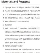
Materials and Reagents
- 15 and 50 ml conical tubes
- Petri dishes (90 mm in diameter)
- pHYG3 plasmid (http://chlamycollection.org/plasmid/phyg3/) or plasmid carrying other selection markers
- C. reinhardtii cell wall-less strain such as dw15 (cw15, nit1, mt+; http://chlamycollection.org/strain/cc-4619-cw15-nit1-mt-dw15-1/)
- Agar (Caisson Laboratories, Phytoblend)
- TAP medium (Harris, 1989)
- Restriction enzyme for linearizing the plasmid DNA, such as PvuII (New England Biolabs)
- Hygromycin (Life Technologies) or other selection means
- TAP agar plates (see Recipes)
- Top agar (see Recipes)
Equipment
- Common bench-top vortexer
- Spectrophotometer for optical density (OD) measurement, Z2 Coulter Counter (Beckman Coulter) or a hemocytometer
- 250 ml flask
- Bench-top centrifuge capable of centrifugation at 1,500 x g and accommodating 50 ml conical tubes
- Shaker
- Z2 Coulter Counter or hemocytometer
- Glass beads 425-600 μm (Sigma-Aldrich, catalog number: G8772 )
- Glass tubes or round bottom plastic tubes with or without cap (~10 mm in diameter)
Procedure
文章信息
版权信息
© 2015 The Authors; exclusive licensee Bio-protocol LLC.
如何引用
Tsai, C. and Benning, C. (2015). Insertional Mutagenesis of Chlamydomonas reinhardtii. Bio-protocol 5(24): e1680. DOI: 10.21769/BioProtoc.1680.
分类
植物科学 > 藻类学 > DNA > 诱/突变
分子生物学 > DNA > 诱/突变
您对这篇实验方法有问题吗?
在此处发布您的问题,我们将邀请本文作者来回答。同时,我们会将您的问题发布到Bio-protocol Exchange,以便寻求社区成员的帮助。