

Sample Preparation for Correlative Light and Electron Microscopy (CLEM) Analyses in Cellular Microbiology
用于细胞微生物学中关联光学和电子显微镜技术(CLEM)分析的样本制备
发布: 2015年10月05日第5卷第19期 DOI: 10.21769/BioProtoc.1612 浏览次数: 17809
评审: Fanglian HeAnonymous reviewer(s)
参见作者原研究论文
The authors used this protocol in:
Sep 2014


Abstract
Dynamic processes in cells are usually monitored by live cell fluorescence microscopy. Unfortunately, this method lacks the ultrastructural information about the structure of interest (SOI). Currently, electron microscopy (EM) is the best tool to achieve highest spatial resolution. In addition, correlative light and electron microscopy (CLEM) analysis of the same structure allows combining authentic live cell imaging with the resolution power of EM. Additionally the reference space of the SOI is revealed. Our CLEM analyses of HeLa cells allow tracing the morphology and dynamic behavior of intracellular micro-compartments in living cells and their ultrastructure and subcellular organization in a highly resolved manner.
Keywords: Sample preparation (样品的制备)Materials and Reagents
- General lab equipment: Gloves, lab coat, pipettes, 15 and 50 ml centrifuge tubes (e.g., BD Biosciences, Falcon®), microcentrifuge tubes (e.g., Eppendorf), plastic Pasteur pipettes, beakers
- 10 cm plastic Petri dish or a comparable vessel
- 3.5 cm plastic Petri dish or a comparable vessel (Acetone-resistant)
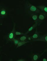
- Eukaryotic cells as biological sample (here: transfected HeLa cells expressing LAMP1-GFP)
- Cell-specific culture medium: DMEM (Biochrom AG, catalog number: FG 0445 ) + 10% Fetal Bovine Serum (FCS) (Thermo Fisher Scientific, GibcoTM, catalog number: 10270 )
- Cell-specific imaging medium: MEM (Biochrom AG, catalog number: F 0475 ) + 30 mM HEPES
- HEPES = 4-(2-hydroxyethyl)-1-piperazineethanesulfonic acid (Carl Roth GmbH, catalog number: HN77.5 )
- Glutaraldehyde [CH2(CH2CHO)2], 25% in H2O (Electron Microscopy Sciences, catalog number: E16221 )
- Glycine, ultra pure (Biomol GmbH, catalog number: 04943.1 )
- Large gelatin capsules, size 13 (Electron Microscopy Sciences, catalog number: 70114 )
- Calcium chloride (CaCl2) (Sigma-Aldrich, catalog number: C 5670 )
- Osmium tetroxide (OsO4), 0.1 g ampules (Electron Microscopy Sciences, catalog number: 19134 )
- Ruthenium red [[(NH3)5RuORu(NH3)4ORu(NH3)5]Cl6] (AppliChem GmbH , catalog number: A34880001 )
- Potassium hexacyanoferrate(III) [K3[Fe(CN)6]] (Sigma-Aldrich, catalog number: 244023 )
- Ethanol p.a. grade (very pure chemical)
- Acetone p.a. grade (very pure chemical)
- EPON 812 (SERVA Electrophoresis GmbH, catalog number: 21045.02 )
- DDSA = Dodecenylsuccinic anhydride (SERVA Electrophoresis GmbH, catalog number: 20755 )
- MNA = Methylnadic anhydride (SERVA Electrophoresis GmbH, catalog number: 29452.03 )
- DMP-30 = 2, 4, 6-Tris(dimethylamino-methyl)phenol (SERVA Electrophoresis GmbH, catalog number: 36975 )
- Uranyl acetate dihydrate [UO2(CH3COO)2.2H2O] (Electron Microscopy Sciences, catalog number: 22400 )
- Lead(II) nitrate [Pb(NO3)2] (Sigma-Aldrich, catalog number: 31137 )
- Trisodium citrate dihydrate (Na3C6H5O7.2H2O) (Carl Roth, catalog number: 3580.1 )
- 1 N Sodium hydroxide (NaOH), carbonate-free (Electron Microscopy Sciences, catalog number: 21170-01 )
- 65% Nitric acid (HNO3)
- Autoclaved ultrapure H2O (MilliQ)
- Liquid nitrogen
- Graded ethanol series (50%, 70%, 80%, 90%, 95%, 100%)
- Mixes of acetone and EPON (3:1 and 1:3 mix)
- 1 M HEPES buffer (see Recipes)
- 2 M CaCl2 in H2O (see Recipes)
- 10% glycine in 0.2 M HEPES buffer (see Recipes)
- 1% ruthenium red in H2O (see Recipes)
- 15% potassium hexacyanoferrate(III) in H2O (see Recipes)
- 2x fixative (see Recipes)
- Post-fixative (see Recipes)
- EPON resin (see Recipes)
- 2% uranyl acetate in H2O (see Recipes)
- Lead citrate according to Reynolds (see Recipes)
- 3% nitric acid (HNO3) (see Recipes)
Equipment
- Cell culture equipment: Cell culture incubator (37 °C, 5% CO2, 90% humidity) and clean bench
- MatTek Glass Bottom Culture Dishes, 35 mm, uncoated, Glass No. 2, gridded (Glass Bottom Dishes, MatTek Corporation, catalog number: P35G-2-14-CGRD )
Attention: Glass No. 2 may be too thick for some objectives with high numerical aperture (NA), but this is the only format available and worked in our application. - Confocal laser-scanning microscope (CLSM) Leica SP5 equipped with an incubation chamber (homemade) maintaining 37 °C and humidity during live cell imaging (Attention: CO2-containing atmosphere can be omitted if using imaging medium buffered with 30 mM HEPES. For carbonate-buffered cell culture media, the atmosphere should contain 5% CO2). The microscope is operated with software package LAS-AF for setting adjustment, image acquisition and image processing
- Several objectives, such as 10x (HC PL FL 10x, NA 0.3, DIC, dry), 20x (HC PL APO CS 20x, NA 0.7, DIC, dry), 40x (HCX PL APO CS 40x, NA 1.25-0.75, DIC, oil immersion) and 100x objective (HCX PL APO CS 100x, NA 1.4-0.7, DIC, oil immersion)
- The polychroic mirror TD 488/543/633 for the three channels GFP/RFP/DIC (Leica Microsystems)
- Several objectives, such as 10x (HC PL FL 10x, NA 0.3, DIC, dry), 20x (HC PL APO CS 20x, NA 0.7, DIC, dry), 40x (HCX PL APO CS 40x, NA 1.25-0.75, DIC, oil immersion) and 100x objective (HCX PL APO CS 100x, NA 1.4-0.7, DIC, oil immersion)
- Fume hood
- Ice or cold metal block
- Scalpel
- Oven, heatable to 60 °C
- Dewar vessel for liquid nitrogen
- Stereo microscope
- Dark permanent marker
- Vice
- Jigsaw
- Universal specimen holder for EPON blocks (Leica Microsystems, catalog number: 16701761 )
- Double edge carbon steel blades (Plano, catalog number: 121-9 )
- Ultramicrotome (Leica EM UC6)
- Diamant knife ultra 45 °C, 2 mm (DIATOME, catalog number: DU4520 )
- Formvar-coated EM cooper slit grids 2 x 1 mm (homemade coating) (Electron Microscopy Sciences, catalog number: G2010-Cu )
- Forceps for EM grids, type 7 stainless steel (Plano GmbH, catalog number: T5039 )
- Storage box for grids (Electron Microscopy Sciences, catalog number: G71138 )
- Large forceps for liquid nitrogen and small forceps for embedding procedure
- Automated staining device (nanofilm surface analysis ultrastainer)
- TEM (ZEISS, model: EFTEM 902 A ), operated at 80 kV and equipped with a 2K wide-angle slow-scan CCD camera (Teacher Retirement System of Texas) with software ImageSP (Teacher Retirement System of Texas, model: image SysProg)
Software
- ImageJ, http://rsbweb.nih.gov/ij/, Photoshop 5.5 (Adobe), or higher or similar software for stitching and overlay of images
- Imaris (Bitplane), ImageJ, ZEN (ZEISS), LASAF (Leica Microsystems), or similar software for processing fluorescence images
Procedure
文章信息
版权信息
© 2015 The Authors; exclusive licensee Bio-protocol LLC.
如何引用
Readers should cite both the Bio-protocol article and the original research article where this protocol was used:
- Liss, V. and Hensel, M. (2015). Sample Preparation for Correlative Light and Electron Microscopy (CLEM) Analyses in Cellular Microbiology. Bio-protocol 5(19): e1612. DOI: 10.21769/BioProtoc.1612.
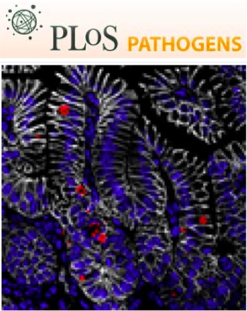
- Krieger, V., Liebl, D., Zhang, Y., Rajashekar, R., Chlanda, P., Giesker, K., Chikkaballi, D. and Hensel, M. (2014). Reorganization of the endosomal system in Salmonella-infected cells: the ultrastructure of Salmonella-induced tubular compartments. PLoS Pathog 10(9): e1004374.
分类
细胞生物学 > 细胞成像 > 电子显微镜
细胞生物学 > 细胞成像 > 活细胞成像
您对这篇实验方法有问题吗?
在此处发布您的问题,我们将邀请本文作者来回答。同时,我们会将您的问题发布到Bio-protocol Exchange,以便寻求社区成员的帮助。