

Pectin Nanostructure Visualization by Atomic Force Microscopy
采用原子力显微镜对果胶纳米结构进行可视化
发布: 2015年09月20日第5卷第19期 DOI: 10.21769/BioProtoc.1598 浏览次数: 9894
评审: Samik BhattacharyaMasahiro MoritaAnonymous reviewer(s)
参见作者原研究论文
The authors used this protocol in:
Oct 2014
Advertisement


Abstract
Pectins, complex polysaccharides rich in galacturonic acid, are a major component of primary plant cell walls. These macromolecules regulate cell wall porosity and intercellular adhesion, being important in the control of cell expansion and differentiation through their effect on the rheological properties of the cell wall. In fruits, pectin disassembly during ripening is one the main event leading to textural changes and softening. Changes in pectic polymer size, composition and structure have been studied by conventional techniques, most of them relying on bulk analysis of a population of polysaccharides but studies of detailed structure of isolated polymer chains are scarce (Paniagua et al., 2014). Atomic force microscopy (AFM) is a versatile and powerful technique able to analyze force measurements, as well as to visualize roughness of biological samples at nanoscale (Morris et al., 2010). Using this technique, recent research has found a close relationship between pectin nanostructural complexity and texture and postharvest behavior in several fruits (Liu and Cheng, 2011; Cybulska et al., 2014; Posé et al., 2015). Here, we describe an AFM procedure to topographically visualize pectic polymers from fruit cell wall extracts that has successfully been used in the study of strawberry ripening (Posé et al., 2012; Posé et al., 2015). Thus, from AFM images the 3D structural analysis of isolated chains (length, height, and branch pattern) can be resolved at high magnification and with minimal sample preparation. A full description of AFM fundamentals and the different sampling modes are described in Morris et al. (2010).
Keywords: Pectin (果胶)Materials and Reagents
- Tri-distilled butanol (VWR International, catalog number: 20810.323 )
- Pectin fractions from cell wall extracts
Notes:- Cell wall extraction protocol is described in Posé et al. (2013).
- Pectin fractions from cell wall material are obtained by sequential extractions with CDTA buffer followed by sodium carbonate buffer, to solubilize a cell wall fraction enriched in ionically and covalently bound pectins respectively, as described in Posé et al. (2013) (see Recipes).
- Both pectin fractions (i.e., one extracted with CDTA and the other with sodium carbonate) were extensively dialyzed and stored until required at -20 ºC as aqueous solutions. Important: in order to prevent possible aggregation, any freeze-drying step must be avoided. It is recommended to aliquot the samples to avoid freeze-thawed successive cycles.
- Cell wall extraction protocol is described in Posé et al. (2013).
- Ammonium bicarbonate (FLUKA, catalog number: 09830 )
Note: Currently, it is “Sigma-Aldrich, catalog number: 09830”. - Trans-1,2-diaminocyclohexane-N,N,N'N'-tetra-acetic acid monohydrate (Sigma-Aldrich, catalog number: D 319945 )
- Sodium carbonate (Sigma-Aldrich, catalog number: S2127 )
- NaBH4 (Sigma-Aldrich, catalog number: D 452882 )
- 10 mM ammonium bicarbonate buffer (pH 8) (see Recipes)
- CDTA buffer (see Recipes)
- Sodium carbonate buffer (see Recipes)
Equipment
- Acoustic-isolated and temperature-controlled room (Figure 1A-1)
- Low-power light microscope, equipped with a television camera, was used to roughly position the AFM probe onto the top of the sample (Figure 1A-2)
- Atomic Force Microscope (East Coast Scientific Limited, Cambridge, UK) (Figure 1A-3; Figure 2A). Any AFM of suitable resolution can be used, although the details of the software and liquid cell will vary
- Anti-vibration table under the AFM microscope (Figure 1A-4)
- Photodiode amplifier (Figure 1A-5)
- Oscilloscope (Figure 1A-6; Figure 3)
- Digital control system composed by computer, DAC (digital-to-analog converter) box, laser driver and high voltage amplifier. (for more details see Morris et al., 2010) (Figure 1A-7)
- PYREX® Culture Tubes with Rubber Liner Screw Caps (Thomas Scientific, catalog number: 9212C21 ) and Teflon-lined caps
Note: The screw-capped glass tubes were acid washed using a 1% hydrochloric acid solution overnight then rinsed with water. - Sheets of mica (Elektron Technology, Agar Scientific, model: G250 ) cleaved with adhesive tape (3M, model: Magic Tape ) (Video 1)
- Short tip variety AFM probe model contact cantilevers (Budget Sensors SiNi, Bulgaria)
Note: The tip is mounted on the edge of a V-shaped cantilever, the typical geometry used for topographical imaging (Figure 1B). - Tip holder and open bucket liquid cell (Figure 1C; Vdeo 2)
- Basic equipment: pipettes, vortex, sonicator bath, heating block
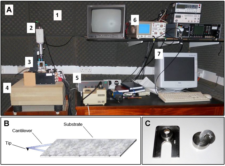
Figure 1. Atomic force microscopy (AFM) equipment. A. Photograph including an overview of AFM room set-up. The numeric labels are in accordance with the equipment list description. B. Scheme of a sharp tip located at the free end of a cantilever. C. Detail of tip holder (left) and liquid cell (right).
Software
- AFM software supplied with the instrument (SPM 6.01, ECS, Cambridge, UK)
- For the length measurements, images were converted to TIFF files using Paint Shop Pro v5.00 software (http://web.archive.org/web/19980514080113/http://jasc.com/)
- Image contrast and 3D effects were optimized using Gwyddion software v2.32
- AFM images were analyzed off-line using Image J v1.43u software (http://imagej.nih.gov/ij/index.html)
- Gwyddion is free and open source software, covered by GNU (General Public License) (http://gwyddion.net/)
Procedure
文章信息
版权信息
© 2015 The Authors; exclusive licensee Bio-protocol LLC.
如何引用
Posé, S., Paniagua, C., Kirby, A. R., Gunning, A. P., Morris, V. J., Quesada, M. A. and Mercado, J. A. (2015). Pectin Nanostructure Visualization by Atomic Force Microscopy. Bio-protocol 5(19): e1598. DOI: 10.21769/BioProtoc.1598.
分类
植物科学 > 植物生物化学 > 糖类
植物科学 > 植物细胞生物学 > 细胞成像
细胞生物学 > 细胞成像 > 固定细胞成像
您对这篇实验方法有问题吗?
在此处发布您的问题,我们将邀请本文作者来回答。同时,我们会将您的问题发布到Bio-protocol Exchange,以便寻求社区成员的帮助。