

Morphological Quantification of Nuclei and Mitochondria in Serial Block-face Scanning Electron Microscopy Images
串行块面扫描电子显微成像检测细胞核和线粒体的形态特征
发布: 2015年09月20日第5卷第18期 DOI: 10.21769/BioProtoc.1593 浏览次数: 9564
评审: Anonymous reviewer(s)
参见作者原研究论文
The authors used this protocol in:
Jul 2014
Advertisement


Abstract
Serial Block-face Scanning Electron Microscopy (SBF-SEM or 3D-EM) is a powerful tool to study biological structure in ultrastructural level. Quantification of cellular ultrastructure is useful to providing biological information. This technique requires not only high quality of tissue fixation and ideal sample embedding to preserve structures, but also delicate 3D image scanning and post-processing of images. We have adapted previous method to optimize the EM technique to detect and study cellular ultrastructure. Here we present the method to embed samples for 3D-EM technique and to quantify the morphological parameters of nucleus and mitochondria.
Part I. Tissue embedding for 3D-EM images
Materials and Reagents
- Adult mouse brain / spinal cord
- 4% Paraformaldehyde (PFA) (Sigma-Aldrich, catalog number: P6148 )
- Glutaraldehyde solution (GA) (Sigma-Aldrich, catalog number: G5882 )
- Sodium Cacodylate Buffer (0.4 M, pH 7.2) (Electronic Microscopy Science, catalog number: 11654 )
- Phosphate buffered saline (PBS) (1x Solution, Fisher BioReagents) (Fisher Scientific, catalog number: BP2438-4 )
- Potassium ferricyanide (III) (Sigma-Aldrich, catalog number: 702587 )
- Thiocarbohydrazide (TCH) (Electronic Microscopy Science, catalog number: 21900 )
- Osmium tetroxide 4% solution (Electronic Microscopy Science, catalog number: 19150 )
- Uranyl acetate (UA) (Electron Microscopy Sciences, catalog number: 22400 )
- Lead Nitrate (Crystalline/Certified ACS), Fisher Chemical (Fisher Scientific, catalog number: L62-100 )
- L-aspartic acid solution (Sigma-Aldrich, catalog number: A9256 )
- Ethanol, 200 proof (100%), USP, DeconTM Labs (Fisher Scientific, catalog number: 07-678-004 )
- Acetone (Electronic Microscopy Science, catalog number: 10010)
- EMbed-812 kit (Electronic Microscopy Sciences, catalog number: 14120 )
- EMS Molded Flat Embedding Mold (Electronic Microscopy Sciences, catalog number: 70905-01 )
Equipment
- Vibratome (Leica, catalog number: Leica VT1000S )
- Zeiss Sigma VP SEM (Zeiss) with Gatan 3View (Gatan)
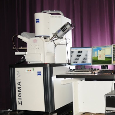
Figure 1. Zeiss Sigma VP SEM (Zeiss) with Gatan 3View (Gatan)
Procedure
文章信息
版权信息
© 2015 The Authors; exclusive licensee Bio-protocol LLC.
如何引用
Lu, H., Ohno, N. and Ransohoff, R. M. (2015). Morphological Quantification of Nuclei and Mitochondria in Serial Block-face Scanning Electron Microscopy Images. Bio-protocol 5(18): e1593. DOI: 10.21769/BioProtoc.1593.
分类
细胞生物学 > 细胞成像 > 固定组织成像
您对这篇实验方法有问题吗?
在此处发布您的问题,我们将邀请本文作者来回答。同时,我们会将您的问题发布到Bio-protocol Exchange,以便寻求社区成员的帮助。