

Visualization of ex vivo Neutrophil Extracellular Traps by Fluorescence Microscopy
荧光显微镜法显示体外中性粒细胞胞外杀菌网

发布: 2015年08月05日第5卷第15期 DOI: 10.21769/BioProtoc.1550 浏览次数: 12243
评审: Ivan ZanoniAchille BroggiMarco Di Gioia
参见作者原研究论文
The authors used this protocol in:

Nov 2014
Advertisement


Abstract
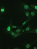
Neutrophil extracellular traps (NETs) are extracellular DNAs decorated with nuclear and granular proteins such as histones, neutrophil elastase or myeloperoxidase. They exhibit fibrous mesh-like, web-like, or string-like structures. Here, we describe our protocol regarding visualization of ex vivo NETs released from neutrophils activated by lipopolysaccharide (LPS) using fluorescence microscopy.
Materials and Reagents
- Whole blood from wild-type C57/BL6 mice (Japan SLC, Inc.)
- Whole blood from human volunteers
- LPS (Escherichia coli, serotype 0111:B4) (Sigma-Aldrich, catalog number: L4391 )
- PolymorphprepTM (Axis Shield PoC AS, catalog number: 1114683 )
- RPMI 1640 medium (no phenol red) (Life Technologies, catalog number: 32404-014 )
- Fetal bovine serum (Life Technologies, catalog number: 12483-020 )
- ACK (Ammonium-chloride-potassium) lysing buffer (Lonza, catalog number: 10-548E )
- SYTOX Green (Life Technologies, InvitrogenTM, catalog number: S7020 )
Equipment
- Glass Pasteur pipets (Iwaki brand, Asahi Techno Glass Corporation)
- 96-well plates (TPP Techno Plastic Products AG)
- CO2 Incubator (SANYO)
- Fluorescence microscopy (Olympus, model: IX71 )
Procedure
文章信息
版权信息
© 2015 The Authors; exclusive licensee Bio-protocol LLC.
如何引用
Tanaka, K., Shimura, T., Toiyama, Y., Okugawa, Y., Inoue, Y., Araki, T., Mohri, Y., Mizoguchi, A. and Kusunoki, M. (2015). Visualization of ex vivo Neutrophil Extracellular Traps by Fluorescence Microscopy. Bio-protocol 5(15): e1550. DOI: 10.21769/BioProtoc.1550.
分类
免疫学 > 免疫细胞功能 > 嗜中性粒细胞
细胞生物学 > 细胞成像 > 荧光
细胞生物学 > 细胞成像 > 活细胞成像
您对这篇实验方法有问题吗?
在此处发布您的问题,我们将邀请本文作者来回答。同时,我们会将您的问题发布到Bio-protocol Exchange,以便寻求社区成员的帮助。