

Electroporation of Embryonic Chick Eyes
鸡胚胎视网膜DNA电转移


发布: 2015年06月20日第5卷第12期 DOI: 10.21769/BioProtoc.1498 浏览次数: 14433
评审: Arsalan DaudiAnonymous reviewer(s)
参见作者原研究论文
The authors used this protocol in:

May 2014


Abstract
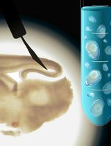
The chick embryo has prevailed as one of the major models to study developmental biology, cell biology and regeneration. From all the anatomical features of the chick embryo, the eye is one of the most studied. In the chick embryo, the eye develops between 26 and 33 h after incubation (Stages 8-9, Hamburger and Hamilton, 1951). It originates from the posterior region of the forebrain, called the diencephalon. However, the vertebrate eye includes tissues from different origins including surface ectoderm (lens and cornea), anterior neural plate (retina, iris, ciliary body and retinal pigmented epithelium) and neural crest/head mesoderm (stroma of the iris and of the ciliary body as well as choroid, sclera and part of the cornea). After gastrulation, a single eye field originates from the anterior neural plate and is characterized by the expression of eye field transcriptional factors (EFTFs) that orchestrate the program for eye development. Later in development, the eye field separates in two and the optic vesicles form. After several inductive interactions with the lens placode, the optic cup forms. At Stages 14-15, the outer layer of the optic cup becomes the retinal pigmented epithelium (RPE) while the inner layer forms the neuroepithelium that eventually differentiates into the retina. One main advantage of the chick embryo, is the possibility to perform experiments to over-express or to down-regulate gene expression in a place and time specific manner to explore gene function and regulation. The aim of this protocol is to describe the electroporation techniques at Stages 8-12 (anterior neural fold and optic vesicle stages) and Stages 19-26 (eye cup, RPE and neuroepithelium). We provide a full description of the equipment, materials and electrode set up as well as a detailed description of the highly reproducible protocol including some representative results. This protocol has been adapted from our previous publications Luz-Madrigal et al. (2014) and Zhu et al. (2014).
Keywords: Electroporation (电穿孔)Materials and Reagents
- Fertilized specific pathogen free (SPF) (Charles River Laboratories) or white Leghorn chicken eggs (Note 1)
- 10x Hank's balanced salt solution (HBSS) (Thermo Fisher Scientific, catalog number: 14185-052 )
- Fast green FCF (Sigma-Aldrich, catalog number: F7258 )
- Indian Ink Type A (Pelikan)
- Tris (hydroxymethyl) aminomethane (suitable for cell culture) (Sigma-Aldrich, catalog number: 252859 )
- EDTA (Ethylenediaminetetraacetic acid), suitable for cell culture (Sigma-Aldrich, catalog number: E6758 )
- pCAG- GFP (Addgene plasmid, catalog number: 11150 ) or pEGFP-N1 (Takara Bio Company, Clontech)
- Plasmid and RCAS-DNA [the name RCAS stands for Replication-Competent ASLV long terminal repeat (LTR) with a Splice acceptor] (Note 6)
- Morpholinos (Note 7)
- NaCl (Fisher Scientific, catalog number: BP358-1 ) (MW 58.44 g/mol)
- CaCl2 anhydrous (Acros Organics, catalog number: AC34961-5000 ) (MW 110.98 g/mol)
- KCl (Fisher Scientific, catalog number: P217-500 ) (MW 74.55 g/mol)
- Na2HPO4 Dibasic anhydrous (Fisher Scientific, catalog number: S374-500 ) (MW 141.96 g/mol)
- KH2PO4 Dibasic Anhydrous (Fisher Scientific, catalog number: P290-500 ) (MW 174.18 g/mol)
- Ringer’s solution (see Recipes)
- 10x Hank's balanced salt solution (HBSS) (see Recipes)
- Fast green FCF (see Recipes)
- 1 M Tris (see Recipes)
- 0.5 M EDTA (see Recipes)
- TE buffer for plasmid solutions (see Recipes)
Equipment
- Beveled-edge watch glass (to hold the egg during the electroporation) (Thermo Fisher Scientific, catalog number: 15-355 )
- 200 μl tips sterile (Corning Incorporated)
- Capillary tubing borosilicate for microinjection (1.0 mm OD, 0.5 mm ID/fiber) [Frederick Haer & Co (FHC), catalog number: 30-30-1 ]
- 1 ml syringe (Thermo Fisher Scientific)
- Pre-pulled beveled glass needles 50 mm long, 20 µm tip diameter and sharpened 10 to 12° (these glass needles are made following the instructions of the Micropipette Puller and Micropipette Beveler- Figure 1d-e)
- 35 mm plastic tissue culture plates (Corning Incorporated)
- 3/4 inch wide clear tape (Scotch, 3 M)
- Micro dissecting tweezers #55 and #5 (Roboz, catalog numbers: RS-4984 and RS-4978 )
- ECM 830 High Throughput Electroporation System (Figure 1a), a Square Wave Pulse generator for in vitro and in vivo electroporation with remote operation Footswitch (BTX, Harvard Apparatus, SKU: ECM_830 _for_In_Vivo_Applications)
- Microinjector (Figure 1b), MicroJect 1000A (BTX, Harvard Apparatus, SKU: 45- 0750 ) with foot Switches to inject and fill and a stainless steel pipette holder (Figure 1c)
- Micropipette beveler (Sutter Instrument Company, model: BV-10 ) (Figure 1d)
- Vertical micropipette puller (Sutter Instrument Company, model: P-30 ) (Figure 1e)
- Nitrogen tank Compressed 2.2 UN1066 NI NI230PP 230CF PP (CAGA580) (Weiler Welding)
- Stereo zoom microscope (Motic, model: SMZ-168 , catalog number: 1100200500322) or equivalent
- Tungsten halogen light source (series equipped with Fiber Optic, model: 8375 ) (Fostec ACE)
- Rotating incubator (45 of angle rotation every hour) calibrated at 37-39 °C (99 to 103 °F) and relative humidity of 50-55% (83-87 °F or 28-31 °C, on wet bulb thermometer) (e.g. Breeding Technology, 1202E Classic Sportsman, https://www.gqfmfg.com/store/front.asp)
- Incubator Thermal Air Hova-Bator (https://www.gqfmfg.com/store/front.asp)
- 200 μl micropipette
- Blue-light filter (12.5 mm diameter, ~100% transmittance up to 500 nm) (Edmond Optics, catalog number: 52-530 )
- Electrodes (Note 8)
- Stage-8-12 electrodes (for set up see Figure 2)
- Platinum/Iridium (Pt/Ir) Microelectrodes unit of 3 (Frederick Haer&Co, catalog number: UEPMEEVENNND ), use the following Metal Microelectrode Spec Sheet (http://www.fh-co.com/uploads/files/ue-spec-2013.pdf) for ordering
- Extreme-Temperature Polyimide Tubing (0.007" ID, 1/16", OD, 0.08" Wall, 1'L,Clear Amber, McMasterCarr, catalog number: 5707K12 )
- Plastic holder (made utilizing a 20 µl pipet tip) with ~2 mm diameter and ~3 mm length (see Figure 2 c1-2)
- Bend-and-Stay 302/304 Stainless Steel Wire (0.032" diameter, 1' Long, McMASTER- CARR, catalog number: 6517K66 )
- Precision Miniature Stainless Steel Tubing, 304 Stainless Steel, 15 Gauge, 0.072" OD, 0.05" ID, .011" Wall (McMASTER-CARR, catalog number: 8988K31 )
- White Delrin® Acetal Resin Rod, 3/8" Diameter (McMASTER-CARR, catalog number: 8572K53 ). This Resin Rod is modified with a press fit to incorporate internally the Stainless Steel Tubing (material #v) and to fit in the polycarbonate tube that functions as a hand holder (material #vii) (Figure 1d).
- Impact-Resistant Polycarbonate Round Tube (McMASTER-CARR, catalog number: 8585K11 )
- 1 m of 26 gauge (stranded, aluminum wire)
- Connector; BNC; Nickel Plated Brass; 20; Gold Plated Beryllium Copper; PVC (Pomona, catalog number: 4969 )
- Flow Mix 60 sec (Epoxy, 1250 psi Strength, part number: 21445 ) (Devcon)
- Platinum/Iridium (Pt/Ir) Microelectrodes unit of 3 (Frederick Haer&Co, catalog number: UEPMEEVENNND ), use the following Metal Microelectrode Spec Sheet (http://www.fh-co.com/uploads/files/ue-spec-2013.pdf) for ordering
- ectrodes (for set up see Figure 3)
- One genetrode kit (5 mm, Gold plated thick electrode - L-shaped, in ovo gene) (Harvard Apparatus, model: 512, catalog number: 45-0115 )
- One platinum/iridium microelectrode unit of 3 (Frederick Haer & Co, catalog number: UEPMEEVENNND), see part “i” from Stage-8-12 electrodes for ordering instructions.
- Tygon® microbore tubing ( 0.010" ID x 0.030"OD, 100 ft/roll) (Cole-Parmer, catalog number: EW-06419-00 ) (Characteristics: Extremely flexible, non-toxic; nonpyrogenic; biocompatible) (Formulation Tygon, catalog number: ND-100-80 )
- Extreme-Temperature Polyimide Tubing .0089" ID, .0104", OD, .0008" Wall (1 L, Clear Amber) (McMaster-Carr, catalog number: 51085K42 )
- One Capillary tubing borosilicate for Microinjection, 1.0 mm OD, 0.5 mm ID/fiber (Frederick Haer&Co, catalog number: 30-30-1)
- One 1 ml pipet tip (Corning Incorporated)
- 50 cm aluminum wire (stranded), 26 gauge
- Connector; BNC; Nickel Plated Brass; 20; Gold Plated Beryllium Copper; PVC (Pomona, catalog number: 4969)
- Flow mix 60 sec (Epoxy, 1250 psi Strength, part number: 21445) (Devcon)
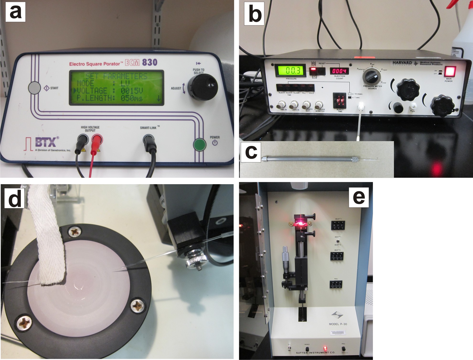
Figure 1. Electroporation equipment. The electroporation equipment consist of the ECM 830 electroporator (a), Microinjector MicroJect 1000A and stainless steel pipette holder (b, c), Micropipette Beveler and Puller necessary to make glass needles (d, e).
- One genetrode kit (5 mm, Gold plated thick electrode - L-shaped, in ovo gene) (Harvard Apparatus, model: 512, catalog number: 45-0115 )
- Stage-8-12 electrodes (for set up see Figure 2)
Procedure
文章信息
版权信息
© 2015 The Authors; exclusive licensee Bio-protocol LLC.
如何引用
Luz-Madrigal, A., Grajales-Esquivel, E. and Del Rio-Tsonis, K. (2015). Electroporation of Embryonic Chick Eyes. Bio-protocol 5(12): e1498. DOI: 10.21769/BioProtoc.1498.
分类
神经科学 > 发育 > 形态建成
发育生物学 > 形态建成
您对这篇实验方法有问题吗?
在此处发布您的问题,我们将邀请本文作者来回答。同时,我们会将您的问题发布到Bio-protocol Exchange,以便寻求社区成员的帮助。