

Intracellular Cytokine Staining (ICS) on Human Lymphocytes or Peripheral Blood Mononuclear Cells (PBMCs)
人淋巴细胞或外周血单核细胞(PBMC)的细胞因子胞内染色(ICS)

发布: 2015年04月05日第5卷第7期 DOI: 10.21769/BioProtoc.1442 浏览次数: 28633
评审: Anonymous reviewer(s)
Advertisement


Abstract
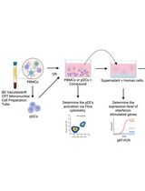
Production of cytokines plays an important role in the immune response. Cytokines are involved in many different pathways including the induction of many anti-viral proteins by IFN gamma, the induction of T cell proliferation by IL-2 and the inhibition of viral gene expression and replication by TNF alpha. Cytokines are not preformed factors but are rapidly produced and secreted in response to cellular activation. Intracellular cytokine detection by flow cytometry has emerged as the premier technique for studying cytokine production at the single-cell level. It detects the production and accumulation of cytokines within the endoplasmic reticulum after cell stimulation, allowing direct TH1 versus TH2 determination. It can also be used in combination with other flow cytometry protocols for immunophenotyping using cell surface markers or with MHC multimers to detect an antigen specific response, making it an extremely flexible and versatile method. This capability, combined with the high throughput nature of the instrumentation, gives intracellular cytokine staining an enormous advantage over existing single-cell techniques such as ELISPOT, limiting dilution, and T cell cloning. The principle steps of intracellular cytokine staining is as follows:
1. Cells are activated for a few hours using either a specific peptide or a non-specific activation cocktail;
2. An inhibitor of protein transport (e.g. Brefeldin A) is added to retain the cytokines within the cell;
3. Next, EDTA is added to remove adherent cells from the activation vessel;
4. After washing, antibodies to cell surface markers can be added to the cells;
5. The cells are then fixed in paraformaldehyde and permeabilized;
6. The anti-cytokine antibody is added and the cells can be analyzed by flow cytometer.
Materials and Reagents
- PBMC (fresh or thawed frozen)
- RPMI-1640 (Hyclone, catalog number: SH30027.01 )
- FBS (Atlanta Biologicals, catalog number: S11150 )
- 100x Pen-strep-Glutamine (Hyclone, catalog number: SV30082.01 )
- Benzonase (Sigma-Aldrich, catalog number: B7651 )
- PBS (10x stock) (Rockland, catalog number: MB-008 )
- Sodium azide (10% w/v solution) (Teknova, catalog number: S0209 )
- PMA (Sigma-Aldrich, catalog number: P8139 )
- Ionomycin (Calbiochem®, catalog number: 407952 )
- Dynabeads Human T Activator CD3/CD28 (Life Technologies, InvitrogenTM, catalog number: 111.32D )
- Brefeldin A (Sigma-Aldrich, catalog number: B7651)
- 1,000x monensin (BioLegend, catalog number: 420701 )
- 0.5 M EDTA (Sigma-Aldrich, catalog number: E-5134 )
- LIVE/DEAD® fixable red dead cell stain (Life Technologies, InvitrogenTM, catalog number: L23102 )
- 10x FACS lysing solution (BD Biosciences, catalog number: 349202 )
- 10x FACS permeabilizing solution 2 (BD Biosciences, catalog number: 347692 )
- Fluorochrome-linked surface markers [e.g. CD3-V500 (BD Biosciences, catalog number: 561416 ); CD8-V450 (BD Biosciences, catalog number: 560348 ); CD4-PerCP-Cy5.5 (BD Biosciences, catalog number: 341654 )]
- Fluorochrome-linked cytokine antibodies [e.g. IFN gamma-FITC (BD Biosciences, catalog number: 340449 ); IL-17 PE (BD Biosciences, catalog number: 560438 ); IL-2 PE-Cy7 (BD Biosciences, catalog number: 560707 ); IL-22-APC (R&D systems, catalog number: IC7821A); TNF- Alexa fluor 700 (BD Biosciences, catalog number: 557996 )]
- BD CompBeads [(anti-mouse Igκ, anti-rat Igκ, or anti-rat/hamster Igκ; BD Biosciences), for creating single-color compensation controls (BD Biosciences, catalog number: 560707)]
- Immunoglobulin capture beads for single-color compensation (e.g., BD Biosciences, catalog number: 560497 )
- Complete RPMI (see Recipes)
- FACS buffer (see Recipes)
Equipment
- 96- deep well V-bottom plates (Corning, catalog number: 3960 ) (1 ml washes in a 2 ml well volume)
- Falcon round-bottom FACS tubes
- Magnet for Dynabead separation (BD Biosciences, IMag, catalog number: 552311 )
- 37 °C water bath
- Biosafety cabinet
- Centrifuge
- CO2 incubator at 37 °C
- Calibrated pipettes
- ViCell (Beckman Coulter) or Hemocytometer cell counter
Procedure
文章信息
版权信息
© 2015 The Authors; exclusive licensee Bio-protocol LLC.
如何引用
Gupta, S. and Maecker, H. T. (2015). Intracellular Cytokine Staining (ICS) on Human Lymphocytes or Peripheral Blood Mononuclear Cells (PBMCs). Bio-protocol 5(7): e1442. DOI: 10.21769/BioProtoc.1442.
分类
免疫学 > 免疫细胞功能 > 细胞因子
免疫学 > 免疫细胞染色 > 流式细胞术
您对这篇实验方法有问题吗?
在此处发布您的问题,我们将邀请本文作者来回答。同时,我们会将您的问题发布到Bio-protocol Exchange,以便寻求社区成员的帮助。