

A Protocol to Measure the Extent of Cell-to-cell Movement of RNA Viruses in Planta
一种测定RNA病毒在植株细胞间运动的方法


发布: 2014年10月20日第4卷第20期 DOI: 10.21769/BioProtoc.1269 浏览次数: 8358
评审: Hiromasa SaitohTie LiuArsalan Daudi
参见作者原研究论文
The authors used this protocol in:
Mar 2014
Advertisement


Abstract
Here, we present a simple and rapid protocol to measure the extent of cell-to-cell movement of RNA viruses in planta. To do that, the green fluorescent protein (GFP) gene was incorporated into the genome of Melon necrotic spot virus (MNSV) as a coat protein (CP) fusion protein using the Thosea asigna virus 2A catalytic peptide (TaV 2a) (Serra-Soriano et al., 2014). TaV 2a allows the co-translational cleavage of the fusion protein resulting in the independent expression of both proteins (Kim et al., 2011). Viral infection was initiated by agro-infiltration of Cucumis melo leaves. At 6-7 days post-infiltration, fluorescent infection foci images were taken with a fluorescent stereo microscope and infection areas were measured using FIJI software.
Keywords: Plant virusMaterials and Reagents
- 3-4 weeks old Cucumis melo L. subsp. melo cv. Galia plants
- Agrobacterium tumefaciens (A. tumefaciens) strain C58C1, or similar, transformed with the binary vector harboring the GFP-tagged viral genome
In our case, pMNSV(Al)/GFP encoding GFP-tagged Melon necrotic spot virus (MNSV) genome under the control of the Cauliflower mosaic virus (CaMV) 35S promoter and the potato proteinase inhibitor terminator (PoPit) (Figure 1).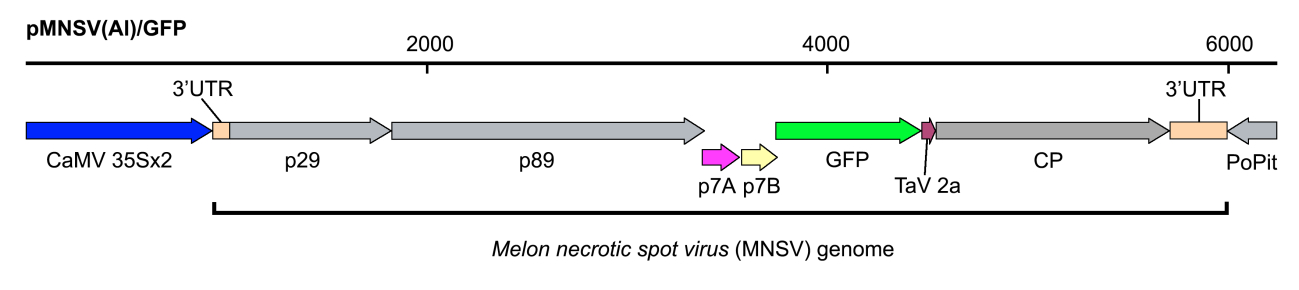
Figure 1. Schematic representation of the recombinant infectious clone pMNSV(Al)/GFP used in this assay - Yeast extract (Difco, catalog number: 212750 )
- Tryptone (Difco, catalog number: 211705 )
- Sodium chloride (NaCl) (Panreac Applichem, catalog number: 131659 )
- Acetosyringone (Sigma-Aldrich, catalog number: D134406 )
- MES (Sigma-Aldrich, catalog number: M8250 )
- Magnesium chloride (MgCl2) (Sigma-Aldrich, catalog number: M9272 )
- Antibiotics
- LB medium (see Recipes)
- Agrobacterium infiltration buffer (see Recipes)
Equipment
- 28 °C growing chamber
- 15 ml culture tubes
- Swinging centrifuge rotor for 15 ml tubes
- 1.5 ml tubes (standard Eppendorf tubes or similar)
- BioPhotometer plus (Eppendorf, catalog number: 6132000008 )
- 1 ml syringes without needle
- Plant growing chamber
- Leica MZ12 fluorescent stereo microscope
Software
- Adobe Photoshop CS5 or higher
- ImageJ, FIJI or similar software
- MS Excel
Procedure
文章信息
版权信息
© 2014 The Authors; exclusive licensee Bio-protocol LLC.
如何引用
Navarro, J. A., Serra-Soriano, M. and Pallás, V. (2014). A Protocol to Measure the Extent of Cell-to-cell Movement of RNA Viruses in Planta. Bio-protocol 4(20): e1269. DOI: 10.21769/BioProtoc.1269.
分类
植物科学 > 植物免疫 > 信号感知与传递
植物科学 > 植物免疫 > 病害生物测定
微生物学 > 微生物-宿主相互作用 > 体内实验模型 > 植物
您对这篇实验方法有问题吗?
在此处发布您的问题,我们将邀请本文作者来回答。同时,我们会将您的问题发布到Bio-protocol Exchange,以便寻求社区成员的帮助。