

Centrifuge Microscopy to Analyze the Sedimentary Movements of Amyloplasts
离心显微镜检查淀粉体的沉淀
发布: 2014年09月05日第4卷第17期 DOI: 10.21769/BioProtoc.1229 浏览次数: 9438
参见作者原研究论文
The authors used this protocol in:
Nov 2013
Advertisement


Abstract
A centrifuge microscope (CMS) functionally consists of a centrifuge producing a centrifugal force (hypergravity condition) and a microscope making an enlarged image of an object. This combination of equipment allows live-cell imaging during centrifugation. We have developed a new CMS (NSK Ltd.) to observe movements of the plant organelles such as amyloplasts, under hypergravity conditions (Toyota et al., 2013). This CMS is distinct from previously designed CMSs in terms of spatio-temporal resolution, ease of use and compactness. Here, we show a quick protocol to prepare a specimen of Arabidopsis inflorescence stem, use the CMS, obtain imaging data and analyze them using a single tracking method.
Materials and Reagents
- Arabidopsis thaliana inflorescence stems
- MS salt mixture (Wako Pure Chemical Industries, catalog number: 392-00591 )
- 1% (w/v) sucrose
- 0.05% (w/v) MES
- 0.1% (w/v) agar
- Growth media (see Recipes)
Equipment
- Fine tweezers
- Scissors
- Razor blade (Electron Microscopy Sciences, catalog number: 72000 )
- Kimwipes
- Aluminum chamber (custom built) (NSK Ltd.)
- Silicone rubber (thickness: 0.5 mm) (AS ONE Corporation, catalog number: 6-611-01 )
- Round cover glass (diameter: 12 mm) (Matsunami Glass, catalog number: CO12001 )
- CMS system (Figure 1, not commercially available) (NSK Ltd., http://www.nsk.com/)
CMS is a newly designed compact centrifuge microscope, 30 cm in height and 20 cm in diameter. CMS consists of a direct-drive motor (NSK Ltd., MEGATORQUE MOTOR™, model: M-PS1006KN002 ) and optics including a 50x objective lens with a working distance of 18 mm (SLMPLN 50x, 0.35 NA, OLYMPUS), LED light source (SCHOTT MORITEX Corporation, model: MEBL-CW25 ) and a CCD camera (SENSOR TECHNOLOGY, model: STC172C ).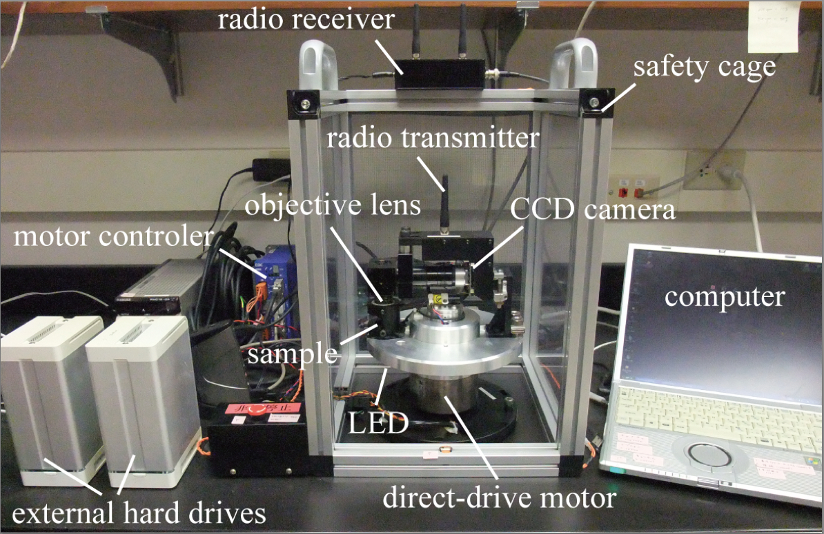
Figure 1. Overview of the CMS system - Windows computer [minimum computer requirements: Windows® XP or later, Pentium M 778 (1.6 GHz), RAM 1024 MB or higher]
Note: To install the software below, Windows computers are highly recommended.
Software
- MEGATORQUE MOTOR™ controller (EDC megaterm software) (NSK Ltd., http://www.nsk.com/)
Note: This is free software to control the motor and is available only for Windows. - Video capture software (COREL, http://www.corel.com/)
Note: You can use any video capture soft/hardware that converts analog video signal into digital signal and stores the data in a computer. - G-Track spot-tracking software (G-Angstrom, http://www.g-angstrom.com/eng/products/index.php)
Note: This is a piece of commercial software to trace fluorescence/bright spots and available only for Windows.
Procedure
文章信息
版权信息
© 2014 The Authors; exclusive licensee Bio-protocol LLC.
如何引用
Toyota, M., Ikeda, N., Tasaka, M. and Morita, M. T. (2014). Centrifuge Microscopy to Analyze the Sedimentary Movements of Amyloplasts. Bio-protocol 4(17): e1229. DOI: 10.21769/BioProtoc.1229.
分类
植物科学 > 植物细胞生物学 > 细胞成像
植物科学 > 植物细胞生物学 > 组织分析
细胞生物学 > 细胞成像 > 活细胞成像
您对这篇实验方法有问题吗?
在此处发布您的问题,我们将邀请本文作者来回答。同时,我们会将您的问题发布到Bio-protocol Exchange,以便寻求社区成员的帮助。