

Analysis of Mycobacterial Protein Secretion
分枝杆菌分泌蛋白的分析

发布: 2014年06月20日第4卷第12期 DOI: 10.21769/BioProtoc.1159 浏览次数: 14182
评审: Fanglian HeRon Saar-DoverAnonymous reviewer(s)
参见作者原研究论文
The authors used this protocol in:
Oct 2013
Advertisement


Abstract
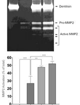
Mycobacterium tuberculosis (Mtb) is the causative agent of tuberculosis. Analysis of proteins secreted by Mtb has been of interest to the field of tuberculosis research since certain secreted proteins interact with the host to promote virulence, while others may be important antigens or serve as biomarkers of infection. Here, we describe a protocol to prepare whole cell extracts (WCE) and short term culture filtrate (CF) from Mtb or the vaccine strain Mycobacterium bovis- bacillus Calmatte- Guérin (BCG) (Mehra et al., 2013). These are both slow growing mycobacteria, but the same basic procedure can easily be adapted to analyze secreted proteins from rapidly growing mycobacteria, such as Mycobacterium smegmatis (Msmeg), a non-pathogenic species commonly used in the laboratory. The fractions obtained can be analyzed by western blotting to examine proteins of interest or by mass spectrometry if antibodies are not available or to examine the entire secretome. Genetic knockout mutants for the gene of interest serve as a negative control. Additionally, levels of a cytosolic protein such as the chaperone GroEL or the pyruvate dehydrogenase E2 component sucB (Rv2215/dlaT) should be assessed in the CF fraction to rule out the possibility that a positive signal in CF is due to bacterial lysis (see Figure 1). By varying the growth conditions of the strain, this in vitro secretion assay can be used to examine conditions that alter the secretome. We are thankful to Magnus Stiegedal for helpful tips on TCA (trichloroacetic acid) precipitation.
Keywords: Mycobacteria culture (结核分枝杆菌培养)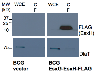
Figure 1. Western analysis of secretion of EsxH by BCG. BCG containing an empty vector control and EsxG-EsxH-FLAG expression construct (FLAG tag at C terminal of EsxH) were analyzed for presence EsxH by anti-FLAG western in WCE and CF prepared as described in the protocol. DlaT was used as a loading control to indicate the degree of bacterial lysis.
Materials and Reagents
Note: All work with live Mtb must be performed in a Biosafety Level 3 (BSL3) facility.
- Middlebrook 7H9 Broth (Difco, catalog number: 271310 )
- Tween-80 (Sigma-Aldrich, catalog number: P4780 )
- Glycerol (Sigma-Aldrich, catalog number: G5516 )
- Albumin-dextrose-catalase (ADC) (BD, catalog number: 212352 )
- Oleic-albumin-dextrose-catalase (OADC) (BD, catalog number: 212351 )
- Potassium phosphate (monobasic) (KH2PO4) (Sigma-Aldrich, catalog number: P9791 )
- L-asparagine monohydrate (Sigma-Aldrich, catalog number: A8381 )
- Citric acid monohydrate (Sigma-Aldrich catalog number: C1909 )
- Ferric ammonium citrate (Sigma-Aldrich catalog number: F5879 )
- Zinc sulfate monohydrate (ZnSO4.H2O) (Sigma-Aldrich, catalog number: 96495 )
- Magnesium sulfate heptahydrate (MgSO4.7H2O) (Sigma-Aldrich, catalog number: 230391 )
- Chelex 100 resin (Bio-Rad Laboratories, catalog number: 142-2822 )
- 100x Halt Protease Inhibitor single use cocktail (Pierce, catalog number: 1860932 )
- 100% trichloroacetic acid (TCA) (pre-chilled prior to use) (Sigma-Aldrich, catalog number: 49010 )
- Acetone (pre-chilled prior to use) (Sigma-Aldrich, catalog number: 32201 )
- Phosphate buffered saline (PBS) (Life Technologies, Gibco®, catalog number: 10010-023 )
- Bromophenol Blue, sodium salt (US Biological, catalog number: 12370 )
- Tris (MP Biomedicals, catalog number: 02194855 )
- Sodium Dodecyl Sulfate (SDS) (US Biological, catalog number: 18220 )
- Ethylenediaminetetraacetic acid (EDTA) (Sigma-Aldrich, catalog number: E6758 )
- β-mercaptoethanol (2-ME) (Sigma-Aldrich, catalog number: M6250 )
- 7H9 complete media (see Recipes)
- Sauton’s media (see Recipes)
- Chelated sauton’s media (an alternative minimal media for mycobacterial growth) (see Recipes)
- Protein extraction buffer (see Recipes)
- 5x SDS-PAGE sample buffer (see Recipes)
Equipment
- Autoclave
- Steriflip-GV filter units (0.22 µM pore size) (Millipore, catalog number: SE1M179M6 )
- 20 ml syringes (BD, catalog number: 302830 )
- 0. 22 µM syringe filter units (33 mm) (Millipore, catalog number: SLGV033RS )
- Disposable Sterile Filter system (1L, 0.22 µm pore size) (Corning, catalog number: 09761104 )
- 0.1 mm zirconia/silica beads (Bio Spec Products, catalog number: 11079101z )
- 30 ml square media bottles (Nalgene®, catalog number: NE/2019-0030 )
- 125 ml square media bottles (Nalgene®, catalog number: NE/2019-0125 )
- 50 ml falcon tubes (Corning, catalog number 430290 )
- 15 ml falcon tubes (Corning, catalog number: 430052 ) with plug seal caps
Note: These 50 ml tubes are compatible with organic solvents and high speed centrifugation. Falcon tubes with these features can be used from different vendors. - Microtubes (2 ml screw cap with O rings) (SARSTEDT AG, catalog number: 72.693 )
- Spectrophotometer
- Centrifuge with swinging bucket rotor for spinning down bacterial cultures (for example, Beckman Coulter, model: Allegra X-15R ; bench top centrifuge with SX4750 rotor)
Notes:- Msmeg and BCG should be handled according to institutional standards of practice for biosafety.
- Mtb cultures should be handled in biosafety level 3 facilities according to institutional standards of practice.
- Centrifuging BCG and Mtb requires appropriate aerosol containment.
- Msmeg and BCG should be handled according to institutional standards of practice for biosafety.
- Beckman Aerosolve® canisters to contain aerosols during centrifugation of mycobacterial cultures (e.g. Beckman Coulter, catalog number: BK359232 )
- 37 °C shaking incubator
- Aerosol containment units for shaking BCG and Mtb liquid cultures in the shaking incubator
Note: Incubator should be placed in BSL3 facility for Mtb cultures. - High speed centrifuge for 50 ml polypropylene falcons used for TCA precipitation of CF (e.g. Beckman Coulter centrifuge with JLA16.2 rotor)
- 50 ml falcon adaptors for rotor JLA16.2
- Bead beater (Bio Spec Products, model: Minibead beater 16; http://www.biospec.com/product/34/mini_beadbeater/)
- Standard table top centrifuge with refrigeration
- Heating block for Eppendorf tubes (set to 95 °C)
- pH meter
Acronyms
- Mtb: Mycobacterium tuberculosis
- BCG: Mycobacterium bovis bacillus Calmatte- Guérin
- Msmeg: Mycobacterium smegmatis
- WCE: Whole cell extract
- CF: Culture filtrate
- DlaT: Rv2215/pyruvate dehydrogenase E2 component sucB protein of mycobacteria
- TCA: Trichloroacetic acid
- BSC: Biosafety cabinet
- OD600: Absorbance or Optical Density at wavelength of 600 nm
Procedure
文章信息
版权信息
© 2014 The Authors; exclusive licensee Bio-protocol LLC.
如何引用
Readers should cite both the Bio-protocol article and the original research article where this protocol was used:
- Mehra, A. and Philips, J. A. (2014). Analysis of Mycobacterial Protein Secretion . Bio-protocol 4(12): e1159. DOI: 10.21769/BioProtoc.1159.
- Mehra, A., Zahra, A., Thompson, V., Sirisaengtaksin, N., Wells, A., Porto, M., Koster, S., Penberthy, K., Kubota, Y., Dricot, A., Rogan, D., Vidal, M., Hill, D. E., Bean, A. J. and Philips, J. A. (2013). Mycobacterium tuberculosis type VII secreted effector EsxH targets host ESCRT to impair trafficking. PLoS Pathog 9(10): e1003734.
分类
微生物学 > 微生物生物化学 > 蛋白质 > 分离和纯化
微生物学 > 微生物细胞生物学 > 细胞分离和培养
您对这篇实验方法有问题吗?
在此处发布您的问题,我们将邀请本文作者来回答。同时,我们会将您的问题发布到Bio-protocol Exchange,以便寻求社区成员的帮助。