

Deflagellation and Regeneration in Chlamydomonas
衣藻的鞭毛脱落和再生
发布: 2014年06月20日第4卷第12期 DOI: 10.21769/BioProtoc.1155 浏览次数: 15088
评审: Ru Zhang
参见作者原研究论文
The authors used this protocol in:

Jan 2013


Abstract
Eukaryotic cilia/flagella are one of the only cellular structures that can be removed without injuring cells, can be highly purified for biochemical analysis, and, in many cells, can be completely reassembled within 90 minutes. Following amputation, the expression of many flagellar genes is up-regulated, and many are packaged and associated with intraflagellar transport (IFT) particles for transport to flagellar bases and into growing flagella. Studies of deciliation and ciliary growth provide insight to mechanisms that regulate microtubule assembly and length, mechanisms that regulate the transport of soluble cytoplasmic proteins into the ciliary compartment and their assembly into microtubules, and mechanisms that regulate trafficking of membrane proteins and lipids to the plasma membrane or to ciliary bases and their movement into and out of the cilium. These are important for motility and for signal transduction.
Deciliation methods for many cells have been developed and most require extracellular calcium ions and activation of signaling pathways that regulate microtubule severing (Quarmby, 2009). Deciliation occurs at the distal end of the basal bodies and, as soon as axonemes are severed, the membrane reseals and basal bodies begin to regenerate cilia.
Chlamydomonas is an ideal organism with which to study ciliary regeneration. Cells are easily and inexpensively cultured, flagellar amputation and regeneration is uniform in all cells in a population and growth can be assayed by observing fixed or living cells with a phase contrast microscope equipped and a 40x objective lens. Flagellar regeneration on individual living cells can be observed using paralyzed mutants immobilized in agarose. Because deflagellation leaves cells intact, the released flagella can be purified without contamination with cellular debris. The most reliable deciliation and regeneration method is the pH shock method developed by Reference 5 (also see References 4 and 11). Other methods are reviewed by Quarmby, (2009). The pH shock method is primarily used for Chlamydomonas but can be used for deciliation and regeneration of Tetrahymena cilia (Gaertig et al., 2013).
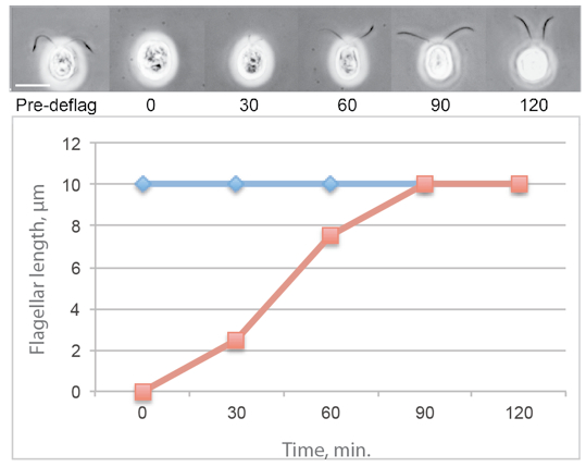
Figure 1. Typical flagellar regeneration curve showing phase contrast images of Chlamydomonas cells photographed before deflagellation and during regeneration. The average flagellar lengths of a population of regenerating cells is shown in red and the average flagellar lengths on a population of nondeflagellated cells is in blue.
Materials and Reagents
- Cells
Chlamydomonas cells can be obtained from a variety of sources and pure strains of a variety of flagellar mutants can be obtained from the Chlamydomonas Resource Center (http://chlamycollection.org/contact-us/). - Media and cell culture (see Notes)
- 0.5 N acetic acid
- 0.5 M NaOH
- 2% glutaraldehyde in M medium or Lugols iodine (see Recipes)
Note: For electron microscopy, one should use fresh glutaraldehyde from sealed ampuoles. To measure flagellar lengths, the age of the glutaraldehyde is not critical.
Optional:
- 2.5% low EEO agar (Thermo Fisher Scientific, FisherBiotechTM, catalog number: BP160-100 )
Note: It is used to observe flagellar growth or maintenance on individual living cells.
- 5 mM (final concentration) colchicine (to inhibit microtubule assembly)
Note: These experiments are carried out in phosphate-buffered Minimal medium. Avoid Tris-containing buffers because they may inhibit the effects of colchicine (Margulis et al., 1969).
- 10 µg/ml (final concentration) Cycloheximide
Note: It is used to inhibit protein synthesis.
- VALAP (see Recipes)
Note: It is used to support coverslips to examine living cells without inducing deflagellation by coverslip pressure.
Equipment
- pH meter calibrated for pH 4-7
Note: Some gel-filled electrodes are not accurate across this pH range.
- Centrifuge and tubes
Note: For small scales, a clinical centrifuge and conical tubes.
- Phase contrast microscope or brightfield microscope
Note: If cells are fixed with Lugol’s iodine, a 40x lens is ideal for flagellar length measurements.
- Orbital shaker
- Magnetic stirrer
- Stir bar
Software
- Image J (http://imagej.nih.gov/ij/)
Procedure
文章信息
版权信息
© 2014 The Authors; exclusive licensee Bio-protocol LLC.
如何引用
Dentler, W. (2014). Deflagellation and Regeneration in Chlamydomonas. Bio-protocol 4(12): e1155. DOI: 10.21769/BioProtoc.1155.
分类
植物科学 > 藻类学 > 细胞分析
细胞生物学 > 细胞运动 > 细胞运动性
细胞生物学 > 细胞成像 > 活细胞成像
您对这篇实验方法有问题吗?
在此处发布您的问题,我们将邀请本文作者来回答。同时,我们会将您的问题发布到Bio-protocol Exchange,以便寻求社区成员的帮助。