- EN - English
- CN - 中文
Fluorescence Microscopy Analysis of Drug Effect on Autophagosome Formation
荧光显微法分析药物对自噬体形成的影响
发布: 2014年04月05日第4卷第7期 DOI: 10.21769/BioProtoc.1089 浏览次数: 11968
评审: Anonymous reviewer(s)

相关实验方案

用于脉冲追踪成像,猝发脉冲追踪成像和基于纳米显微技术的细胞裂解物检测的融合SNAP病毒蛋白的按需标记
Roland Remenyi [...] Mark Harris
2019年02月20日 6977 阅读
Abstract
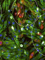
The autophagy protein, LC3 represents a reliable characteristic marker for autophagosomal structures. The initial LC3 is processed by the cysteine protease autophagy-related gene 4 (Atg4) at its C terminus in order to create LC3-I generally localized in the cytoplasm. Afterwards LC3-I is conjugated with phosphatidylethanolamine (PE) to become LC3-PE or LC3-II predominantly localised on the autophagosomal membranes (outer and inner). Autolysosomal content of LC3-II is very low as upon autophago/lysosomal fusion it is either cleaved off from the outer membrane by Atg4 or degraded together with the inner membrane by the lysosomal activity. Therefore GFP-LC3 and mCherry-GFP-LC3 might be visualized by conventional or confocal fluorescence microscopy (FM). In this situation mCherry-GFP-LC3 or GFP-LC3 cytoplasmic pool is visualized as a homogeneously dispersed signal and mCherry-GFP-LC3-II or GFP-LC3-II containing autophagosomes are detected as punctae formations. The number of punctae may be used as marker of autophagosomal abundance. In general we recommend counting the average number of GFP-LC3 punctae per cell.
Keywords: Autophagy (自噬)Materials and Reagents
- Cell lines of interest (HepG2, HUH7, CMK, K562 etc.) stably expressing GFP-LC3
We recommend the following commercially available plasmids: pBABEpuro GFP-LC3 (plasmid 22405) and pBABE-puro mCherry-EGFP-LC3B (plasmid 22418) generated by Jayanta Debnath from Addgene to be inserted into retroviral constructs and used for cell transduction
- Eagle's minimal essential medium (EMEM) (ATCC, catalog number: 30-2003 ) containing 10% fetal bovine serum (FBS) with 100 U/100 μg/ml penicillin/streptomycin (Life Technologies, Gibco®, catalog number: 15140-122 )
- RPMI 1640 with L-glutamine (Lonza, catalog number: BE12-702F ) containing 10% fetal bovine serum (FBS) with 100 U/100 μg/ml penicillin/streptomycin
- Fetal Bovine Serum (FBS) (Biochrom, catalog number: S0615 )
- Dulbecco’s Phosphate Buffered Saline (PBS) (Biochrom, catalog number: L1825 )
- 1x 0.05% Trypsin-EDTA (phenol red) (Life Technologies, catalog number: 25300 )
- Hanks Balanced Salt Solution (HBSS) (Life Technologies, Gibco®, catalog number: 14025 ) containing 6 mM glucose (starvation medium)
- Rapamycin from Streptomyces hygroscopicus (1-5 µmol/L) (Sigma-Aldrich, catalog number: R0395 )
- PP242 hydrate (1-5 µmol/L) (Sigma-Aldrich, catalog number: P0037 )
- 3-methyladenine (3-MA) (3-10 mmol/L) (Sigma-Aldrich, catalog number: M9281 )
- Wortmannin (30-100 nmol/L) (Sigma-Aldrich, catalog number: W3144 )
- LY294002 (7-20 µmol/L) (Sigma-Aldrich, catalog number: L9908 )
- Nocodazole (12-50 µmol/L) (Sigma-Aldrich, catalog number: M1404 )
- Vinblastine (12-50 µmol/L) (Sigma-Aldrich, catalog number: V1377 )
- Ammonium chloride (NH4Cl) (10-20 mmol/L) (Sigma-Aldrich, catalog number: A0171 )
- Hydrohychloroquine sulphate (HCQ) (5-10 µmol/L) (Sigma-Aldrich, catalog number: H0915 )
- Chloroquine (CQ) (5-10 µmol/L) (Sigma-Aldrich, catalog number: C6628 )
- Dimethyl sulfoxide DMSO (Sigma-Aldrich, catalog number: D8418 )
Equipment
- 37 °C, 5% CO2 humidified incubator
- Centrifuge
- Olympus IX81 instrument and analySIS (Soft Imaging System GmbH) or analogous equipment
Procedure
文章信息
版权信息
© 2014 The Authors; exclusive licensee Bio-protocol LLC.
如何引用
Stankov, M., Panayotova-Dimitrova, D., Leverkus, M. and Behrens, G. (2014). Fluorescence Microscopy Analysis of Drug Effect on Autophagosome Formation. Bio-protocol 4(7): e1089. DOI: 10.21769/BioProtoc.1089.
分类
微生物学 > 抗微生物试验 > 自体吞噬试验
细胞生物学 > 细胞成像 > 荧光
您对这篇实验方法有问题吗?
在此处发布您的问题,我们将邀请本文作者来回答。同时,我们会将您的问题发布到Bio-protocol Exchange,以便寻求社区成员的帮助。
Share
Bluesky
X
Copy link


