往期刊物2014

卷册: 4, 期号: 12

left

Dec

20

Dec

5

Nov

20

Nov

5

Oct

20

Oct

5

Sep

20

Sep

5

Aug

20

Aug

5

Jul

20

Jul

5

Jun

20

Jun

5

May

20

May

5

Apr

20

Apr

5

Mar

20

Mar

5

Feb

20

Feb

5

Jan

20

Jan

5

right

癌症生物学

溶酶体稳定性分析

Lysosomal Stability Assay

溶酶体稳定性分析

NP Nikolaj H. T. Petersen
TK Thomas Kirkegaard
MJ Marja Jäättelä
17394 Views
Jun 20, 2014
This assay makes use of the dye Acridine Orange (AO) to determine the stability of lysosomes in living cells upon exposure to a confocal microscope laser. AO is a lipophilic amine that readily diffuses into cells (Figure 1). Inside the cell it enters the acidic lysosomal compartment where it is protonated and sequestered, shifting its emission spectrum towards a longer wavelength (i.e. red). Once inside the lysosomes, the metachromatic AO sensitizes the lysosomal membrane to photo-oxidation by blue light (Brunk et al., 1997). Upon light-induced loss of the lysosomal pH gradient and subsequent leakage of AO into the cytosol, the emission spectrum of AO shifts from red to green (Figure 2). Hence, loss of lysosomal integrity can be measured as a ‘loss of red dots’ or as a quantitative rise in green fluorescence (Petersen et al., 2010; Kirkegaard et al., 2010; Petersen et al., 2013).Figure 1. Acridine Orange Figure 2. Snapshots visualizing the U2OS cells at various steps of the recording procedure (Petersen et al., 2010)

细胞生物学

骨髓淋巴样红细胞(EML)中的红血球和噬中性细胞分化实验流程

EML Erythroid and Neutrophil Differentiation Protocols

骨髓淋巴样红细胞(EML)中的红血球和噬中性细胞分化实验流程

Cristina  Pina Cristina Pina
CF Cristina Fugazza
TE Tariq Enver
11718 Views
Jun 20, 2014
Erythroid-Myeloid-Lymphoid cells (EML) are a multipotent haematopoietic cell line of mouse bone marrow origin capable of long-term maintenance in vitro in the presence of SCF (stem cell factor) (Tsai et al., 1994). The self-renewal capacity of the EML cell line is conferred by the presence of a dominant-negative retinoic acid receptor (RAR) originally delivered by retroviral transduction (Tsai et al., 1994), which arrests cells at an early progenitor stage blocked from normal progression into myeloid differentiation. The presence of the RAR trans-gene does not interfere with erythroid differentiation, and it is possible to capture a low percentage of early erythroid, but not myeloid, committed cells in maintenance cultures (Pina et al., 2012; Ye et al., 2005).Cells can be driven into granulocytic/neutrophil differentiation through the use of high doses of retinoic acid (RA), which overcomes the differentiation block. It should be noted that these pharmacological doses of RA are not compatible with erythroid differentiation, and it is hence not viable to obtain robust erythroid and myeloid differentiation in the same assay. Indeed, colonies scored as mixed-lineage in CFC assays are a mixture of undifferentiated and erythroid cells (Tsai et al., 1994). Nevertheless, robust single-lineage erythroid and neutrophil differentiation can be obtained in liquid culture under defined cytokine conditions, as specified below.

免疫学

骨髓源嗜酸性细胞培养

Bone Marrow Derived Eosinophil Cultures

骨髓源嗜酸性细胞培养

TL Thomas X. Lu
Marc E.  Rothenberg Marc E. Rothenberg
13729 Views
Jun 20, 2014
Eosinophils are multifunctional effector cells implicated in the pathogenesis of a variety of diseases including asthma, eosinophil gastrointestinal disorders and helminth infection. Mouse bone marrow derived progenitor cells can be differentiated into eosinophils following IL-5 exposure. These bone marrow derived eosinophils are fully differentiated at the end of a 14 day culture based on morphology and expression of molecular markers.

微生物学

分枝杆菌分泌蛋白的分析

Analysis of Mycobacterial Protein Secretion

分枝杆菌分泌蛋白的分析

Alka  Mehra Alka Mehra
JP Jennifer A. Philips
13386 Views
Jun 20, 2014
Mycobacterium tuberculosis (Mtb) is the causative agent of tuberculosis. Analysis of proteins secreted by Mtb has been of interest to the field of tuberculosis research since certain secreted proteins interact with the host to promote virulence, while others may be important antigens or serve as biomarkers of infection. Here, we describe a protocol to prepare whole cell extracts (WCE) and short term culture filtrate (CF) from Mtb or the vaccine strain Mycobacterium bovis- bacillus Calmatte- Guérin (BCG) (Mehra et al., 2013). These are both slow growing mycobacteria, but the same basic procedure can easily be adapted to analyze secreted proteins from rapidly growing mycobacteria, such as Mycobacterium smegmatis (Msmeg), a non-pathogenic species commonly used in the laboratory. The fractions obtained can be analyzed by western blotting to examine proteins of interest or by mass spectrometry if antibodies are not available or to examine the entire secretome. Genetic knockout mutants for the gene of interest serve as a negative control. Additionally, levels of a cytosolic protein such as the chaperone GroEL or the pyruvate dehydrogenase E2 component sucB (Rv2215/dlaT) should be assessed in the CF fraction to rule out the possibility that a positive signal in CF is due to bacterial lysis (see Figure 1). By varying the growth conditions of the strain, this in vitro secretion assay can be used to examine conditions that alter the secretome. We are thankful to Magnus Stiegedal for helpful tips on TCA (trichloroacetic acid) precipitation.
基于SYBR Green荧光染料的实时逆转录聚合酶链式反应(RT-PCR)试验检测新生 H7N9 病毒

A SYBR Green-based Real Time RT-PCR Assay for Detection of the Emerging H7N9 Virus

基于SYBR Green荧光染料的实时逆转录聚合酶链式反应(RT-PCR)试验检测新生 H7N9 病毒

ZZ Zheng Zhu
Lunbiao  Cui Lunbiao Cui
9594 Views
Jun 20, 2014
Most recently a novel avian-origin influenza A (H7N9) virus emerged in China and has been associated with lots of human infection and fatal cases. Molecular diagnostic methods are thus urgently needed in public health laboratories. We developed a SYBR green-based one-step real time reverse transcription-PCR (RT-PCR) to detect the novel H7N9 virus.
链霉菌属物种潮湿抑制发育孢子的氧气呼吸率测定

Determination of Oxygen Respiration Rates in Wetted Developmentally Arrested Spores of Streptomyces Species

链霉菌属物种潮湿抑制发育孢子的氧气呼吸率测定

Marco  Fischer Marco Fischer
Dörte  Falke Dörte Falke
RS R. Gary Sawers
8577 Views
Jun 20, 2014
Streptomyces species produce spores, which, while not as robust as endospores of Bacillus or Clostridium species, are capable of surviving for months or even years (Hopwood, 2006). During this time these spores remain viable, surviving by slowly degrading internal stores of carbon compounds, such as the carbohydrate trehalose. To enable metabolism to continue they must have access to an electron acceptor that allows the removal of the reducing equivalents that accumulate through metabolic activity. The most commonly used acceptor is oxygen. We describe the quantitative measurement of oxygen respiration rates by developmentally arrested spores of the streptomycete Streptomyces coelicolor (Fischer et al., 2013).
应用完整微生物细胞或孢子进行硝酸快速还原分析

Rapid Nitrate Reduction Assay with Intact Microbial Cells or Spores

应用完整微生物细胞或孢子进行硝酸快速还原分析

Marco  Fischer Marco Fischer
Dörte  Falke Dörte Falke
RS R. Gary Sawers
8498 Views
Jun 20, 2014
Many microorganisms have the capacity to use nitrate as a respiratory electron acceptor. Reduction of nitrate is catalyzed by a multi-subunit nitrate reductase that is often located associated with the cytoplasmic membrane and has its active site oriented toward the cytoplasm. This means that nitrate must be transported into the cell and often this occurs concomitantly with the export of the reduced nitrite product. Often nitrate and nitrite transport are coupled through the action of a nitrate: nitrite antiporter. Microbial cells, spores and mycelium harbour intracellular storage compounds such as trehalose or glycogen that, upon metabolism, function as endogenous electron donors for nitrate reduction. It is also possible to use glucose supplied exogenously as a substrate for nitrate reduction. The method described here allows the direct analysis of nitrate reduction by whole cell material without the requirement for artificial electron donors. This method is also applicable to the study of spores, particularly those of Streptomyces species (Fischer et al., 2013). The paper by Fischer et al. 2013 provides examples of datasets for the method presented below.

植物科学

原生质体制备和细胞死亡的鉴定

Protoplast Preparation and Determination of Cell Death

原生质体制备和细胞死亡的鉴定

Antoine  Danon Antoine Danon
18443 Views
Jun 20, 2014
The protoplasts assay constitutes a powerful tool that allows an easy uptake of active agents and a precise quantification of cell death induction in different populations. Our study showed that the basal level of cell death in our controls is low and stable throughout the length of our experiments (Danon et al., 2005; Pineau et al., 2013). In addition, the data obtained from the protoplast assay are applicable to intact seedlings, where it is possible to see differences in the intensity of necrotic lesions (Danon et al., 2006) even if those differences are not as easily and clearly quantifiable as with the protoplast assay.
衣藻的鞭毛脱落和再生

Deflagellation and Regeneration in Chlamydomonas

衣藻的鞭毛脱落和再生

WD William Dentler
14603 Views
Jun 20, 2014
Eukaryotic cilia/flagella are one of the only cellular structures that can be removed without injuring cells, can be highly purified for biochemical analysis, and, in many cells, can be completely reassembled within 90 minutes. Following amputation, the expression of many flagellar genes is up-regulated, and many are packaged and associated with intraflagellar transport (IFT) particles for transport to flagellar bases and into growing flagella. Studies of deciliation and ciliary growth provide insight to mechanisms that regulate microtubule assembly and length, mechanisms that regulate the transport of soluble cytoplasmic proteins into the ciliary compartment and their assembly into microtubules, and mechanisms that regulate trafficking of membrane proteins and lipids to the plasma membrane or to ciliary bases and their movement into and out of the cilium. These are important for motility and for signal transduction.Deciliation methods for many cells have been developed and most require extracellular calcium ions and activation of signaling pathways that regulate microtubule severing (Quarmby, 2009). Deciliation occurs at the distal end of the basal bodies and, as soon as axonemes are severed, the membrane reseals and basal bodies begin to regenerate cilia. Chlamydomonas is an ideal organism with which to study ciliary regeneration. Cells are easily and inexpensively cultured, flagellar amputation and regeneration is uniform in all cells in a population and growth can be assayed by observing fixed or living cells with a phase contrast microscope equipped and a 40x objective lens. Flagellar regeneration on individual living cells can be observed using paralyzed mutants immobilized in agarose. Because deflagellation leaves cells intact, the released flagella can be purified without contamination with cellular debris. The most reliable deciliation and regeneration method is the pH shock method developed by Reference 5 (also see References 4 and 11). Other methods are reviewed by Quarmby, (2009). The pH shock method is primarily used for Chlamydomonas but can be used for deciliation and regeneration of Tetrahymena cilia (Gaertig et al., 2013).
细胞器的蛋白质合成

In organello Protein Synthesis

细胞器的蛋白质合成

Malgorzata Kwasniak-Owczarek Malgorzata Kwasniak-Owczarek
Hanna  Janska Hanna Janska
13219 Views
Jun 20, 2014
In organello protein synthesis method allows the analysis of mitochondrial translation products. The principle of this method relies on incubation of isolated intact mitochondria with radiolabeled amino acids such as 35S methionine. After protein synthesis, the radiolabeled translation products are subsequently separated by SDS polyacrylamide gel electrophoresis and analysed by autoradiography. For in organello analysis of protein synthesis, the isolated intact mitochondria must retain their bioenergetics capacity, and in consequence be fully functional and able to perform coupled respiration. This in turn requires a quick and gentle purification of mitochondria during their isolation.
拟南芥幼苗中荧光素酶活性的测定

Determination of Luciferase Activity in Arabidopsis seedling

拟南芥幼苗中荧光素酶活性的测定

Mohan  TC Mohan TC
GC Gabriel Castrillo
Antonio  Leyva Antonio Leyva
12627 Views
Jun 20, 2014
There are different direct and indirect methods available to study gene expression in plant systems. In this protocol we describe a modified expression assay using transgenic plants expressing the luciferase gene under the control of phosphate transporter PHT1;1 promoter. This assay was originally optimized for analyzing the repression of PHT1;1 promoter in response to arsenate As (V) which can be adapted to study the kinetics of transcriptional regulation of any gene in response to biotic or environmental stimuli measuring Luciferase activity in Arabidopsis thaliana.
拟南芥MTV1 的pull-down实验

MTV1 Pull-down Assay in Arabidopsis

拟南芥MTV1 的pull-down实验

Michael  Sauer Michael Sauer
12529 Views
Jun 20, 2014
This protocol is an example of how to analyse suspected interactions between proteins using a pull-down assay (Sauer et al., 2013). A bait protein of interest (in this case, MTV1 of Arabidopsis thaliana) is fused to a GST tag and expressed in bacteria. The protein is isolated and allowed to bind to a matrix of glutathione-conjugated agarose beads via the GST-tag. Unspecifically binding proteins from the bacterial lysate are removed from the matrix. A native plant protein extract is then passed over the matrix and binding between the bait GST-MTV1 and prey proteins can occur. Extensive washes remove unspecifically bound proteins and finally, bait and prey proteins are released from the beads. Immunoblot analysis is then used to identify the proteins that bound to GST-MTV1. Importantly, a negative control consisting of the GST-tag alone is analysed in parallel to exclude the possibility that prey protein binding to the GST-MTV1 bait was due to the GST-tag.
纤毛和鞭毛囊泡(外皮层)的纯化

Ciliary and Flagellar Membrane Vesicle (Ectosome) Purification

纤毛和鞭毛囊泡(外皮层)的纯化

WD William Dentler
9977 Views
Jun 20, 2014
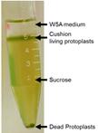
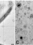
Eukaryotic cilia/flagella are ideal organelles for the analysis of membrane trafficking, membrane assembly, and the functions of a variety of signal transduction molecules. Cilia are peninsular organelles and the membrane lipids, membrane proteins, and microtubular-associated components are selectively transported into cilia through the region formed by the basal body/transition region and tightly associated ciliary membrane. Cilia can be isolated from many organisms without disrupting cells and many will rapidly regenerate cilia (with the ciliary membrane lipids and proteins) to replace those that are released. Despite their ease of isolation, we have relatively little understanding of the mechanisms that regulate lipid and protein transport into ciliary membranes (Pazour and Bloodgood, 2008; Bloodgood, 2009; Bloodgood, 2012).Chlamydomonas flagella shed membrane vesicles, also called ectosomes (Wood et al., 2013) from flagellar tips and these vesicles can be purified from the culture medium without damaging or deflagellating cells (McLean et al., 1974; Bergman et al., 1975; Snell, 1976; Kalshoven et al., 1990). Based on a comparison of biotinylated proteins on the shed vesicles with biotinylated proteins isolated from purified flagella and cell bodies, the ectosomes contain most, but not all, flagellar surface proteins and none of the major cell body proteins (Dentler, 2013). Although ectosomes have only been purified from Chlamydomonas cells, preliminary evidence indicates that similar vesicles are released from Tetrahymena cilia (Dentler, unpublished). Flagellar (and ciliary) membranes or membrane proteins also can be released from purified flagella/cilia. Most membrane proteins can be solubilized by extracting purified cilia with nonionic detergent [Triton X-100 or X-114 or Nonidet P-40 (NP-40)] and pelleting the microtubules (axonemes). However, not all membranes are released by detergent (Dentler, 1980) and the supernatant also contains all of the flagellar proteins that are not attached to the microtubules. Intact membrane vesicles can be released from flagella by agitation of flagella, often with low concentrations of nonionic detergents or freeze-thawing (Witman et al., 1972; Snell, 1976; Dentler, 1980; Dentler, 1995; Bloodgood and May, 1982; Pasquale and Goodenough, 1987; Iomini et al., 2006; Huang et al., 2007). Once released, they can be purified from axonemes by differential centrifugation.Each of these methods may enrich for different populations of axonemal and membrane proteins and lipids. The different solubility of membranes may reveal local differences in lipid or protein composition (Bloodgood, 2009). The ectosomes contain most but not all surface proteins found on purified Chlamydomonas flagella (Dentler, 2013). The ectosomes vesicles may be enriched in different soluble flagellar proteins than those trapped as vesicles are released from purified flagella. The detergent-solubilized “membrane+matrix” will contain all soluble membrane proteins as well as all of the soluble proteins in the flagellar compartment. In this paper, a method to purify ectosomes vesicles released from the tips of living Chlamydomonas cells is presented as are two methods to release flagellar membrane vesicles and proteins from purified flagella.

系统生物学

白色念珠菌的染色质免疫共沉淀测序(ChIP-Seq)

ChIP-Seq in Candida albicans

白色念珠菌的染色质免疫共沉淀测序(ChIP-Seq)

Sadri  Znaidi Sadri Znaidi
CP Caroline Proux
SW Sandra Weber
SD Simon Drouin
FR François Robert
MR Martine Raymond
JC Jean-Yves Coppée
Cd Christophe d’Enfert
18202 Views
Jun 20, 2014
Systems biology approaches can be used to study the regulatory interactions occurring between many components of the biological system at the whole-genome level and decipher the circuitries implicated in the regulation of cellular processes, including those imparting virulence to opportunistic fungi. Candida albicans (C. albicans) is a leading human fungal pathogen. It undergoes morphological switching between a budding yeast form and an elongated multicellular hyphal form. This transition is required for C. albicans’ ability to cause disease and is regulated through highly interconnected regulatory interactions between transcription factors (TFs) and target genes. The chromatin immunoprecipitation (ChIP)-High-throughput sequencing (Seq) technology (ChIP-Seq) is a powerful approach for decoding transcriptional regulatory networks. This protocol was optimized for the preparation of ChIP DNA from filamenting C. albicans cells followed by high-throughput sequencing to identify the targets of TFs that regulate the yeast-to-hyphae transition.