往期刊物2014

卷册: 4, 期号: 7

left

Dec

20

Dec

5

Nov

20

Nov

5

Oct

20

Oct

5

Sep

20

Sep

5

Aug

20

Aug

5

Jul

20

Jul

5

Jun

20

Jun

5

May

20

May

5

Apr

20

Apr

5

Mar

20

Mar

5

Feb

20

Feb

5

Jan

20

Jan

5

right

生物化学

UPF1蛋白单核苷酸分离紫外线交联和免疫沉淀(iCLIP)

Individual-nucleotide-resolution UV Cross-linking and Immunoprecipitation (iCLIP) of UPF1

UPF1蛋白单核苷酸分离紫外线交联和免疫沉淀(iCLIP)

David  Zünd David Zünd
Oliver  Mühlemann Oliver Mühlemann
14392 Views
Apr 5, 2014
The fate of mRNA, in particular its stability, localization and rate of translation is regulated by RNA binding proteins assembling to messenger ribonucleoprotein (mRNP) complexes. To investigate the transcriptome-wide RNA binding sites of UPF1, the core factor of nonsense-mediated mRNA decay (NMD), we performed individual-nucleotide-resolution UV cross-linking and immunoprecipitation (iCLIP) (Zund et al., 2013) followed by high-throughput sequencing. The presented protocol is optimized to investigate the RNA-binding sites of UPF1 and is based on previously described studies (Konig et al., 2010; Konig et al., 2011; Hafner et al., 2010). We want to thank the Group of Mihaela Zavolan (Swiss Institute of Bioinformatics, Basel, Switzerland) and Jernej Ule (Medical Research Council Laboratory of Molecualar Biology, Cambridge, UK) for technical support in setting up these experiments.
Mini-μ 载体-表达细胞中的UPF1 RNA 免疫沉淀

UPF1 RNA Immunoprecipitation from Mini-μ Construct–expressing Cells

Mini-μ 载体-表达细胞中的UPF1 RNA 免疫沉淀

David  Zünd David Zünd
Oliver  Mühlemann Oliver Mühlemann
10965 Views
Apr 5, 2014
UPF1, an RNA helicase and a core factor of nonsense-mediated mRNA decay (NMD), interacts with RNA independently of the sequence context. To investigate the influence of translation on the association of UPF1 with specific reporter transcripts, UPF1 RNA immunoprecipitations (RIPs) are performed from Hela cells that either express a normally translated immunoglobulin-µ (Ig-µ) reporter (mini µ) or a version with a stable stem loop in the 5' UTR (SL mini µ) that efficiently inhibit translation initiation (Zund et al., 2013). Both the cloning of the SL mini µ reporter construct and the UPF1 RIP experiment are described in detail.

癌症生物学

不同肿瘤阶段成纤维细胞的分离和永生化

Isolation and Immortalization of Fibroblasts from Different Tumoral Stages

不同肿瘤阶段成纤维细胞的分离和永生化

Fernando  Calvo Fernando Calvo
SH Steven Hooper
ES Erik Sahai
19351 Views
Apr 5, 2014
Tumour microenvironment and cancer-associated fibroblasts in particular exhibit tumour promoting abilities that are not present in their normal counterparts (Calvo et al., 2013; Hanahan and Coussens, 2012). Therefore, functional and molecular characterization of the modifications occurring in fibroblasts during tumour progression is essential to fully understand their role in tumour progression. Previous studies have addressed this issue using human fibroblasts and comparing normal and adjacent fibroblasts to tumour-associated fibroblasts (Kalluri and Zeisberg, 2006). However, these studies are hampered by the intrinsic variability of human samples (e.g. pairing, age, genomic landscape, etc). In order to overcome these issues, we used a fully characterised mouse breast cancer model, MMTV-PyMT (Guy et al., 1992; Lin et al., 2003). MMTV-PyMT transgenic mice express the Polyoma Virus middle T antigen under the direction of the mouse mammary tumor virus promoter/enhancer. This is a multifocal luminal breast cancer model that goes through well defined and characterised stages (namely, hyperplasia, adenoma, carcinoma and invasive carcinoma). Interestingly, this model has a 100% incidence, is very desmoplastic (presenting high concentration of fibroblasts) and gives raise to spontaneous metastasis in the lung with 80-94% incidence. Importantly, at least for the inguinal mammary glands (glands 4 and 9), the different tumoral stages are well correlated to the age of the mouse: hyperplasia arising at 6 weeks of age, adenoma between 6-8 weeks of age, carcinoma and invasive carcinoma from 8 weeks onwards. This model allowed us to confidently isolate fibroblasts from different tumoral stages and carefully characterise their functional and molecular properties (Calvo et al., 2013).

免疫学

从人小肠组织分离细胞

Isolation of Cells from Human Intestinal Tissue

从人小肠组织分离细胞

Heli  Uronen-Hansson Heli Uronen-Hansson
EP Emma Persson
PN Petra Nilsson
WA William Agace
17274 Views
Apr 5, 2014
The intestinal lamina propria contains a dense network of T cells, dendritic cells (DCs) and macrophages, which play an important role in local innate and adaptive immune responses. We have recently identified distinct subsets of DCs (Persson et al., 2013) and macrophages (Bain et al., 2013) in the human intestine. In addition, we have studied T cells in healthy and diseased intestine. Here, we describe two methods for isolating these cell populations: 1) enzymatic treatment and 2) migration based isolation. The enzymatic method can be used to isolate T cells, DC and macrophages, whereas the migration based ‘walk-out’ protocol is suitable for DC isolation, as these cells migrate out from the tissues.
α2β1-整合素的群集和内化实验方案

α2β1-integrin Clustering and Internalization Protocol

α2β1-整合素的群集和内化实验方案

VM Varpu Marjomäki
MK Mikko Karjalainen
PU Paula Upla
ES Elina Siljamäki
NR Nina Rintanen
PT Paula Turkki
9229 Views
Apr 5, 2014
α2β1-integrin clustering experiment can be used to trigger internalization of α2β1-integrin. When clustering is performed with sequential administration of primary and fluorescent secondary antibodies, the entry kinetics of integrin can be followed into the cell. The idea is first to allow binding of primary antibodies (recognizing the extracellular epitope) to the α2β1-integrins and then to cluster the α2β1-integrin-bound primary antibodies together by the means of the secondary antibody. Binding is done on ice so that the α2β1-integrins will not internalize before both sets of antibodies are bound. Clustering is known to trigger α2β1-integrin internalization efficiently from the cell surface to the cytoplasm. In this protocol we used antibody-induced clustering of α2β1-integrin in order to quantitate the amount of internalized α2β1-integrins in comparison to cell surface-associated α2β1-integrin.

微生物学

使用Cyto-ID染色原代细胞并用流式细胞法分析其自噬活性

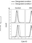
Flow Cytometric Analysis of Autophagic Activity with Cyto-ID Staining in Primary Cells

使用Cyto-ID染色原代细胞并用流式细胞法分析其自噬活性

MS Metodi Stankov
Diana  Panayotova-Dimitrova Diana Panayotova-Dimitrova
ML Martin Leverkus
Jan-Henning  Klusmann Jan-Henning Klusmann
Georg  Behrens Georg Behrens
26606 Views
Apr 5, 2014
Flow cytometry allows very sensitive and reliable high-throughput analysis of autophagic flux. This methodology permits to screen cells in flow and capture multi-component images. Using this technology autophagic flux may be analysed accurately in both suspension as well as adherent cells upon trypsinization independent of how heterogeneous the autophagosomal content might be. The method is based on Cyto-ID staining of autophagic compartments (pre-autophagosomes, autophagosomes, and autophagolysosomes) in live cells using Cyto-ID® Autophagy Detection Kit. Autophagic compartments are intermediate constituents of a dynamic lysosomal degradation process and their intracellular abundance at a particular time point is a function of the established equilibrium between their generation and degradation. Determination of autophagic flux facilitates the discrimination between early induction of autophagosome formation and late inhibition of autophagosome maturation as both results in an ultimate increase in autophagosomal presence. Cyto-ID assay is based on the usage of a specific dye that selectively stains autophagic compartments and therefore allows determination of autophagic flux as accumulation of stained compartments in basic or activated conditions [rapamycin (1-5 µmol/L), PP242 (1-5 µmol/L) or Hanks’ Balanced Salt Solution containing 6 mmol/L glucose (starvation medium)] after blockage of autophagolysosomal degradation using lysosomotropic compounds such as ammonium chloride (NH4Cl) (10-20 mmol/L) or chloroquine (CQ) (5-10 µmol/L). ΔMFI Cyto-ID = MFI Cyto-ID (+CQ/NH4Cl) - MFI Cyto-ID (-CQ/NH4Cl).
采用LC3B-GFP荧光对自噬活性进行流式细胞检测分析

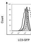
Flow Cytometric Analyses of Autophagic Activity using LC3-GFP fluorescence

采用LC3B-GFP荧光对自噬活性进行流式细胞检测分析

MS Metodi Stankov
Diana  Panayotova-Dimitrova Diana Panayotova-Dimitrova
ML Martin Leverkus
Georg  Behrens Georg Behrens
13382 Views
Apr 5, 2014
Flow cytometry allows very sensitive and reliable high-throughput analysis of autophagic flux. This methodology permits to screen cells in flow and capture multi-component images. Using this technology autophagic flux may be analysed accurately in both suspension as well as adherent cells upon trypsinization independent of how heterogeneous the LC3 punctae content might be. The method is based on the fact that intra-cellularly expressed LC3-GFP serves as a potential autophagic substrate for degradation. Therefore changes in total intracellular LC3-GFP fluorescence intensity is used as an indicator of cellular autophagic activity in living cells. Increased autophagic flux is expected to result in a progressive delivery of LC3-GFP to autolysosome where this substrate undergoes degradation. Therefore, enhanced autophagic flux is detected as a decreased total cellular GFP signal. On the other hand an inhibition of autophagic flux independent of the stage (autophagosome formation, maturation or acidification) leads to accumulation of undegraded LC3-GFP and may be detected as an enhanced intracellular GFP signal. (Caution: This methodology is based on the assumption that LC3-GFP is expressed constitutively by the model system. Data from analysis of substances or conditions influencing cellular LC3-GFP expression should be interpreted with care.)
荧光显微法分析药物对自噬体形成的影响

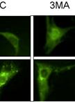
Fluorescence Microscopy Analysis of Drug Effect on Autophagosome Formation

荧光显微法分析药物对自噬体形成的影响

MS Metodi Stankov
Diana  Panayotova-Dimitrova Diana Panayotova-Dimitrova
ML Martin Leverkus
Georg  Behrens Georg Behrens
11251 Views
Apr 5, 2014
The autophagy protein, LC3 represents a reliable characteristic marker for autophagosomal structures. The initial LC3 is processed by the cysteine protease autophagy-related gene 4 (Atg4) at its C terminus in order to create LC3-I generally localized in the cytoplasm. Afterwards LC3-I is conjugated with phosphatidylethanolamine (PE) to become LC3-PE or LC3-II predominantly localised on the autophagosomal membranes (outer and inner). Autolysosomal content of LC3-II is very low as upon autophago/lysosomal fusion it is either cleaved off from the outer membrane by Atg4 or degraded together with the inner membrane by the lysosomal activity. Therefore GFP-LC3 and mCherry-GFP-LC3 might be visualized by conventional or confocal fluorescence microscopy (FM). In this situation mCherry-GFP-LC3 or GFP-LC3 cytoplasmic pool is visualized as a homogeneously dispersed signal and mCherry-GFP-LC3-II or GFP-LC3-II containing autophagosomes are detected as punctae formations. The number of punctae may be used as marker of autophagosomal abundance. In general we recommend counting the average number of GFP-LC3 punctae per cell.
天然BAD-1与肝素琼脂糖的结合

Native BAD-1 Binding to Heparin-agarose

天然BAD-1与肝素琼脂糖的结合

TB T. Tristan Brandhorst
7184 Views
Apr 5, 2014
BAD-1 is an adhesin created by the dimorphic fungus Blastomyces dermatitidis, the causative agent of blastomycosis. We have determined that it has an affinity for heparin, which may explain its impact on virulence and human immune function as a number of cells related to immune function have heparin like moieties on their surfaces. This assay allows a quantification of binding between soluble BAD-1 and immobilized heparin.

分子生物学

电泳迁移率实验

Electrophoresis Mobility Shift Assay

电泳迁移率实验

MN Masaru Nakata
Masaru  Ohme-Takagi Masaru Ohme-Takagi
17631 Views
Apr 5, 2014
Protein (transcription factors and/or transcription cofactors)-binding to DNA is a critical event in regulation of transcription. Electrophoresis Mobility Shift Assay (EMSA), also known as gel shift assay, is a useful tool to detect protein- or protein complex-DNA/RNA interaction and to evaluate DNA binding specificity of transcription factors in vitro. Here we describe a simple method for EMSA with fluorescent dye-bound oligo DNA probes and recombinant protein expressed in bacterial cells. Using fluorescent dye instead of radioisotope enables easy handling and long-term storage of labelled-probes without reduction of detection sensitivity.

植物科学

花青素含量的定量测定

Quantification of Anthocyanin Content

花青素含量的定量测定

MN Masaru Nakata
Masaru  Ohme-Takagi Masaru Ohme-Takagi
28752 Views
Apr 5, 2014
Anthocyanins are a class of flavonoids and important plant pigments. They attract insects to pollinate flowers, protect plants from UV irradiation, and act as antimicrobial agents against herbivores and pathogens. Biosynthesis of anthocyanin is stimulated by diverse developmental signals and environmental stresses including drought, wounding, pathogen infection and insect attack. Plant hormones such as jasmonates, a stress-related plant hormone, also induce accumulation of anthocyanins. Sensitivity of plants to these stress stimuli can be measured by accumulation of anthocyanins. Here we describe a simple method for measurement of anthocyanins in Arabidopsis thaliana seedlings. Amount of anthocyanins are calculated only from absorbances at 530 and 657 nm of crude extract.
种皮钌红染色试验

Seed Coat Ruthenium Red Staining Assay

种皮钌红染色试验

HM Heather E. McFarlane
DG Delphine Gendre
TW Tamara L. Western
19488 Views
Apr 5, 2014
The goal of this protocol is to assay for defects in synthesis/secretion/release of seed coat mucilage by ruthenium red staining of mature whole seeds.The mucilage secretory cells of the Arabidopsis seed coat synthesize and secrete a large quantity of primarily pectinaceous mucilage to a ring-shaped apical domain during their differentiation. This makes them an excellent model system to identify genes involved in both cell wall synthesis and secretion (et al., 2000). When wild-type seeds are incubated in ruthenium red stain, hydrated mucilage is extruded from epidermal cells and a ‘halo’ of red-stained mucilage is observed surrounding the seed (Western et al., 2000). Reduced mucilage staining may result from defects in cell wall biosynthesis, secretion, or impaired release upon hydration.
拟南芥幼苗中同源重组频率的测定

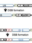
Measuring Homologous Recombination Frequency in Arabidopsis Seedlings

拟南芥幼苗中同源重组频率的测定

Marisa  Rosa Marisa Rosa
OS Ortrun Mittelsten Scheid
12575 Views
Apr 5, 2014
Somatic homologous recombination (SHR) is a major pathway of DNA double-strand break (DSB) repair, in which intact homologous regions are used as a template for the removal of lesions. Its frequency in plants is generally low, as most DSB are removed by non-homologous mechanisms in higher eukaryotes. Nevertheless, SHR frequency has been shown to increase in response to various chemical and physical agents that cause DNA damage and/or alter genome stability (reviewed in March-Díaz and Reyes, 2009). We monitor the frequency of SHR in transgenic Arabidopsis seedlings containing recombination substrates with two truncated but overlapping parts of the β-glucuronidase (GUS) reporter gene (Orel et al., 2003; Schuermann et al., 2005). Upon an SHR event, a functional version of the transgene can be restored (Figure 1A). A histochemical assay applicable to whole plantlets allows the visualization of cells in which the reporter is restored, as the encoded enzyme converts a colorless substrate into a blue compound. This type of reporter has been extensively used to identify gene products required for regulating SHR levels in plants. We analyze plants stimulated for SHR by treatments with DNA damaging agents (bleocin, mitomycin C and UV-C) and compare them to non-treated plants.
拟南芥幼苗的DNA损伤敏感性实验

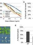
DNA Damage Sensitivity Assays with Arabidopsis Seedlings

拟南芥幼苗的DNA损伤敏感性实验

Marisa  Rosa Marisa Rosa
OS Ortrun Mittelsten Scheid
10780 Views
Apr 5, 2014
We describe fast and reproducible sensitivity assays to quantify the response of Arabidopsis seedlings of different genotypes to a wide range of DNA damaging agents. We apply (1) γ-irradiation, which produces DNA breaks, (2) bleocin, a radiomimetic drug, (3) mitomycin C, a DNA intrastrand cross-linker, (4) hydroxyurea, an inhibitor of DNA synthesis and (5) UV-C, which causes mainly photoproducts. The “true leaf assay” and the “UV resistance assay” are based on easily determined phenotypes as readouts. Using a set of diverse damaging agents combined with different readouts allows establishing relative sensitivity/resistance compared to a reference line, e.g. wild type, determining the most effective type of induced damage and the potential repair pathway affected.

干细胞

3D 乳腺集落形成细胞培养方法

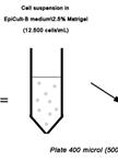
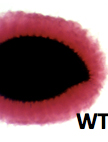
3D Mammary Colony-Forming Cell Assay

3D 乳腺集落形成细胞培养方法

Giusy  Tornillo Giusy Tornillo
Sara  Cabodi Sara Cabodi
16611 Views
Apr 5, 2014
The mammary epithelium consists of multiple phenotypically and functionally distinct cell populations, which are organized as a hierarchy of stem cells, progenitors and terminally differentiated cells. Identification of the mechanisms regulating the growth and differentiation of mammary stem and progenitor cells is of great interest not only to better understand the mammary gland development but also to clarify the origins of breast cancer, as these cells seem to be the likely targets of malignant transformation within the mammary epithelium. Hence, a variety of approaches have been developed for quantifying and studying these specific mammary cell subsets. Given their high proliferative capacity, mammary progenitor cells are able to form colonies in vitro in low-density cultures. Here we describe how to perform a three dimensional (3D) Mammary Colony-Forming Cell (Ma-CFC) Assay, an in vitro functional assay suitable for the detection and analysis of mammary progenitor cells in feeder-free culture conditions. Briefly, this protocol involves the seeding of mammary single cells, at clonal density, onto a semi-solid matrix (Matrigel), thus allowing mammary progenitors to proliferate and give rise to discrete 3D colonies. The number and the cell composition of the resulting colonies will vary according to the frequency and the differentiation potential of the progenitors, respectively.