往期刊物2013

卷册: 3, 期号: 20

left

Dec

20

Dec

5

Nov

20

Nov

5

Oct

20

Oct

5

Sep

20

Sep

5

Aug

20

Aug

5

Jul

20

Jul

5

Jun

20

Jun

5

May

20

May

5

Apr

20

Apr

5

Mar

20

Mar

5

Feb

20

Feb

5

Jan

20

Jan

5

right

癌症生物学

软琼脂锚着非依赖性分析

Soft Agar Anchorage-independent Assay

软琼脂锚着非依赖性分析

LW Li-Ting Wang
SH Shih-Hsien Hsu
14649 Views
Oct 20, 2013
Chronic inflammation drives initiation of hepatocellular carcinoma (HCC), but the underlying mechanisms linking inflammation and tumor formation remain obscure. In this study, soft agar anchorage-independent assay were used to determine tumor transform activity of hepatoma cells with ISX over expression or knockdown in vitro.
异种移植肿瘤增生分析

Xenograft Tumor Growth Assay

异种移植肿瘤增生分析

LW Li-Ting Wang
SH Shih-Hsien Hsu
13507 Views
Oct 20, 2013
Chronic inflammation drives initiation of hepatocellular carcinoma (HCC), but the underlying mechanisms linking inflammation and tumor formation remain obscure. In this study, Xenograft tumor assay was used to determine the tumorigenic activity of hepatoma cells with ISX over expression on nude mice in vivo.
流动状态下T淋巴细胞对肿瘤细胞的粘着强度分析

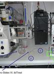
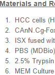
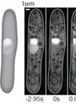
Protocol for T-cell Adhesion Strength on Tumor Cells under Flow Conditions

流动状态下T淋巴细胞对肿瘤细胞的粘着强度分析

Marie  Boutet Marie Boutet
KF Katarzyna Franciszkiewicz
Audrey Le  Floc’h Audrey Le Floc’h
Fathia  Mami-Chouaib Fathia Mami-Chouaib
9998 Views
Oct 20, 2013
This method allows evaluating the relative adhesion strength between T lymphocytes and specific adherent target cells using a shear force in flow chambers. It is based on the measure of the resistance of conjugates formed between T cells and adherent tumor cells to shear stress in a microfluidic system. For this purpose, T cells, stained with a CellTracker probe, are added into flow channels containing a monolayer of adherent target cells and their progressive detachment under a constant shear stress is then recorded using a fluorescent microscope.

细胞生物学

通过双分子荧光互补(BiFC)试验进行体内蛋白质之间的相互作用的可视化分析

Bimolecular Fluorescence Complementation (BiFC) Assay for Direct Visualization of Protein-Protein Interaction in vivo

通过双分子荧光互补(BiFC)试验进行体内蛋白质之间的相互作用的可视化分析

Hsien-Tsung Lai Hsien-Tsung Lai
Cheng-Ming  Chiang Cheng-Ming Chiang
23325 Views
Oct 20, 2013
Bimolecular Fluorescence Complementation (BiFC) assay is a method used to directly visualize protein-protein interaction in vivo using live-cell imaging or fixed cells. This protocol described here is based on our recent paper describing the functional association of human chromatin adaptor and transcription cofactor Brd4 with p53 tumor suppressor protein (Wu et al., 2013). BiFC was first described by Hu et al. (2002) using two non-fluorescent protein fragments of enhanced yellow fluorescent protein (EYFP), which is an Aequorea victoria GFP variant protein, fused respectively to a Rel family protein and a bZIP family transcription factor to investigate interactions between these two family members in living cells. The YFP was later improved by introducing mutations to reduce its sensitivity to pH and chloride ions, thus generating a super-enhanced YFP, named Venus fluorescent protein, without showing diminished fluorescence at 37 °C as typically observed with EYFP (Nagai et al., 2006). The fluorescence signal is regenerated by complementation of two non-fluorescent fragments (e.g., the Venus N-terminal 1-158 amino acid residues, called Venus-N, and its C-terminal 159-239 amino acid residues, named Venus-C; see Figure 1A and Gully et al., 2012; Ding et al., 2006; Kerppola, 2006) that are brought together by interaction between their respective fusion partners (e.g., Venus-N to p53, and Venus-C to the PDID domain of human Brd4; see Figure 1B and 1C). The intensity and cellular location of the regenerated fluorescence signals can be detected by fluorescence microscope. The advantages of the proximity-based BiFC assay are: first, it allows a direct visualization of spatial and temporal interaction between two partner proteins in vivo; second, the fluorescence signal provides a sensitive readout for detecting protein-protein interaction even at a low expression level comparable to that of the endogenous proteins; third, the intensity of the fluorescence signal is proportional to the strength of protein-protein interaction (Morell et al., 2008); and fourth, the BiFC signals are derived from intrinsic protein-protein interaction, rather than from extrinsic fluorophores that may not reflect true protein-protein interaction due to their nonspecific association with cellular macromolecules or subcellular compartments. However, some limitations of BiFC include slow maturation (T1/2 ~ 1 hour) of an eventually stable BiFC complex (Hu et al., 2002), making it unsuitable for real-time observation of transient interaction that disappears prior to BiFC detection, and enhanced BiFC background at high expression levels due to fusion-independent association between two non-fluorescent fragments association. BiFC signals generated by in vivo protein-protein interaction can be validated by amino acid mutation introduced at the protein-protein contact surfaces. This imaging technique has been widely used in different cell types and organisms (Kerppola, 2006).
采用荧光漂白恢复法(FRAP)测定接合蛋白质的动力学

Measurement of Junctional Protein Dynamics Using Fluorescence Recovery After Photobleaching (FRAP)

采用荧光漂白恢复法(FRAP)测定接合蛋白质的动力学

Rashmi  Priya Rashmi Priya
GG Guillermo A. Gomez
13566 Views
Oct 20, 2013
Fluorescence Recovery After Photobleaching (FRAP) (Lippincott-Schwartz et al., 2003; Reits and Neefjes, 2001) was employed to determine dynamic properties of proteins localized at the ephitelial zonula adherens (ZA) (Kovacs et al., 2011; Otani et al., 2006). The proteins of interest were expressed in cells using a knockdown and reconstitution approach in which endogenous proteins were depleted by RNA interference (RNAi) and replaced by expression of an RNAi-resistant gene fused to GFP (Priya et al., 2013; Smutny et al., 2010; Smutny et al., 2011; Vitriol et al., 2007). By choosing expression levels of GFP-tagged proteins that were comparable to endogenous levels, we minimized transient overexpression artifacts due to overcoming regulatory mechanisms that directly affect protein dynamics (Goodson et al., 2010). Using this approach, junctional E-cadherin-GFP or GFP-Ect2 were subjected to FRAP analysis in small areas corresponding to the ZA using confocal microscopy (Priya et al., 2013; Ratheesh et al., 2012; Gomez et al., 2005; Trenchi et al., 2009). Although in principle this approach is similar in every case, bleaching conditions, acquisition parameters and analysis details might differ depending on the time scale of the recovery process (Lippincott-Schwartz et al., 2003). In this protocol we will describe the experimental procedure to perform FRAP experiments and how to optimize bleaching and acquisition conditions for optimal measurements of protein dynamics at cell-cell junctions.
裂殖酵母细胞核的荧光光漂白技术(FRAP)

Fluorescence Recovery After Photobleaching (FRAP) in the Fission Yeast Nucleus

裂殖酵母细胞核的荧光光漂白技术(FRAP)

Petrina  Delivani Petrina Delivani
MC Mariola R. Chacón
BS Britta Schroth-Diez
Iva M. Tolić-Nørrelykke Iva M. Tolić-Nørrelykke
10559 Views
Oct 20, 2013
We use fluorescence recovery after photobleaching (FRAP) to calculate the diffusion coefficient of GFP in the nucleoplasm of fission yeast. The FRAP method can be generally used to measure the mobility of proteins inside the cell or its organelles. In our experiment we only measured the diffusion of GFP inside the nucleoplasm of fission yeast mitotic cells. However, if GFP is fused to a protein, the mobility of the protein of interest can be calculated following the GFP signal in the bleached area. We did not, however, address this in our experiments; therefore other sources could be searched for this topic. To compare FRAP and FLIP, both techniques can be used to measure protein mobility inside a cell. However, with FRAP, the diffusion of a protein is measured in the region of interest (ROI), to observe the recovering of fluorescence in this area. In FLIP, fluorescence recovery is measured in an area different from where the bleaching was done, to observe whether the tagged protein is able to move into that area, which would become darker, gaining the bleached proteins. The major difference here is that for FRAP a single bleaching event is sufficient, while FLIP requires a number of bleaching steps, in order to avoid reflux of fluorescent protein in the same region.Technically FRAP in the nucleus und FRAP in the cytoplasm has no difference. However, we measured a difference between the diffusion coefficient inside the nucleus (D = 5.6 ± 2.8 μm2/s) and in the cytoplasm (D = 8.6 ± 2.2 μm2/s). This is due to the different compositions inside these compartments, consisting of differing amounts of proteins, DNA and RNA.
胚胎心肌细胞的肥大分析和量化

Hypertrophy Analysis and Quantification in Embryonic Cardiomyocites

胚胎心肌细胞的肥大分析和量化

GL Guillermo Luxán
JP José Luis de la Pompa
9617 Views
Oct 20, 2013
Myocardial growth goes from proliferation to hypertrophy during development. The measurement of the relative cell area provides information of cardiomyocyte hypertrophy, which is ideal for studying myocardial development.
从外周血液中分离全单核细胞

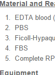
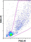
Isolation of Whole Mononuclear Cells from Peripheral Blood

从外周血液中分离全单核细胞

BM Beatriz Martínez-Poveda
GL Guillermo Luxán
JP José Luis de la Pompa
8056 Views
Oct 20, 2013
Whole mononuclear cells from peripheral blood are an easy to obtain and useful population of cells where protein and expression patterns of genes can be studied in patients.
结肠组织免疫电镜分析

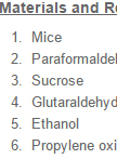
Colon Tissue Immunoelectron Microscopy

结肠组织免疫电镜分析

MI Megumi Iwano
Akio  Tsuru Akio Tsuru
KK Kenji Kohno
7455 Views
Oct 20, 2013
The method described here is intended to study intracellular localization of proteins in colon cells. This protocol was used to localize IRE1β in the endoplasmic reticulum membrane. We used anti-IRE1β antibody raised in guinea pig for this purpose. We also studied the location of BiP (also known as GRP78), with the antibody raised in rabbit. Both antibodies used with appropriate gold particle-conjugated secondary antibodies gave good results. Primary mouse antibodies are not recommended because secondary anti-mouse antibodies also react with the internal mouse epitopes.

免疫学

T淋巴细胞与树突细胞以及T淋巴细胞之间的体外簇集试验

In vitro T Cell–DC and T Cell–T Cell Clustering Assays

T淋巴细胞与树突细胞以及T淋巴细胞之间的体外簇集试验

AG Audrey Gérard
15029 Views
Oct 20, 2013
To get activated, T cells need to find their cognate antigen at the surface of an antigen-presenting cell (APC). Recognition of cognate antigen in the context of the MHC (Major histocompatibility complex) by the TCR (T-Cell Receptor) results in long lasting interactions between T cells and APCs. Subsequently, T cells form homotypic interactions with each other, which is seen as a hallmark of T cell activation. This protocol describes a method to analyze T-APC and T-T conjugation.

微生物学

表面多糖提取和量化

Surface Polysaccharide Extraction and Quantification

表面多糖提取和量化

CB Cedric Arthur Brimacombe
John  Thomas Beatty John Thomas Beatty
18959 Views
Oct 20, 2013
Gram-negative bacterial cells possess two membranes - the inner cytoplasmic membrane and the outer membrane. The two membranes are distinct in their composition; the inner membrane is composed of a phospholipid bilayer, whereas the outer membrane (OM) is composed of an asymmetrical bilayer, with the outer leaflet containing lipopolysaccharide (LPS) (Raetz and Whitfield, 2002). Surface polysaccharides, such as LPS O-antigen, or capsular polysaccharide, are often tightly associated with the OM (Whitfield, 2006). This tight association can be used to generate a rough quantification of surface polysaccharides of Gram-negative bacterial cells, as the OM can easily be dissociated from cells without associated cell lysis (Brimacombe et al., 2013). The following method describes how to quickly extract and quantify OM-associated polysaccharides.
靶细胞中HIV-1 逆转录的细胞分级分离和量化分析

Cell Fractionation and Quantitative Analysis of HIV-1 Reverse Transcription in Target Cells

靶细胞中HIV-1 逆转录的细胞分级分离和量化分析

Vaibhav B  Shah Vaibhav B Shah
CA Christopher Aiken
10350 Views
Oct 20, 2013
This is a protocol to detect HIV-1 reverse transcription products in cytoplasmic and nuclear fractions of cells infected with VSV-G-pseudotyped envelope-defective HIV-1. This protocol can also be extended to HIV-1 with regular envelope.
白色念珠菌中组蛋白H4乙酰化和组蛋白H2A磷酸化的免疫印迹分析

Immunoblot Analysis of Histone H4 Acetylation and Histone H2A Phosphorylation in Candida albicans

白色念珠菌中组蛋白H4乙酰化和组蛋白H2A磷酸化的免疫印迹分析

Michael  Tscherner Michael Tscherner
Karl Kuchler Karl Kuchler
9245 Views
Oct 20, 2013
Posttranslational modifications of histones are required for different processes including transcription, replication and DNA damage repair. This protocol describes the preparation of a whole-cell extracts for the fungal pathogen Candida albicans. Furthermore, the extract is used to detect lysine acetylation of histone H4 as well as serine 129 phosphorylation of histone H2A by immunoblot analysis.

神经科学

[3H]-螺旋哌丁苯与多巴胺D2、D3 和D4受体的饱和结合

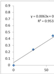
[3H]-Spiperone Saturation Binding to Dopamine D2, D3 and D4 Receptors

[3H]-螺旋哌丁苯与多巴胺D2、D3 和D4受体的饱和结合

JW Jan-Peter van Wieringen
MM Martin C. Michel
10222 Views
Oct 20, 2013
This protocol is intended for use in 96 well plates (1,200 μl wells) but it can similarly be applied to standard test tubes (Levant, 2007). D2, D3, and D4 dopamine receptors are members of the D2-like class of dopamine receptors. They can be studied using the radioligand [3H]-spiperone, which is an antagonist binding to D2, D3, and D4 receptors with comparable affinity. A saturation assay can be used to determine the affinity of a radioligand to a receptor (Kd) and to determine the total number of receptors present in the assay (Bmax). If saturation binding experiments are performed in the absence and presence of a fixed concentration of another, not radiolabeled ligand, it can also be determined whether the other ligand acts in a competitive manner. If the specific radioactivity is low (tritiated) relative to the affinity of the radioligand (e.g. expressed recombinant receptors). To obtain reliable estimates of these parameters at least 6 different concentrations of radioligand must be tested, but particularly when a receptor is first detected in a given tissue or cell type a greater number of concentrations are helpful. The incubation time and temperature are chosen to allow formation of equilibrium between association and dissociation with the receptor for both radioligand and competitor. Each experiment can be divided into different steps such as assay preparation, membrane preparation, incubation, filtration, counting of the samples and data analysis. To minimize experimental error all assays are performed at least in duplicate. Radioligand dilutions should be prepared to cover the desired concentration range. Optimally these concentrations should cover both the high range (corresponding to 5-10x Kd and hence saturation of the receptor) and the low range (around Kd), so that both Kd and Bmax can reliably be estimated without undue extrapolation. At each radioligand concentration total and non-specific binding should be determined; the agent used for the definition of non-specific binding (NSB) should be chemically (different family) and physically (avoid combination of two lipophilic compounds) distinct from the radioligand to avoid artifacts. For discussion of specific benefits of chosen assay conditions see van Wieringen et al. (2013) (copy can be obtained from the author).
[3H]-螺旋哌丁苯与多巴胺D2、D3和 D4受体的竞争性结合

[3H]-Spiperone Competition Binding to Dopamine D2, D3 and D4 Receptors

[3H]-螺旋哌丁苯与多巴胺D2、D3和 D4受体的竞争性结合

JW Jan-Peter van Wieringen
MM Martin C. Michel
8905 Views
Oct 20, 2013
This protocol is intended for use in 96 well plates (1,200 μl wells) but it can similarly be applied to standard test tubes (Levant, 2007). D2, D3, and D4 dopamine receptors are members of the D2-like class of dopamine receptors. They can be studied using the radioligand [3H]-spiperone, which is an antagonist binding to D2, D3, and D4 receptors with comparable affinity. A competition experiment is usually performed to determine the affinity of a compound for a receptor. If multiple subtypes or states of the receptor are present and the competing compound differentiates them, a competition binding experiment can quantify the relative contribution of the two subtypes or states; while resolution of more than two subtypes or states is theoretically possible, in practical terms it is almost never feasible. Thus, radioligand binding to a receptor is quantified in the presence of various concentrations of the unlabelled compound of interest. The concentration of the radioligand in a competition study should be about 2-3 its Kd value as determined in saturation binding; this will allow a sufficient occupancy of the receptor to obtain a strong signal and at the same time avoid that competition becomes too difficult due to high radioligand concentration. The incubation time and temperature are chosen to allow formation of equilibrium between association and dissociation with the receptor for both radioligand and competitor. Of note, a simple competition experiments does not necessarily prove a competitive nature of the interaction between unlabelled drug and receptor. If the specific radioactivity is low (tritiated) relative to the affinity of the radioligand (e.g. expressed recombinant receptors). The number of required competitor concentrations depends on the goal of the experiment. If only a rough estimate of antagonist potency is required, 1-2 concentration per log increment will be sufficient. However, if it is the aim to test for possible subtypes or states of the receptor, 3-5 concentrations per log increment are needed. If possible, the lowest competitor concentrations in the assays should not cause any detectable inhibition, whereas the highest concentrations should completely abolish specific binding. Each experiment can be divided into different steps such as assay preparation, membrane preparation, incubation, filtration, counting of the samples and data analysis. To minimize experimental error all assays are performed at least in duplicate. Additionally, duplicates of total binding and non-specific binding should be included in the assay; the agent used for the definition of non-specific binding (NSB) should be chemically (different family) and physically (avoid combination of two lipophilic compounds) distinct from the radioligand to avoid artifacts. For discussion of specific benefits of chosen assay conditions see van Wieringen et al., (2013) (copy can be obtained from the author).

植物科学

高通量检测病毒感染植株中siRNA

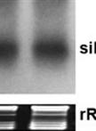
A High Resolution Short Interfering RNA (siRNA) Detection Method from Virus-infected Plants

高通量检测病毒感染植株中siRNA

Vinay  Panwar Vinay Panwar
Guus  Bakkeren Guus Bakkeren
12770 Views
Oct 20, 2013
Plant viruses are strong inducers as well as targets of RNA silencing. In plants RNA silencing acts as a natural defense mechanism against viral infection and is associated with accumulation of virus-specific small interfering RNAs (siRNAs). The continuing discoveries, increasing awareness and interest in the regulatory roles of non-coding small RNAs have raised the need for methods that can reliably detect and quantitate the expression levels of small RNAs. Northern blot analysis of small RNAs involving the separation of RNA molecules using polyacrylamide gel electrophoresis (PAGE) has remained a popular and valuable analytical method to validate small RNAs. Northern blot analysis consist of resolving RNAs by gel electrophoresis, followed by transferring and fixing to nylon membranes as well as detecting by hybridization using radioactive probes. The following protocol provides a method for isolation and detection of small RNAs from virus-infected plants and was successfully used in Panwar et al. (2013a), Panwar et al. (2013b).
一种适用于利用96孔板测定海藻糖酶活性的方法

Trehalase Activity in Arabidopsis thaliana Optimized for 96-well Plates

一种适用于利用96孔板测定海藻糖酶活性的方法

Hilde Van  Houtte Hilde Van Houtte
Patrick Van  Dijck Patrick Van Dijck
12186 Views
Oct 20, 2013
Trehalose is a nonreducing disaccharide. It is a common sugar in bacteria, fungi and yeast, where it functions as a carbon source and stress protectant. In contrast, plants, although encoding large trehalose biosynthesis gene families, contain only small amounts of trehalose. The intermediate compound of trehalose, trehalose-6-phosphate (T6P), is a signaling molecule in plants, regulating metabolism, growth, and development. Most plants contain only a single trehalase, the enzyme that specifically hydrolyzes trehalose into two glucose molecules. High trehalase activity has been suggested to be part of the defense mechanism in plants hosting mycorrhizal fungi, rhizobia, and the plant pathogen Plasmodiophora brassica. Recently, it was shown in Arabidopsis thaliana that high trehalase activity is associated with an increase in drought stress tolerance and that trehalase fulfills an important role in stomatal regulation. Here we describe a protocol for measuring trehalase activity in Arabidopsis tissues, optimized for 96-well plates. Dialyzed protein extracts will be incubated with trehalose, followed by the quantitation of the released glucose using glucose oxidase-peroxidase.

干细胞

小鼠降结肠和空肠薄切片的免疫标记

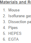
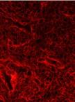
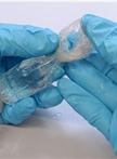
Immunolabelling of Thin Slices of Mouse Descending Colon and Jejunum

小鼠降结肠和空肠薄切片的免疫标记

JB Julien Bellis
ID Isabelle Duluc
JF Jean-Noël Freund
Jan R.  De Mey Jan R. De Mey
9526 Views
Oct 20, 2013
This protocol describes a method for efficient immunolabelling of thin tissue slices containing a few rows of intact intestinal crypts, which yields large numbers of them being oriented favorably for recording stacks of optical sections aligned with the crypt long axis (Bellis et al., 2012). The latter can then be used for cell positional analysis, 3D-reconstruction and -analysis. The simple epithelium lining the small intestine is organized into contiguous crypts of Lieberkühn (Potten, 1998; Barker et al., 2012; De Mey and Freund, 2013) several of which making up a crypt/villus unit. Each crypt is a multicellular proliferation unit with a tight hierarchical organization. Under steady state conditions, the epithelium is continuously and rapidly renewed, driven by divisions of multipotent intestinal SCs near the crypt base and cell removal from the villus tip. Techniques for analyzing the organization of the crypts play an important role in the field. Maximal efficiency is obtained by using optical sections obtained from confocal scanning and/or Nomarski optics passing through the center of the longitudinal crypt axis to view the crypt as two cell columns of hierarchical lineage starting from cells positioned at or near the crypt base. This enables positional analysis of certain cellular capacities like performing DNA synthesis, undergoing mitosis and apoptosis (Caldwell et al., 2007; Fleming et al., 2007; Quyn et al., 2010), responding to injury (Potten et al., 1997), or expressing genes (Barker et al., 2012; Bjerknes et al., 2012; Itzkovitz et al., 2012). Our protocol has allowed us to demonstrate that some divisions are asymmetric with respect to cell fate and the occurrence of oriented cell division (OCD) in 80% of the proliferating cells in the upper stem cell and transit amplifying zones. It has further revealed planar cell polarities which are important for crypt homeostasis and stem cell biology and alterations in apparently normal crypts and microadenomas of mice carrying germline Apc mutations shedding new light on the first stages of progression towards colorectal cancer.