往期刊物2013

卷册: 3, 期号: 15

left

Dec

20

Dec

5

Nov

20

Nov

5

Oct

20

Oct

5

Sep

20

Sep

5

Aug

20

Aug

5

Jul

20

Jul

5

Jun

20

Jun

5

May

20

May

5

Apr

20

Apr

5

Mar

20

Mar

5

Feb

20

Feb

5

Jan

20

Jan

5

right

生物化学

在大肠杆菌K12菌株中进行蛋白原位免疫标记

Immunolabeling of Proteins in situ in Escherichia coli K12 Strains

在大肠杆菌K12菌株中进行蛋白原位免疫标记

NB Nienke Buddelmeijer
MA Mirjam Aarsman
Td Tanneke den Blaauwen
12578 Views
Aug 5, 2013
This protocol was developed to label proteins in bacterial cells with antibodies conjugated to a fluorophore for fluorescence microscopy imaging. The procedure is optimized to minimize morphological changes and also to minimize the amount of antibodies needed for the staining. The protocol can also be used with primary antibodies conjugated to a fluorophore. The method has been verified extensively (van der Ploeg et al., 2013), but it should be noted that one case in Caulobacter crescentus (Hocking et al., 2012) has been reported in which the localization of a protein changed upon fixation by formaldehyde/glutaraldehyde. However, the localization of the same protein in E. coli did not change.

癌症生物学

制备小鼠肺上皮细胞

Generation of Mouse Lung Epithelial Cells

制备小鼠肺上皮细胞

AK Andrea L. Kasinski
FS Frank J. Slack
13618 Views
Aug 5, 2013
Although in vivo models are excellent for assessing various facets of whole organism physiology, pathology, and overall response to treatments, evaluating basic cellular functions, and molecular events in mammalian model systems is challenging. It is therefore advantageous to perform these studies in a refined and less costly setting. One approach involves utilizing cells derived from the model under evaluation. The approach to generate such cells varies based on the cell of origin and often the genetics of the cell. Here we describe the steps involved in generating epithelial cells from the lungs of KrasLSL-G12D/+;p53LSL-R172/+ mice (Kasinski and Slack, 2012). These mice develop aggressive lung adenocarcinoma following cre-recombinase dependent removal of a stop cassette in the transgenes and subsequent expression of Kra-G12D and p53R172. While this protocol may be useful for the generation of epithelial lines from other genetic backgrounds, it should be noted that the Kras; p53 cell line generated here is capable of proliferating in culture without any additional genetic manipulation that is often needed for less aggressive backgrounds.
逆转录病毒介导小鼠胚胎成纤维细胞的恶性转化

Retrovirus Mediated Malignant Transformation of Mouse Embryonic Fibroblasts

逆转录病毒介导小鼠胚胎成纤维细胞的恶性转化

Huei San Leong Huei San Leong
Marnie Blewitt Marnie Blewitt
10980 Views
Aug 5, 2013
Cellular transformation is a widely used method to artificially induce cells to form tumours in vivo. Here, we describe the methodology for malignant transformation of mouse embryonic fibroblasts (MEFs) for transplantation into immunodeficient nude mice, as used in Leong et al. (2013). The two-step process involves: 1) down-regulation of Trp53 expression using a short hairpin RNA (shRNA); and 2) overexpression of the oncogenic HRasV12 protein. Reduction of Trp53 expression leads to cell immortalisation, and the subsequent overexpression of oncogenic HRasV12 results in malignant transformation of a cell.

细胞生物学

A549 细胞中的表皮生长因子(GCF)受体内吞作用分析

Epidermal Growth Factor (EGF) Receptor Endocytosis Assay in A549 Cells

A549 细胞中的表皮生长因子(GCF)受体内吞作用分析

SR Sabrina Rizzolio
LT Luca Tamagnone
18626 Views
Aug 5, 2013
The following endocytosis assay has been optimized to assess EGF-stimulated EGFR endocytosis; but could be modified to assess other ligand-stimulated endocytosis of plasma membrane receptors (for which fluorochrome-conjugated ligands are available to track their receptor internalization). In brief, cells are treated with fluorescent EGF at 4 °C to allow binding to the receptor, but not internalization; then, endocytosis is allowed at 37 °C for different timepoints. For the setting up of this protocol we are really indebted to Dr. Letizia Lanzetti (Lanzetti et al., 2000).
血脑障壁和胎盘屏障透过率的分析

Assay of Blood Brain Barrier and Placental Barrier Permeability

血脑障壁和胎盘屏障透过率的分析

SA Saurabh Kumar Agnihotri
PS Poonam Singh
BK Balawant Kumar
PS Pankaj Singh
SJ Swatantra Kumar Jain
MC Mahesh Chandra Tewari
SK Sadan Kumar
Monika Sachdev Monika Sachdev
RT Raj Kamal Tripathi
12144 Views
Aug 5, 2013
Evans blue dye solution was used to observe the effect of a viral protein on two pre-defined barriers of the body i.e. the blood brain barrier (BBB) and the placental barrier (PB). This dye has strong affinity for serum albumin and does not cross these barriers under natural conditions. As all the dye gets bound to albumin, all the neural tissues and embryonic tissues remain unstained. When the BBB and PB are compromised due to the breach of these barriers, albumin-bound Evans blue enters the CNS and the placenta.
绒毛外滋养层细胞的迁移和侵袭试验

Extravillous Trophoblast Migration and Invasion Assay

绒毛外滋养层细胞的迁移和侵袭试验

MA Magdalena Angelova
HM Heather L. Machado
KS Kenneth F. Swan
CM Cindy Morris
DS Deborah E. Sullivan
11033 Views
Aug 5, 2013
Extravillous trophoblast (EVT) migration and invasion through the decidualized endometrium is essential to successful placentation. SGHPL-4 cells, an EVT cell line derived from first trimester placenta, is a widely used model of cytotrophoblast differentiation into an invasive phenotype. Here we describe a quantitative cell migration assay that can be modified to also measure cell invasion. SGHPL-4 cells were seeded into BD Fluoroblok cell culture inserts constructed with an 8 μm porous membrane and allowed to migrate towards epidermal growth factor, a known chemoattractant for EVTs. To assess EVT invasion, Fluoroblok inserts were first coated with Matrigel, a basement membrane matrix. SGHPL-4 cells were labeled with calcein AM and cells that had invaded and/or migrated across the membrane were quantified by a bottom-reading fluorescence plate reader. The advantage of the Fluoroblok inserts over other migration/invasion assays is that they allow nondestructive detection of migrated cells.

免疫学

完整脾脏的流式细胞分析

Whole Spleen Flow Cytometry Assay

完整脾脏的流式细胞分析

CY Cathy S. Yam
AH Adeline M. Hajjar
35613 Views
Aug 5, 2013
In the Whole Spleen Flow Cytometry Assay, we used splenocytes directly ex vivo for stimulation with a variety of TLR ligands. The splenocytes were stimulated for a total of 4 hours, then stained for intracellular cytokines. We then examined cytokine production via flow cytometry. This allowed us to compare the responses of minimally manipulated primary macrophages/monocytes and conventional dendritic cells.
采用PHA活化和Neon电穿孔法转染人幼稚CD4+T淋巴细胞

Transfection of Human Naive CD4+ T Cells with PHA Activation and Neon Electroporation

采用PHA活化和Neon电穿孔法转染人幼稚CD4+T淋巴细胞

Amy Palin Amy Palin
DL David B. Lewis
17340 Views
Aug 5, 2013
Transfection of primary T cells can be challenging. This protocol describes a method to transfect primary human naive CD4+ T cells with an AP-1 luciferase reporter using low-level activation by phytohemagglutinin (PHA) and electroporation, as published (Palin et al., 2013). This technique is a modification of one previously described by our group (Cron et al., 2013). Anyone wishing to transfect murine T cells should consult the publication by Cron et al., 2013. This technique may be adapted for other primary T cell types by optimizing the Neon electroporation conditions, as described in the text. Other luciferase or GFP reporters may be used, and will require optimization of the stimulation conditions for that particular reporter.
肺髓过氧化物酶活性试验

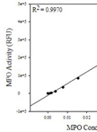
Pulmonary Myeloperoxidase Activity

肺髓过氧化物酶活性试验

TO Tammy Regena Ozment
10587 Views
Aug 5, 2013
Neutrophils are considered one of the first responders of the innate immune response. Their primary activities are to migrate to sites of infection by chemotaxis and trans-migration across the endothelium (Gaines et al., 2005). Once at the site of infection, they phagocytize microbes and kill them. Critical to the neutrophil’s ability to kill microbes are the multiple degradative enzymes contained within granules. The activity of these enzymes is non-specific, and therefore, neutrophils also contribute to tissue damage at the site of infection (Gaines and Berliner, 2005). Measurement of neutrophil infiltration into tissues is one way to gauge the severity of infection, inflammation, and tissue damage (Ayala et al., 2002). Myeloperoxidase is found in the primary granules of neutrophils and is an effective measure of neutrophil infiltration into tissues (Gaines and Berliner, 2005).

微生物学

采用凝胶迁移实验(EMSA)分析Rgg蛋白与DNA的结合

EMSA Analysis of DNA Binding By Rgg Proteins

采用凝胶迁移实验(EMSA)分析Rgg蛋白与DNA的结合

BL Breah LaSarre
MF Michael J. Federle
14513 Views
Aug 5, 2013
In bacteria, interaction of various proteins with DNA is essential for the regulation of specific target gene expression. Electrophoretic mobility shift assay (EMSA) is an in vitro approach allowing for the visualization of these protein-DNA interactions. Rgg proteins comprise a family of transcriptional regulators widespread among the Firmicutes. Some of these proteins function independently to regulate target gene expression, while others have now been demonstrated to function as effectors of cell-to-cell communication, having regulatory activitiesthat that are modulated via direct interaction with small signaling peptides. EMSA analysis can be used to assess DNA binding of either type of Rgg protein. EMSA analysis of Rgg protein activity has facilitated in vitro confirmation of regulatory targets, identification of precise DNA binding sites via DNA probe mutagenesis, and characterization of the mechanism by which some cognate signaling peptides modulate Rgg protein function (e.g. interruption of DNA-binding in some cases).
采用荧光光谱法检测环磷酸腺苷与膜的相互作用

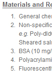
CAMP-Membrane Interactions Using Fluorescence Spectroscopy

采用荧光光谱法检测环磷酸腺苷与膜的相互作用

RS Ron Saar-Dover
YS Yechiel Shai
8302 Views
Aug 5, 2013
The molecular mechanism by which peptide antibiotics (also referred as cationic antimicrobial peptides-CAMPs) penetrate through the bacterial wall barrier, interact with, and disrupt their membrane is complex. It depends mainly on the peptide properties (structure, length, charge and hydrophobicity), on the characteristics of the cell wall matrix and the membrane itself. Here, we present two fluorescence spectroscopic techniques, one for tracking the interaction of CAMPs with membranes, and the other for evaluating the ability of a peptide to cross the bacterial cell-wall and reach the membrane. The fluorescence approach is relatively simple, highly sensitive, non-invasive and allows time-scale investigation. It can be applied to lipid vesicles or intact bacteria. For membrane model systems such as liposomes, it allows to determine the binding kinetics of a peptide to vesicle and to assess the depth of penetration. By using bacterial strains carrying different mutations in their cell wall components, but not in their membrane, we can investigate how a specific element may affect the cell wall permeability to CAMPs (Saar-Dover et al., 2012). In order to track the peptide-membrane interaction we conjugate a lipid environmentally sensitive NBD (7-nitrobenz-2-oxa-1, 3-diazole-4-yl) fluorophore to peptides. NBD fluorescence can increase up to approximately 10-fold upon interaction with membranes. Its high excitation wavelength (467 nm) and the high quantum yield reduce significantly the contribution of light scattering. NBD-labeled peptides exhibit fluorescence emission maxima around 540 nm in hydrophilic solution (Shai, 1999). However, upon interaction with lipid component such as the bacterial membrane, relocation of the NBD group into a more hydrophobic environment results in an increase in its fluorescence intensity and a blue shift of the emission maxima (Chattopadhyay and London, 1987). The first property is used to determine the binding constant of the peptide to the membrane. The second property is exploited to evaluate the depth of penetration (Merklinger et al., 2012; Zhao and Kinnunen, 2002). Here, we will focus on how to determine the binding constant. The advantage of the NBD moiety conjugation is that it allows the use of experimental conditions in which the lipid: peptide molar ratio range from 15,000:1. The addition of NBD does not change the biological function of most of the peptide, as was found for different antimicrobial peptides such as paradaxin (Rapaport and Shai, 1992), dermaseptins (Pouny et al., 1992), cecropins (Gazit et al., 1994) and cathelicidin LL-37 (Oren et al., 1999). However, pre-examination must be done for each newly investigated peptide.

分子生物学

从鲜冻组织中激光捕获显微解剖人宫颈上皮细胞后分离总RNA

Total RNA Isolation after Laser-capture Microdissection of Human Cervical Squamous Epithelial Cells from Fresh Frozen Tissue

从鲜冻组织中激光捕获显微解剖人宫颈上皮细胞后分离总RNA

SW Saskia M Wilting
RS Renske DM Steenbergen
10909 Views
Aug 5, 2013
As most tissue specimens contain a mixture of different cell types including epithelial cells, stromal cells and immune cells, selection of the cells of interest is of utmost importance for the accurate determination of gene/microRNA expression. Laser capture microdissection enables the researcher to obtain homogeneous ultrapure cell selections from heterogeneous starting material. The following protocol was optimized for the isolation of total RNA from cervical (premalignant) squamous epithelial cells from fresh frozen biopsy specimens.

神经科学

小鼠脑组织匀浆的亚细胞分离

Subcellular Fractionation of Mouse Brain Homogenates

小鼠脑组织匀浆的亚细胞分离

Ditte Olsen Ditte Olsen
CG Camilla Gustafsen
13967 Views
Aug 5, 2013
This subcellular fractionation protocol is used for separation of cellular organelles based on their density. We have designed and optimized the protocol for separation of subcellular compartments of brain homogenates with focus on the localization and trafficking of transmembrane proteins, but we have also successfully used this protocol for fractionation of other types of tissue. The protocol has two major steps 1) preparation of homogenate from dissected tissue and 2) separation of organelles by centrifugation of homogenates using a continuous sucrose gradient.
体内神经发生

In vivo Neurogenesis

体内神经发生

Désirée R. M.  Seib Désirée R. M. Seib
Ana Martin-Villalba Ana Martin-Villalba
13523 Views
Aug 5, 2013
This protocol shows how to characterize the dynamics of hippocampal neurogenesis in the adult mouse by describing preparation of brain tissue, immunofluorescence of brain sections and confocal stereotactic cell counting.
神经元形态分析

Neuronal Morphology Analysis

神经元形态分析

Désirée R. M.  Seib Désirée R. M. Seib
Ana Martin-Villalba Ana Martin-Villalba
12093 Views
Aug 5, 2013
This protocol describes how to visualize neuronal morphology and how to determine neuronal complexity of immature and mature hippocampal neurons in the mouse in vivo including tissue preparation, staining of brain sections and confocal cell analysis.
从小鼠脑部分离神经元生长锥

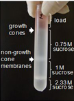
Isolation of Growth Cones from Mouse Brain

从小鼠脑部分离神经元生长锥

Iryna  Leshchyns’ka Iryna Leshchyns’ka
Vladimir Sytnyk Vladimir Sytnyk
11401 Views
Aug 5, 2013
Growth cones are motile structures at the tips of growing neurites, which play an essential role in regulation of growth and navigation of growing axons and dendrites of neurons in the developing nervous system. This protocol describes isolation of growth cones from the brain tissue from young mice. Growth cones isolated using this protocol have been extensively characterized using electron microscopy (Pfenninger et al., 1983) and may be used for any kind of subsequent biochemical and/or functional analyses, including Western blot analysis of protein expression (Westphal et al., 2010), analysis of the activity of growth cone-accumulated enzymes (Leshchyns’ka et al., 2003; Li et al., 2013), and analysis of the endocytosis and exocytosis rates (Chernyshova et al., 2011).

植物科学

玉米籽粒的固定(FAA)、包埋、切片及染色

Maize Kernels – Fixation in FAA, Embedding, Sectioning and Feulgen Staining

玉米籽粒的固定(FAA)、包埋、切片及染色

Aleš  Kladnik Aleš Kladnik
17480 Views
Aug 5, 2013
The protocol describes preparation of young developing maize kernels for microscopical analysis of nuclei in tissue sections. The fixative FAA (formaldehyde, acetic acid and ethanol) is suitable for preservation of nuclear morphology and allows for quantitative staining of DNA with Schiff reagent in Feulgen staining. The fixation and embedding protocol may be used also for various other histology staining procedures, but care must be taken as the cytoplasm usually shrinks a bit using this procedure. The protocol was used for analysis of seed development of various maize lines, mutants and maize relatives (Vilhar et al., 2002; Kladnik et al., 2006; Dermastia et al., 2009; Bernardi et al., 2012).
拟南芥根中铁还原酶活性的测定

Determination of Ferric Chelate Reductase Activity in the Arabidopsis thaliana Root

拟南芥根中铁还原酶活性的测定

Emre Aksoy Emre Aksoy
HK Hisashi Koiwa
15649 Views
Aug 5, 2013
Plants have developed two distinct mechanisms, i.e., strategy I (reduction strategy) and II (chelation strategy), to mobilize insoluble Fe(III) in the rhizosphere and transport it through the plasma membrane. Arabidopsis thaliana and other dicots rely on strategy I. In this strategy, the rhizosphere is first acidified by a PM-localized H+-ATPase, AHA2. Then, FERRIC CHELATE REDUCTASE 2 (FRO2) reduces Fe(III) to soluble Fe(II). Finally, the reduced Fe is taken up by a high-affinity transporter, IRON-REGULATED TRANSPORTER 1 (IRT1). Root ferric chelate reductase activity can be quantified spectrophotometrically by the formation of Purple-colored Fe(II)-ferrozine complex in darkness.
蛋白印迹分析小球藻物种中的叶绿体蛋白HSP70B

Western Blot Analysis of Chloroplast HSP70B in Chlorella Species

蛋白印迹分析小球藻物种中的叶绿体蛋白HSP70B

Stephka Chankova Stephka Chankova
Zhana  Mitrovska Zhana Mitrovska
Nadezhda Yurina Nadezhda Yurina
10396 Views
Aug 5, 2013
Western blotting allows for the specific detection of proteins by an antibody of interest. This protocol utilizes isolation of total proteins protocol for Chlorella vulgaris prior to gel electrophoresis. After electrophoresis, the selected antibodies are used to detect and quantify levels of chloroplast HSP70B.
热击处理莱茵衣藻和小球藻细胞

Heat Shock Treatment of Chlamydomonas reinhardtii and Chlorella Cells

热击处理莱茵衣藻和小球藻细胞

Stephanie Chankova Stephanie Chankova
Zhana  Mitrovska Zhana Mitrovska
Nadezhda Yurina Nadezhda Yurina
9318 Views
Aug 5, 2013
The protocol is very reliable and simple for inducing heat shock in unicellular green algae cells. The main purpose was to compare cellular response of three Chlorella species, isolated from different habitats: Chlorella vulgaris 8/1- thermophilic, Chlorella kesslery- mesophilic and C. vulgaris- extremophilic. Species were isolated from different habitats and differ in their temperature preferences and tolerance. Temperature induced stress response was measured as cell survival, induction of chloroplast HSP70B and DSBs induction and rejoining.