往期刊物2013

卷册: 3, 期号: 14

left

Dec

20

Dec

5

Nov

20

Nov

5

Oct

20

Oct

5

Sep

20

Sep

5

Aug

20

Aug

5

Jul

20

Jul

5

Jun

20

Jun

5

May

20

May

5

Apr

20

Apr

5

Mar

20

Mar

5

Feb

20

Feb

5

Jan

20

Jan

5

right

癌症生物学

内源性MALT1活性的测定

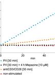
Measurement of Endogenous MALT1 Activity

内源性MALT1活性的测定

DN Daniel Nagel
DK Daniel Krappmann
12149 Views
Jul 20, 2013
MALT1(Mucosa associated lymphoid tissue protein 1) is an important adapter protein for the NF-kB driven lymphocyte activation and the development and survival of distinct B-cell lymphoma entities. In addition MALT1 is a cysteine protease that structurally resembles caspases while having a different substrate preference and mechanism of activation. This paracaspase activity of MALT1 has been shown to be critical for an optimal NF-kB activation and survival of the aggressive ABC-DLBCL (Activated B cell-type of diffuse large B cell lymphoma), which highlights the protease as an attractive therapeutic target for the treatment of distinct B-cell lymphomas and immune diseases like rheumatoid arthritis or multiple sclerosis. In this protocol we describe a fluorogenic cleavage assay, which can be used to measure endogenous and also ectopic MALT1 activity. To this end, cellular MALT1 needs to be precipitated from the lysed cells via antibody immunoprecipitation and subsequently incubated with a fluorogenic substrate peptide. The MALT1 cleavage assay has been developed to directly determine the activity profile of MALT1 in the course of the adaptive immune response as well as in pathological signaling in lymphoid malignancies. In addition, the MALT1 activity assay has been successfully used to monitor cellular MALT1 inhibition with small molecule inhibitors.
 MCF-7 细胞中的雌激素受体(ER)染色质免疫共沉淀

Estradiol Receptor (ER) Chromatin Immunoprecipitation in MCF-7 Cells

MCF-7 细胞中的雌激素受体(ER)染色质免疫共沉淀

Pia Giovannelli Pia Giovannelli
Gabriella Castoria Gabriella Castoria
Antimo Migliaccio Antimo Migliaccio
10566 Views
Jul 20, 2013
Steroid hormone receptors, for example estradiol receptor, act like transcription factors. In the cell, steroids bind to a specific receptor. Upon ligand binding, many steroid receptors dimerize and enter nuclei where they bind specific DNA sequences called Hormone Responsive Elements (HRE) and regulate gene transcription. ER is able to bind DNA sites that are not Estrogen Responsive Elements (ERE) so regulating also the transcription of genes that are not classically controlled by estrogens.

免疫学

采用磁珠从树突细胞中分离吞噬体

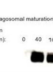
Isolation of Phagosomes from Dendritic Cells by Using Magnetic Beads

采用磁珠从树突细胞中分离吞噬体

Bénédicte Manoury Bénédicte Manoury
10451 Views
Jul 20, 2013
Phagosomes are intracellular organelles in dendritic cells in which pathogens such as viruses, bacteria and parasites are internalised to be proteolysed and killed. Phagosomes are formed by fusion with the plasma membrane, some area of the endoplasmic reticulum as well as the lysosome. This protocol described the purification of phagosomal compartments at different stage of their maturation using magnetic beads.
骨髓源树突细胞中溶酶体pH值的测定

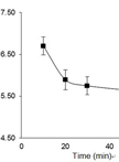
Endosomal pH Measurement in Bone Marrow Derived Dendritic Cells

骨髓源树突细胞中溶酶体pH值的测定

Sophia Maschalidi Sophia Maschalidi
Bénédicte Manoury Bénédicte Manoury
9598 Views
Jul 20, 2013
Endosomes embraces different set of compartments such as early endosomes, intermediate endosomes and late endosomes or lysosomes. They become acidic as they mature. This acidification is generated by the vacuolar membrane proton pump V-ATPase that is recruited in late endosomes. This protocol described the measurement of endosomal pH using dextran molecules labelled with pH sensitive and insensitive dyes.

微生物学

利用荧光底物高通量分析β半乳糖苷酶和β葡萄糖醛酸酶

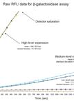
High-throughput β-galactosidase and β-glucuronidase Assays Using Fluorogenic Substrates

利用荧光底物高通量分析β半乳糖苷酶和β葡萄糖醛酸酶

Joshua P. Ramsay Joshua P. Ramsay
15460 Views
Jul 20, 2013
β-galactosidase and β-glucuronidase enzymes are commonly used as reporters for gene expression from gene promoter-lacZ or uidA fusions (respectively). The protocol described here is a high-throughput alternative to the commonly used Miller assay (Miller, 1972) that utilises a fluorogenic substrate (Fiksdal et al., 1994) and 96-well plate format. The fluorogenic substrates 4-Methylumbelliferyl β-D-galactoside (for β-galactosidase assays) (Ramsay et al., 2013) or 4-Methylumbelliferyl β-D-glucuronide (for β-glucuronidase assays) (Ramsay et al., 2011) are cleaved to produce the fluorescent product 4-methylumbelliferone. Cells are permeabilized by freeze-thawing and lysozyme, and the production of 4-methylumbelliferone is monitored continuously by a fluorescence microplate reader as a kinetic assay. The rate of increase in fluorescence is then calculated, from which relative gene-expression levels are extrapolated. Due to the high sensitivity fluorescence-based detection of 4-methylumbelliferone and the high density of time points collected, this assay may offer increased accuracy in the quantification of low-level gene expression. The assay requires small sample volumes and minimal preparation time. The permeabilisation conditions outlined in this protocol have been optimised for Gram-negative bacteria (specifically Escherichia coli and Serratia), but is likely suitable for other organisms with minimal optimisation.
从绿脓杆菌中提取并量化环鸟苷二磷酸

Extraction and Quantification of Cyclic Di-GMP from Pseudomonas aeruginosa

从绿脓杆菌中提取并量化环鸟苷二磷酸

Ankita Basu Roy Ankita Basu Roy
Olga E.  Petrova Olga E. Petrova
Karin Sauer Karin Sauer
14514 Views
Jul 20, 2013
Cyclic di-GMP (c-di-GMP) has emerged as an important intracellular signaling molecule, controlling the transitions between planktonic (free-living) and sessile lifestyles, biofilm formation, and virulence in a wide variety of microorganisms. The following protocol describes the extraction and quantification of c-di-GMP from Pseudomonas aeruginosa samples. We have made every effort to keep the protocol as general as possible to enable the procedure to be applicable for the analysis of c-di-GMP levels in various bacterial species. However, some modifications may be required for the analysis of c-di-GMP levels in other bacterial species.
试剂对支原体滑脱和粘附作用的抑制效应检测

Analyzing Inhibitory Effects of Reagents on Mycoplasma Gliding and Adhesion

试剂对支原体滑脱和粘附作用的抑制效应检测

Taishi  Kasai Taishi Kasai
Makoto  Miyata Makoto Miyata
11565 Views
Jul 20, 2013
Dozens of Mycoplasma species bind to solid surfaces and glide in the direction of the membrane protrusion at a pole. In gliding, Mycoplasma legs catch, pull and release sialylated oligosaccharides fixed on a solid surface. The analyses of inhibitory effects of sialylated compounds on gliding of Mycoplasma can determine the target structure of Mycoplasma for gliding and adhesion.
制备白色念珠菌生物膜用于透射电镜分析

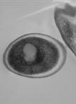
Preparation of Candida albicans Biofilms for Transmission Electron Microscopy

制备白色念珠菌生物膜用于透射电镜分析

HT Heather T. Taff
David R. Andes David R. Andes
10685 Views
Jul 20, 2013
Transmission Electron Microscopy is a form of microscopy that allows for imaging of distinct portions of an individual cell. For Candida albicans biofilms, it is often used to visualize the cell walls of fixed samples of yeast and hyphae. This protocol describes how to grow, harvest, and fix Candida albicans biofilms in preparation for Transmission Electron Microscopy.
HIV-1病毒样颗粒芽殖试验

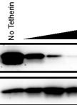
HIV-1 Virus-like Particle Budding Assay

HIV-1病毒样颗粒芽殖试验

NV Nathan H Vande Burgt
LC Luis J Cocka
PB Paul Bates
10597 Views
Jul 20, 2013
Viral replication culminates with the egress of the mature virion from the host cell. This step of the viral life cycle has recently garnered increased attention with the discovery of the cellular restriction factor, Tetherin, which tethers budded virions to the surface of infected cells and inhibits viral spread. The importance of this block in viral infections has been suggested by the discovery of viral antagonists, such as HIV-1 Vpu, which counteract Tetherin. This protocol describes a system to study HIV-1 budding under BSL-2 safety conditions. It takes advantage of the ability of many viral matrix/capsid proteins to generate non-infectious virus-like particles (VLPs) with the expression of a single viral protein (i.e. HIV-1 p24 Gag). This protocol was recently used to characterize the effect of Tetherin isoforms on VLP release in the presence of HIV-1 Vpu (Cocka and Bates, 2012). Simultaneous expression of Tetherin and other viral antagonists can be used to study Tetherin-mediated restriction on viral budding.
使用体内大鼠中心静脉导管制备白色念珠菌生物膜

Preparation of Candida albicans Biofilms Using an in vivo Rat Central Venous Catheter Model

使用体内大鼠中心静脉导管制备白色念珠菌生物膜

HT Heather T. Taff
KM Karen Marchillo
David R. Andes David R. Andes
8911 Views
Jul 20, 2013
In vivo biofilms grown on medical devices are necessary to understand the interactions of the fungal biofilm and the host environment in which it is most commonly found. This protocol describes a way to grow Candida albicans biofilms on the interior lumen of central venous catheters surgically implanted into rats, which mimics quite well the clinical cases of biofilms found on human central venous catheters. These infected catheters can then be studied via a multitude of different experiments, including cell counting by plating, imaging the catheters under light or electron microscopy, or comparing the relative content of in vivo biofilms to in vitro biofilms and planktonic cultures. These biofilms also provide enough high quality RNA for transcriptional profiling.

分子生物学

多核糖体图谱分析

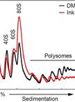
Polysome Profiling Analysis

多核糖体图谱分析

Masahiro  Morita Masahiro Morita
TA Tommy Alain
IT Ivan Topisirovic
NS Nahum Sonenberg
52627 Views
Jul 20, 2013
Polysome profiling is a method that allows monitoring of translation activity of mRNAs in cells and tissues. Once each polysome fractions are collected, the translation activity of each mRNA is analyzed using various molecular biology techniques such as Northern blotting, RT-PCR, microarray or deep-sequencing.
脱氧核糖核酸酶I 足迹法识别蛋白结合位点

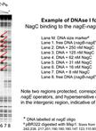
DNase I Footprinting to Identify Protein Binding Sites

脱氧核糖核酸酶I 足迹法识别蛋白结合位点

IG Isabelle Gaugué
DB Dominique Bréchemier-Baey
JP Jacqueline Plumbridge
22806 Views
Jul 20, 2013
DNase I footprinting is used to precisely localise the position that a DNA binding protein, e.g. a transcription factor, binds to a DNA fragment. A DNA fragment of a few hundred bp is labelled at one end and then incubated with the proteins suspected to bind. After a limited digestion with DNase I, the reaction is quenched, DNA is precipitated and analysed on a denaturing polyacrylamide gel. This protocol uses 32P-radioactively labeled DNA.

植物科学

拟南芥中丙二醛、叶绿素脯氨酸、可溶性糖和谷胱甘肽含量的分析

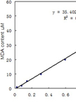
Analysis of Malondialdehyde, Chlorophyll Proline, Soluble Sugar, and Glutathione Content in Arabidopsis seedling

拟南芥中丙二醛、叶绿素脯氨酸、可溶性糖和谷胱甘肽含量的分析

Zhijin  Zhang Zhijin Zhang
Rongfeng Huang Rongfeng Huang
33667 Views
Jul 20, 2013
The protocol has four sub-protocols, which are about the measurement of malondialdehyde, chlorophyll proline, soluble sugar, and glutathione content, respectively, in Arabidopsis seedling by using spectrophotometer. These methods are simple, effective and reproducible, which will help the researchers who are not familiar with these approaches, quickly get reliable results.
玉米胚乳蛋白的提取和分析

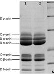
Maize Endosperm Protein Extraction and Analysis

玉米胚乳蛋白的提取和分析

XC Xinze Chen
DY Dongsheng Yao
RS Rentao Song
13291 Views
Jul 20, 2013
Alcohol-solubility is the most characteristic feature of the zein proteins, the major storage protein in maize. Using sodium borate buffer system with added reducing agent, total proteins are isolated, and zein proteins are separated from non-zein proteins. The extraction effect is intuitive on a SDS-PAGE isolation system. In addition, a simple and rapid approach to extract zeins is introduced, taking full advantage of alcohol-solubility of zeins directly.
UDP糖基转移酶动力学常数的测定

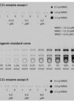
Determination of Enzyme Kinetic Parameters of UDP-glycosyltransferases

UDP糖基转移酶动力学常数的测定

Jörg M. Augustin Jörg M. Augustin
SB Søren Bak
12519 Views
Jul 20, 2013
The determination of enzyme kinetic parameters, such as the Km and kcat values, is an essential part of the characterization of newly discovered enzymes. This protocol describes the determination of enzyme kinetic parameters of the Barbarea vulgaris UDP-glycosyltransferases (UGTs) UGT73C11 and UGT73C13 toward the sapogenins oleanolic acid and hederagenin as sugar acceptor substrates. UGTs catalyze the transfer of glycosyl residues. They generally use uridine sugar nucleotides as their sugar donor substrates, whereas sugar acceptor substrates arise from structurally diverse sets of metabolite classes. This protocol is based on the quantification of 14C-labeled glycosides following thin layer chromatography (TLC)-based separation. The dependence of the measured signal on a universal radioactively-labeled sugar donor substrate allows the potential application of the protocol in combination with a wide range of different sugar acceptor substrates. However, since the here described TLC separation procedure has been optimized for the separation of sapogenins and their glycosides, some modifications may become necessary when investigating other compound classes.Figure 1. Glucosylation reaction catalyzed by UGT73C10-UGT73C13 from Barbarea vulgaris (Augustin et al., 2012). All four enzymes utilize uridine diphosphate glucose (UDP-glc) as glucosyl-moiety donor substrate and different sapogenins such as the oleanane sapogenins oleanolic acid and hederagenin as glucosyl-moiety acceptor substrates.
欧洲山芥皂苷配基的提取和葡糖基化修饰

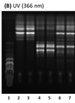
Extraction and Reglucosylation of Barbarea vulgaris Sapogenins

欧洲山芥皂苷配基的提取和葡糖基化修饰

Jörg M. Augustin Jörg M. Augustin
CO Carl Erik Olsen
SB Søren Bak
10059 Views
Jul 20, 2013
Plants produce a vast array of natural compounds. Many of them are not commercially available, and are thus lacking to be tested as substrates for enzymes. This protocol describes the extraction and acidic hydrolysis of metabolites from Barbarea vulgaris with special focus on saponins and their agylcones (sapogenins). It was developed to determine if some B. vulgaris UDP-glucosyltransferases (UGTs) that were shown to glucosylate commercially available sapogenins, would also accept additional sapogenins from this plant as substrate, which are yet chemically uncharacterized and/or commercially unavailable (Figure 1).Figure 1. Glucosylation reaction catalyzed by UGT73C10-UGT73C13 from Barbarea vulgaris (Augustin et al., 2012). All four enzymes utilize uridine diphosphate glucose (UDP-glc) as glucosyl-moiety donor and different sapogenins such as the oleanane sapogenins oleanolic acid and hederagenin as glucosyl-moiety acceptor. Oleanolic acid and hederagenin both naturally occur in G-type B. vulgaris, where they are predominantly found in their 3-O-cellobiosylated form. Additional saponins from G-type B. vulgaris have been identified by Nielsen et al., 2010. However, the majority of saponins and sapogenins that occur in B. vulgaris remain unidentified.