往期刊物2016

卷册: 6, 期号: 20

left

Dec

20

Dec

5

Nov

20

Nov

5

Oct

20

Oct

5

Sep

20

Sep

5

Aug

20

Aug

5

Jul

20

Jul

5

Jun

20

Jun

5

May

20

May

5

Apr

20

Apr

5

Mar

20

Mar

5

Feb

20

Feb

5

Jan

20

Jan

5

right

生物化学

检测小鼠胚胎成纤维细胞调节培养基中的Wnt5

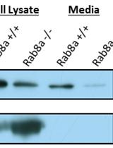
Detection of Wnt5 in Media Conditioned by Mouse Embryonic Fibroblast

检测小鼠胚胎成纤维细胞调节培养基中的Wnt5

JF Juan Flores
Nan Gao Nan Gao
8462 Views
Oct 20, 2016
This protocol describes the procedure of visualizing secreted Wnt5 protein in serum free media via western blotting. This procedure can also be used to visualize other secreted proteins larger than 10,000 daltons. The work presented in this paper visualizes Wnt5 secreted by mouse embryonic fibroblast (MEF), but can be adapted to other cell lines including those transiently transfected by plasmids.

癌症生物学

基于鸡胚绒毛尿囊膜的体内癌症转移模型

The Chick Embryo Chorioallantoic Membrane as an in vivo Model to Study Metastasis

基于鸡胚绒毛尿囊膜的体内癌症转移模型

Piero Crespo Piero Crespo
BC Berta Casar
21598 Views
Oct 20, 2016
Metastasis is a complex process that includes several steps: neoplastic progression, angiogenesis, cell migration and invasion, intravasation into nearby blood vessels, survival in the circulatory system, extravasation followed by homing into distant tissues, the formation of micrometastases, and finally the growth into macroscopic secondary tumors. This complexity makes metastases difficult to investigate and quantify in animal models. The chick embryo is a unique in vivo model that overcomes many limitations for studying the metastatic process, due to the accessibility of the chorioallantoic membrane (CAM), a well-vascularized extra-embryonic tissue located under the eggshell, that is receptive to the xenografting of mammalian tumor cells, including human. Since the chick embryo is naturally immunodeficient at this stage, the CAM can support the engraftment of tumor cells, and their growth therein can faithfully recapitulate most of the characteristics of the carcinogenic process including: growth, invasion, angiogenesis and colonization of distant tissues (Deryugina and Quigley, 2008; Zijlstra et al., 2002). The CAM sustains rapid tumor formation within 5-7 days after cancer cell grafting. This feature provides a unique experimental model for a rapid study of the intravasation and colonization steps of the metastatic cascade. Furthermore, using quantitative PCR to detect species-specific sequences, such as Alu, the chick embryo CAM model can be used to monitor and quantify the presence of the xenografted, ectopic tumor cells in distant tissues. Thus, the chick embryo model has proved a valuable tool for cancer research, in particular for the investigation of molecules and pathways involved in cancer metastasis and to analyze the response of metastatic cancer to potential therapies (Herrero et al., 2015; Casar et al., 2014). In this respect, the use of the rapid and quantitative spontaneous metastasis chick embryo model can provide an alternative approach to conventional mouse model systems for screening anti-cancer agents.
采用免疫荧光显微技术检测哺乳动物细胞分裂后期桥的形成

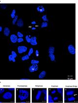
Detection of Anaphase Bridge Formation by Immunofluorescence Microscopy in Mammalian Cells

采用免疫荧光显微技术检测哺乳动物细胞分裂后期桥的形成

Thomas Aschacher Thomas Aschacher
Florian Enzmann Florian Enzmann
12132 Views
Oct 20, 2016
The aim of this protocol is to provide a comprehensive description of the materials, equipment and reproducible methods to detect and analyze anaphase bridges in immunofluorescence microscopy using DAPI to detect cells that failed to completely segregate during mitosis. It describes the process of cell preparation, staining and microscopic settings for detection of anaphase bridges. The protocol has been adapted from our previous publication (Aschacher et al., 2016).
细胞表面蛋白检测法检测受体的内化

Cell Surface Protein Detection to Assess Receptor Internalization

细胞表面蛋白检测法检测受体的内化

Magdalena Czarnecka Magdalena Czarnecka
Joanna Kitlinska Joanna Kitlinska
10068 Views
Oct 20, 2016
The migration of membrane receptors upon exposure to different stimulants/inhibitors is of great importance. Among others, the internalization of membrane receptors affects their accessibility to ligands and cell responsiveness to environmental cues. Experimentally, receptor internalization can be used as a measure of their activation. In our studies, we employed this approach to explore cross-talk between a seven transmembrane domain receptor for neuropeptide Y (NPY), Y5R, and a tyrosine kinase receptor for brain-derived neurotrophic factor (BDNF), TrkB. To this end, we measured the internalization of Y5R upon stimulation with the TrkB ligand, BDNF. Upon treatment with BDNF, the cells were exposed to a membrane impermeable, biotinylation reagent that selectively labels surface proteins. Subsequently, the biotinylated membrane proteins were affinity-purified on columns with avidin resins and analyzed by Western blot. Differences in the fraction of receptors present on the cell surface of control and ligand-treated cells served as a measure of their internalization and response to particular stimuli.
肺内肿瘤细胞与免疫细胞反应的体内成像

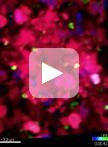
In vivo Imaging of Tumor and Immune Cell Interactions in the Lung

肺内肿瘤细胞与免疫细胞反应的体内成像

Richard N. Hanna Richard N. Hanna
GC Grzegorz Chodaczek
CH Catherine C. Hedrick
9627 Views
Oct 20, 2016
Immunotherapy has demonstrated great therapeutic potential by activating the immune system to fight cancer. However, little is known about the specific dynamics of interactions that occur between tumor and immune cells. In this protocol we describe a novel method to visualize the interaction of tumor and immune cells in the lung of live mice, which can be applied to other organs. In this protocol fluorescent-labeled tumor cells are transferred to recipient mice expressing fluorescently tagged immune cells. Tumor-immune cell interactions in the lung are then imaged by confocal or two photon microscopy. Analysis of tumor interactions with immune cells using this protocol should aid in a better understanding of the importance of these interactions and their role in developing immunotherapies.
肿瘤物质摄取的量化

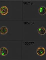
Quantification of Tumor Material Uptake

肿瘤物质摄取的量化

Richard N. Hanna Richard N. Hanna
CH Catherine C. Hedrick
8717 Views
Oct 20, 2016
Extracellular tumor material including exosomes, microvesicles and apoptotic tumor debris may help cancers invade new organs. Enhancing the removal of extracellular tumor material by immune cells represents a novel immunotherapy approach for preventing cancer metastasis. This protocol quantifies the uptake and removal of extracellular tumor material from circulation and tissues by immune cells. In this assay fluorescent tumor cells are transferred into mice, and then immune cells are quantified by either flow cytometry or imaging cytometry for their uptake of tumor material.

细胞生物学

人永生子宫肌层细胞的三维培养系统

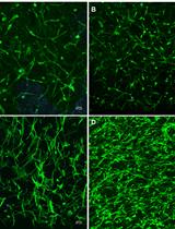
A 3D Culture System of Human Immortalized Myometrial Cells

人永生子宫肌层细胞的三维培养系统

Minnie Malik Minnie Malik
Joy Britten Joy Britten
William H. Catherino William H. Catherino
8427 Views
Oct 20, 2016
Myometrium forms the middle layer of the uterus and is mainly composed of the smooth muscle cells. The cells in vitro are usually grown in a single layer (2-dimensional; 2D) format, whereas in vivo cells are structured in an extracellular matrix scaffolding that allows the cells to communicate and respond to environmental cues. We have developed human myometrium and leiomyoma 3-dimensional (3D) culture, wherein the cells retain their molecular characteristics and respond to environmental cues (Malik and Catherino, 2012; Malik et al., 2014).

免疫学

以HeLa细胞研究蛋白质和膜相互作用的一系列方法:脂质提取和气相色谱定量;挤压法制备大单室脂质体(LUV);蛋白质和脂质体外孵育;采用透射电子显微镜(TM)和浮式离心法在不连续蔗糖梯度范围分析蛋白质和脂质互作;

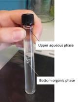
Lipid Extraction from HeLa Cells, Quantification of Lipids, Formation of Large Unilamellar Vesicles (LUVs) by Extrusion and in vitro Protein-lipid Binding Assays, Analysis of the Incubation Product by Transmission Electron Microscopy (TEM) and by Flotation across a Discontinuous Sucrose Gradient

以HeLa细胞研究蛋白质和膜相互作用的一系列方法:脂质提取和气相色谱定量;挤压法制备大单室脂质体(LUV);蛋白质和脂质体外孵育;采用透射电子显微镜(TM)和浮式离心法在不连续蔗糖梯度范围分析蛋白质和脂质互作;

AB Amina Bittame
JL Jodie Lopez
GE Gregory Effantin
Nicolas Blanchard Nicolas Blanchard
Marie-France Cesbron-Delauw Marie-France Cesbron-Delauw
Jean  Gagnon Jean Gagnon
CM Corinne Mercier
16176 Views
Oct 20, 2016
Dissecting the interactions established between proteins and membranes in a given type of cells is not an easy task. Using a cell-free system of large unilamellar vesicles (LUVs) to analyze these interactions may help decipher these interactions and identify potential membrane deformations induced by the proteins incubated with these LUVs. This article describes the protocols for 1) extraction of total lipids from eukaryotic cells using the method developed by Bligh and Dyer (1959), 2) the quantification of glycerophospholipids by gas chromatography after methanolysis, followed by 3) the formation of LUVs by extrusion, 4) protein-lipid binding assay, 5) analysis of the incubation product by transmission electron microscopy (TEM) and by flotation across a discontinuous sucrose gradient and finally, 6) analysis of the proteins by immunoblot and revelation of the glycerophospholipids by iodin fumigation.
耳部炎症和耳部整体染色

Ear Inflammation and Whole-mount Ear Staining

耳部炎症和耳部整体染色

Jia Tong Loh Jia Tong Loh
MG Merry Gunawan
I-hsin Su I-hsin Su
10610 Views
Oct 20, 2016
The recruitment of circulating neutrophils from the bloodstream to the site of inflammation represents one of the earliest events during an innate immune response. During this response, neutrophils tether and roll along the vessel walls before transmigrating across the endothelium into the interstitial space to exert their functions. Here, we describe a protocol for the staining of intravascular and tissue-localized neutrophils following contact sensitization of the skin with croton oil. Visualization of the neutrophilic distribution in skin provides for a better interpretation of the local immune response.
PKC-θ激酶体外活性试验

PKC-θ in vitro Kinase Activity Assay

PKC-θ激酶体外活性试验

Xudong  Wang Xudong Wang
YL Yingqiu Li
9354 Views
Oct 20, 2016
Protein kinase C-θ (PKC-θ), a member of the Ca2+-independent PKC subfamily of kinases, serves as a regulator of T cell activation by mediating the T cell antigen receptor (TCR)- and coreceptor CD28-induced activation of the transcription factors NF-κB and AP-1 and, to a lesser extent, NFAT, and, subsequently, interleukin 2 (IL-2) production and T cell proliferation. In T cells, TCR and CD28 stimulation-induced activation of PKC-θ is the integrated result of diacylglycerol-mediated membrane recruitment, GLK-mediated phosphorylation at activation loop, CD28, Lck, and sumoylation-mediated central immunological synapse localization (Wang et al., 2015; Monks et al., 1997; Kong et al., 2011; Isakov and Altman, 2012; Chuang et al., 2011). Phosphatidylserine (PtdSer) and the phorbol ester Phorbol 12-myristate 13-acetate (PMA, a surrogate of diacylglycerol [DAG]) are the cofactors for the Ca2+-independent PKC subfamily that bind to PKC directly and activate it by changing its conformation (Nishizuka, 1995). A protocol to analyze the PKC-θ kinase activity in vitro is described here. Myelin basic protein is used as the substrate and its phosphorylation is detected by the incorporation of radioactive phosphate into the substrate, which is analyzed by a laser scanner.
评估TCR诱导的PKC-θ类泛素化(Sumoylation)修饰

Assessment of TCR-induced Sumoylation of PKC-θ

评估TCR诱导的PKC-θ类泛素化(Sumoylation)修饰

Xudong  Wang Xudong Wang
ZC Zhilong Chen
QW Qilong Wang
YL Yingqiu Li
6839 Views
Oct 20, 2016
Sumoylation controls many cellular processes. Protein kinase C-θ (PKC-θ), a member of the Ca2+-independent PKC subfamily of kinases, serves as a regulator of T cell activation by mediating the T cell antigen receptor (TCR)- and coreceptor CD28-induced activation of the transcription factors NF-κB and AP-1 and, to a lesser extent, NFAT, and, subsequently, interleukin 2 (IL-2) production and T cell proliferation. We recently proved that TCR-induced sumoylation of PKC-θ is required for its function in T cells (Wang et al., 2015). Here we describe the method to analyze TCR-induced sumoylation of overexpressed or endogenous PKC-θ, which is carried out by immunoprecipitation of PKC-θ followed by immunoblotting with anti-SUMO1 antibody.

分子生物学

通过原核移植生成线粒体-细胞核置换小鼠

Generation of Mitochondrial-nuclear eXchange Mice via Pronuclear Transfer

通过原核移植生成线粒体-细胞核置换小鼠

Robert A.  Kesterson Robert A. Kesterson
LJ Larry W. Johnson
LL Laura J. Lambert
JV Jay L. Vivian
DW Danny R. Welch
Scott W. Ballinger Scott W. Ballinger
11480 Views
Oct 20, 2016
The mitochondrial paradigm for common disease proposes that mitochondrial DNA (mtDNA) sequence variation can contribute to disease susceptibility and progression. To test this concept, we developed the Mitochondrial-nuclear eXchange (MNX) model, in which isolated embryonic pronuclei from one strain of species are implanted into an enucleated embryo of a different strain of the same species (e.g., C57BL/6 and C3H/HeN, Mus musculus), generating a re-constructed zygote harboring nuclear and mitochondrial genomes from different strains. Two-cell embryos are transferred to the ostia of oviducts in CD-1 pseudopregnant mice and developed to term. Nuclear genotype and mtDNA haplotype are verified in offspring, and females selected as founders for desired MNX colonies. By utilizing MNX models, many new avenues for the in vivo study for mitochondrial and nuclear genetics, or mito-Mendelian genetics, are now possible.

神经科学

人工光基因刺激线虫触觉神经实验

Artificial Optogenetic TRN Stimulation of C. elegans

人工光基因刺激线虫触觉神经实验

Ithai Rabinowitch Ithai Rabinowitch
Millet Treinin Millet Treinin
Jihong Bai Jihong Bai
7948 Views
Oct 20, 2016
Optogenetics is a powerful tool for manipulating neuronal activity with high temporal and spatial precision. In the nematode C. elegans optogentics is especially useful and easy to apply. This is because C. elegans is translucent, so its neurons are highly accessible to optic stimulation. In addition, many of its neurons can be exclusively targeted using cell-specific promoters. We have recently taken advantage of optogenetics to deliver artificial patterns of prolonged activation to a class of mechanosensory neurons, called touch receptor neurons (TRNs) in worms that lack touch sensation due to a genetic mutation. Our aim was to examine whether we can counteract the effects of sensory loss by artificially activating the sensory neurons. Here we describe in detail the various components of the protocol that we used. This consists of exposing worms expressing the light-sensitive ion channel Channelrohdopsin 2 (ChR2) in TRNs to long-term random flashes of light.
利用秀丽隐杆线虫评估洋葱伯克霍尔德菌复合体的致病性

Evaluation of Burkholderia cepacia Complex Bacteria Pathogenicity Using Caenorhabditis elegans

利用秀丽隐杆线虫评估洋葱伯克霍尔德菌复合体的致病性

PT Pietro Tedesco
Elia Di Schiavi Elia Di Schiavi
Fortunato Palma Esposito Fortunato Palma Esposito
Donatella de Pascale Donatella de Pascale
7157 Views
Oct 20, 2016
This protocol describes two biological assays to evaluate pathogenicity of Burkholderia cepacia complex (Bcc) strains against the nematode Caenorhabditis elegans. Specifically, these two assays allow one to identify if the under-investigated Bcc strains are able to kill the nematodes by intestinal colonization (slow killing assay, SKA) or by toxins production (fast killing assay, FKA). The principal differences between the two assays rely on the different killing kinetics for worms.

植物科学

采用一步两步式水解法和HPAEC-PAD快速测定植物细胞壁的纤维素、中性糖和糖醛酸

Rapid Determination of Cellulose, Neutral Sugars, and Uronic Acids from Plant Cell Walls by One-step Two-step Hydrolysis and HPAEC-PAD

采用一步两步式水解法和HPAEC-PAD快速测定植物细胞壁的纤维素、中性糖和糖醛酸

TY Trevor Yeats
TV Tamara Vellosillo
NS Nadav Sorek
AI Ana B. Ibáñez
SB Stefan Bauer
17346 Views
Oct 20, 2016
The plant cell wall is primarily composed of the polysaccharides cellulose, hemicellulose and pectin. The structural and compositional complexity of these components are important for determining cell wall function during plant growth. Moreover, cell wall structure defines a number of functional properties of plant-derived biomass, such as rheological properties of foods and feedstock suitability for the production of cellulosic biofuels. A typical characterization of cell wall chemistry in the molecular biology lab consists of a mild acid hydrolysis for the quantification of hemicellulose and pectin-derived monomers and a separate analysis of cellulose by the Updegraff method. We have adopted a streamlined ‘one-step two-step’ hydrolysis protocol that allows for the simultaneous determination of cellulose content, neutral sugars, and uronic acids by high-performance anion-exchange chromatography with pulsed amperometric detection (HPAEC-PAD) of paired samples. In our work, this protocol has largely replaced Updegraff cellulose quantification and hydrolysis with 2 M TFA for the determination of matrix polysaccharide composition at the micro scale.
采用苯胺蓝和Calcofluor White对绿藻类双星藻属和克里藻属的胼胝质和纤维素进行染色

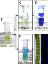
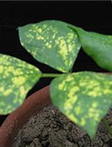
Aniline Blue and Calcofluor White Staining of Callose and Cellulose in the Streptophyte Green Algae Zygnema and Klebsormidium

采用苯胺蓝和Calcofluor White对绿藻类双星藻属和克里藻属的胼胝质和纤维素进行染色

KH Klaus Herburger
AH Andreas Holzinger
13844 Views
Oct 20, 2016
Plant including green algal cells are surrounded by a cell wall, which is a diverse composite of complex polysaccharides and crucial for their function and survival. Here we describe two simple protocols to visualize callose (1→3-β-D-glucose) and cellulose (1→4-β-D-glucose) and related polysaccharides in the cell walls of streptophyte green algae. Untreated or algal cells heated in NaOH are incubated in Calcofluor white (binding to β-glucans including cellulose) or Aniline blue (binding to callose), respectively. Both dyes can be visualized by epifluorescence microscopy.
采用HPLC-UV 结合硫解和MALDI-TOF质谱的方法测定植物组织中缩合单宁的分子结构

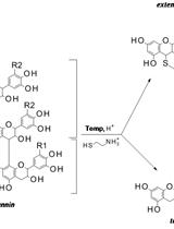
Determination of Molecular Structures of Condensed Tannins from Plant Tissues Using HPLC-UV Combined with Thiolysis and MALDI-TOF Mass Spectrometry

采用HPLC-UV 结合硫解和MALDI-TOF质谱的方法测定植物组织中缩合单宁的分子结构

SB Sauro Bianchi
IK Ivana Kroslakova
Ingo Mayer Ingo Mayer
11554 Views
Oct 20, 2016
Condensed tannins extracted from plant tissues are suitable substitutes for phenolic resins. Their molecular structure, which might influence their chemical reactivity, can be assessed by the use of both HPLC-UV after acid thiolysis and MALDI-TOF mass spectrometry. Thiolysis of plant extracts in acidic methanol with cysteamine hydrochloride results in the release of the monomeric units of the condensed tannin oligomers that can be further quantified by reversed-phase HPLC-UV by comparison with analytical standards. MALDI-TOF mass spectrometry using 2,5-dihydroxybenzoic acid as matrix and K+ as cationization agent highlights the molecular structural characteristics (e.g., monomeric unit sequence) of the tannin oligomers. The methodologies permit the estimation of the mean and the maximum (observable) degree of polymerization, the type of monomeric units and the presence of glycosylation and/or esterification of the tannin oligomers.
黑吉豆中的绿豆黄化花叶印度病毒(MYMIV)感染、sRNA库建立和鉴定microRNA的深度测序

Mungbean Yellow Mosaic India Virus (MYMIV)-infection, Small RNA Library Construction and Deep Sequencing for MicroRNA Identification in Vigna mungo

黑吉豆中的绿豆黄化花叶印度病毒(MYMIV)感染、sRNA库建立和鉴定microRNA的深度测序

Anirban Kundu Anirban Kundu
Sujay Paul Sujay Paul
Amita Pal Amita Pal
GT Genotypic Technology
10452 Views
Oct 20, 2016
This protocol describes small RNA library preparation from Vigna mungo total RNA followed by deep sequencing and analysis for microRNA identification.​
拟南芥CESA1催化结构域的异源表达和纯化

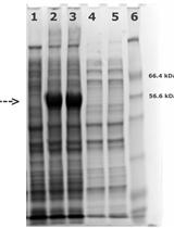
Heterologous Expression and Purification of Catalytic Domain of CESA1 from Arabidopsis thaliana

拟南芥CESA1催化结构域的异源表达和纯化

VV Venu Gopal Vandavasi
HO Hugh O’ Neill
7017 Views
Oct 20, 2016
Heterologous expression of plant cellulose synthase (CESA) and its purification has remained a challenge for decades impeding detailed biophysical, biochemical and structural characterization of this key enzyme. An in-depth knowledge of structure and function of CESA proteins would enable us to better understand the hierarchical structure of the plant cell wall. Here, we report a detailed, and reproducible method of purification of catalytic domain of CESA1 from Arabidopsis thaliana that was recombinantly expressed in Escherichia coli. The method relies on a two stage purification procedure to obtain the catalytic domain in monomer and trimer forms. The biochemical and biophysical data including low resolution structures of the protein have been published (Vandavasi et al., 2016). Currently the crystallization studies of this protein are underway.[Background] Cellulose is the most important structural component of plant cell walls and constitutes the Earth’s largest source of biorenewable material, yet the mechanism of its synthesis by plants is poorly understood. The plant cellulose synthesis complex (CSC), also called a ‘rosette’ because of its hexameric appearance in electron microscope images, is a large multi-subunit transmembrane protein complex responsible for synthesis of cellulose chains and their assembly into microfibrils. The number of cellulose synthase (CESA) proteins in the CSC and the number of cellulose chains in a microfibril have been debated for many years. Structural information about CESA proteins from plants is crucial to provide answers to some of the basic questions regarding the mechanism of cellulose synthesis. However, elucidation of the structure of CESA proteins has proved difficult because they are multi-domain proteins comprised of disordered, globular, and membrane associated domains. As an alternative to pursuing structural studies of CESA holoproteins, we are developing approaches for recombinant expression of individual CESA domains (e.g., N-terminal domain, central-cytosolic domain, C-terminal transmembrane domain) in large quantities suitable for structural studies. The current protocol has been optimized for isolation of the catalytic domain of A. thaliana CESA1 as reported (Vandavasi et al., 2016). Using this protocol, it is possible to control the oligomerization state of the protein enabling structural studies of the monomer and the trimeric form of the protein. The approach described may be broadly applicable to other systems.

干细胞

通过CD45+骨髓细胞的脾内移植来实验性治疗肝纤维化

Experimental Liver Fibrosis and Intrasplenic Transplantation of CD45+ Bone Marrow Cells

通过CD45+骨髓细胞的脾内移植来实验性治疗肝纤维化

Prakash Baligar Prakash Baligar
Sebanta Pokhrel Sebanta Pokhrel
AM Asok Mukhopadhyay
13831 Views
Oct 20, 2016
Liver fibrosis results from the excessive collagen deposition (collagen scar) by activated hepatic stellate cells (HpSCs), leading to the inhibition of normal liver regeneration and function. Fibrogenesis is a complex mechanism involving both the synthesis and degradation of matrix proteins by different cell types, mainly macrophages in the liver. Carbon tetrachloride-induced fibrosis (CCl4) and cirrhosis is one of the oldest, simplest and probably the most widely used toxin-based experimental model for the induction of fibrosis. Here we have explained experimental animal model of liver fibrosis using CCl4, injecting twice a week for a period of 8 weeks. In these fibrotic mice, bone marrow (BM) derived CD45+ cells were transplanted via intrasplenic route after 8 weeks of CCl4 injection, and half of the CCl4 dose was continued till the end of the experiment to know the effect of transplanted cells on liver fibrosis and regeneration. So far, crude bone marrow (BM) cells or mesenchymal stem cells (MSCs) have been used for the treatment of liver fibrosis. Low survival rate, less fibrolytic and profibrogenic properties of MSCs remain the major concerns for inadequate recovery of liver from fibrosis. This led us to investigate BM cells devoid of mesenchymal lineage that is CD45+ cells for the antifibrotic effect as this population consisting of mononuclear cells which are the precursor of macrophages and may involve in the scar degradation process. Cells transplantation can be followed in different ways like intrasplenic infusion, tail vein injection and ectopic cell transplantation in experimental animal models. The survival of the cells after ectopic transplantation is less when compared to tail vein and intrasplenic infusion. Intrasplenic route of transplantation is effective in engraftment and long term survival of the donor cells especially in case of liver disease models. This protocol describes fibrosis mouse model development, intrasplenic route of cell transplantation and tracking of the donor cells after transplantation.