往期刊物2016

卷册: 6, 期号: 17

left

Dec

20

Dec

5

Nov

20

Nov

5

Oct

20

Oct

5

Sep

20

Sep

5

Aug

20

Aug

5

Jul

20

Jul

5

Jun

20

Jun

5

May

20

May

5

Apr

20

Apr

5

Mar

20

Mar

5

Feb

20

Feb

5

Jan

20

Jan

5

right

生物化学

RNA结合蛋白质的识别

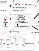
Identification of RNA-binding Proteins

RNA结合蛋白质的识别

KM Kazuya Masuda
TK Tadamitsu Kishimoto
13750 Views
Sep 5, 2016
This protocol describes the extraction of RNA-binding proteins (RBPs) from cell lysates. In order to pull down target RBPs, 5-bromo-UTP (BrUTP)-incorporated RNA probes are used, which are generated by in vitro transcription. The schematic diagram (Flowchart) with procedure is indicated (Figure1 and Figure 2).Figure 1. Schematic diagram of procedure (A-H). Flow chart of experimental procedure is indicated at A-H.Figure 2. Linearization of plasmids by restricted enzyme. The plasmid is cut at restriction sites adjacent to its cloning element.
Sxl-Unr-msl2 核糖核蛋白复合体的蛋白质表达、纯化和结晶

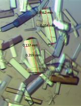
Protein Expression, Purification and Crystallization of the Sxl-Unr-msl2 Ribonucleoprotein Complex

Sxl-Unr-msl2 核糖核蛋白复合体的蛋白质表达、纯化和结晶

Janosch Hennig Janosch Hennig
MS Michael Sattler
9470 Views
Sep 5, 2016
This protocol describes the expression, purification and crystallization of a ternary protein-protein-RNA complex, consisting of the two RNA recognition motifs (RRMs) of Sex-lethal (Sxl), the first of five cold shock domains of Upstream-of-N-Ras (Unr), and an 18-nucleotide region of msl2 mRNA, called the F fragment (Hennig et al., 2014).The biological role of the complex is the translational repression of msl2 mRNA, preventing the formation of the dosage compensation complex and subsequent 2-fold hypertranscription of X-linked genes in Drosophila females. As orthologous RRM-containing proteins and Unr exist in humans, similar complexes potentially also form during translational repression in vertebrates. The protocol describes the in vitro assembly of the complex and its purification followed by crystallization for X-ray crystallography structure determination. Part of the protocol has been published elsewhere (Hennig et al., 2013 and 2014), but some parts are described here in more detail.
小鼠组织转铁蛋白和非转铁蛋白结合铁摄取的测量

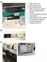
Measurement of Transferrin- and Non-transferrin-bound Iron Uptake by Mouse Tissues

小鼠组织转铁蛋白和非转铁蛋白结合铁摄取的测量

SJ Supak Jenkitkasemwong
Chia-Yu Wang Chia-Yu Wang
Mitchell D. Knutson Mitchell D. Knutson
8592 Views
Sep 5, 2016
Iron in blood plasma is bound to its transport protein transferrin, which delivers iron to most tissues. In iron overload and certain pathological conditions, the carrying capacity of transferrin can become exceeded, giving rise to non-transferrin-bound iron, which is taken up preferentially by the liver, kidney, pancreas, and heart. The measurement of tissue transferrin- and non-transferrin-bound iron (TBI and NTBI, respectively) uptake in vivo can be achieved via intravenous administration of 59Fe-labeled TBI or NTBI followed by gamma counting of various organs. Here we describe a detailed protocol for the measurement of TBI and NTBI uptake by mouse tissues.

细胞生物学

显微镜观察BODIPY 493/503 染色中性脂滴以及用流式细胞仪量化

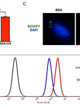
BODIPY 493/503 Staining of Neutral Lipid Droplets for Microscopy and Quantification by Flow Cytometry

显微镜观察BODIPY 493/503 染色中性脂滴以及用流式细胞仪量化

BQ Bo Qiu
MS M. Celeste Simon
61587 Views
Sep 5, 2016
Lipid droplets (LDs) are ubiquitous, dynamic organelles and function as a storage depot for neutral lipids, including triglycerides and cholesterol esters (Walther and Farese, 2012). The movement of lipid species into and out of LDs impacts a variety of cellular processes, such as energy homeostasis, lipid-based signaling, and membrane homeostasis (Greenberg et al., 2011). For example, neutral lipid storage is enhanced upon increased synthesis or uptake of lipid species. On the other hand, extracellular signals can enhance the release of lipid species packaged within neutral LDs. Thus, the investigation of topics involving lipid metabolism may require the assessment of cellular neutral lipid content. In this protocol, we describe the use of the fluorescent neutral lipid dye 4,4-difluoro-1,3,5,7,8-pentamethyl-4-bora-3a,4a-diaza-s-indacene (BODIPY 493/503) to facilitate quantification of neutral lipid content by flow cytometry and observation of LDs by microscopy.

发育生物学

大脑嗅球(OB)移植

Olfactory Bulb (OB) Transplants

大脑嗅球(OB)移植

Maria Savvaki Maria Savvaki
Domna Karagogeos Domna Karagogeos
7679 Views
Sep 5, 2016
Transplantation in mouse brain slices is a powerful tool in order to study axon targeting and migrational events during development. Taking advantage of donors and recipients belonging to different genotypes, this technique allows researchers to assess the contribution of donor and/or recipient tissue by performing various combinations and to study cell-autonomous functions or effects that are influenced by the recipient’s environment (Bastakis, et al., 2015). Here we describe the transplantation procedure on sagittal brain slices containing olfactory bulb (OB). Specifically, we have transplanted the proximal-to-the-cortex part of dorsal OB to the same region on a recipient slice. Transplanted slices can be cultured for up to 3 days before their morphology is disfigured due to growth in 3D. Re-sectioning of these slices allows for a more detailed immunohistochemical analysis.

免疫学

关节中免疫浸润细胞的分离

Isolation of Joint-infiltrating Cells

关节中免疫浸润细胞的分离

Aoi Akitsu Aoi Akitsu
Yoichiro Iwakura Yoichiro Iwakura
10831 Views
Sep 5, 2016
Infiltration of leukocytes into joints is one of the main features of autoimmune inflammatory arthritis. Here, we describe the protocol for isolation of joint-infiltrating cells in mice. This protocol is useful to analyze cell surface antigens and intracellular cytokines by flow cytometry.

微生物学

尖孢镰刀菌细胞壁应激的体外实验

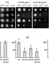
In vitro Cell Wall Stress Assay for Fusarium oxysporum

尖孢镰刀菌细胞壁应激的体外实验

Elena Pérez-Nadales Elena Pérez-Nadales
AD Antonio Di Pietro
10033 Views
Sep 5, 2016
In this protocol we describe a cell wall stress assay for the fungal pathogen F. oxysporum, based on exposure to the two anionic dyes Calcofluor White (CFW) and Congo Red (CR). Both compounds have been used to exert stress upon the fungal cell wall in vitro (Perez-Nadales and Di Pietro, 2015; Perez-Nadales and Di Pietro, 2011; Leach et al., 2012; Heilmann et al., 2013; Garcia et al., 2015). CFW perturbs chitin assembly, whereas CR interferes with β-glucan synthesis, resulting in cell wall-weakening and activation of the cell wall stress response (Ram and Klis, 2006; Kopecka and Gabriel, 1992; Roncero and Duran, 1985). Presumably, the signaling pathways and cell wall changes associated with this response reflect cell wall homeostasis during normal growth as well as cell wall remodeling events in response to stresses encountered during the fungus-host interactions. The conditions for preparation of CFW and CR culture medium specified in this protocol are based on the paper by Ram and Klis entitled “Identification of fungal cell wall mutants using susceptibility assays based on Calcofluor white and Congo red”, published in Nature protocols (Ram and Klis, 2006). This paper established the optimum conditions for preparation of CFW and CR stock solutions and suggested maintaining the culture medium at a constant pH to avoid acidification, protonation and precipitation of these dyes. This cell wall stress assay has been widely used in our group for the characterization of F. oxysporum mutants in mitogen activated protein kinase (MAPK) signaling pathway genes involved in cell wall integrity (Perez-Nadales and Di Pietro, 2015; Perez-Nadales and Di Pietro, 2011; Turra et al., 2014).

神经科学

小鼠环境与对象认知记忆测试

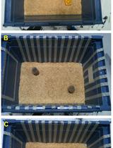
Object-context Recognition Memory Test for Mice

小鼠环境与对象认知记忆测试

SK Sofia Kanatsou
HK Harm Krugers
9913 Views
Sep 5, 2016
The object in-context (OIC) task is a variant of the widely used object recognition (OR) task (Dix and Aggleton, 1999). The OIC task makes use of the fact that rodents have a natural tendency to explore novel environments and objects. The hippocampus appears to play a major role in the OIC task (much more so than in the original OR task), where animals should be able to distinguish between two familiar objects of which one is in a different context from the training trial (Ennaceur and Aggleton,1997; Bermudez-Rattoni et al., 2005; Albasser et al., 2009; Roozendaal et al., 2010; Banks et al., 2014; Bermudez-Rattoni, 2014). Recognition memory encompasses a number of additional components, such as an item's associations with its context, place, etc. (Bussey et al., 1999, 2000). Here, we describe a version of the OIC task in mice, based on earlier reports (Dix and Aggleton, 1999; Eacott and Norman, 2004; Balderas et al., 2008; Barsegyan et al., 2014; Kanatsou et al., 2015a; Kanatsou et al., 2015b).
采用胚胎内脏异位移植分析肠道神经嵴细胞迁移

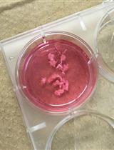
Analysis of Enteric Neural Crest Cell Migration Using Heterotopic Grafts of Embryonic Guts

采用胚胎内脏异位移植分析肠道神经嵴细胞迁移

Rodolphe Soret Rodolphe Soret
Nicolas Pilon Nicolas Pilon
7798 Views
Sep 5, 2016
Hirschsprung disease (HSCR), also named aganglionic megacolon, is a severe congenital malformation characterized by a lack of enteric nervous system (ENS) in the terminal regions of the bowel (Bergeron et al., 2013). As the ENS notably regulates motility in the whole gastrointestinal track, the segment without neurons remains tonically contracted, resulting in functional intestinal obstruction and accumulation of fecal material (megacolon). HSCR occurs when enteric neural progenitors of vagal neural crest origin fail to fully colonize the developing intestines. These “enteric” neural crest cells (ENCCs) have to migrate in a rostro-caudal direction during a fixed temporal window, which is between embryonic day (e) 9.5 and e14.5 in the mouse (Obermayr et al., 2013). Recently, our group generated a new HSCR mouse model called Holstein in which migration of ENCCs is impaired because of increased collagen VI levels in their microenvironment (Soret et al., 2015). Here, we describe the method that allowed us to demonstrate the cell-autonomous nature of this migration defect. In this system adapted from a previously described heterotopic grafting approach (Breau et al., 2006), the donor tissue is a fully colonized segment of e12.5 midgut while the host tissue is an aneural segment of e12.5 hindgut. Extent of ENCC migration in host tissue is assessed after 24 h of culture and is greatly facilitated when donor tissue has a transgenic background such as the Gata4-RFP (Pilon et al., 2008) that allows endogenous labeling of ENCCs with fluorescence. Depending of the genetic background of donor and host tissues, this approach can allow evaluating both cell-autonomous and non-cell-autonomous defects of ENCC migration.

植物科学

干旱胁迫棉花叶片中脯氨酸和丙二醛含量以及抗氧化酶活性的测定

Measurements of Proline and Malondialdehyde Content and Antioxidant Enzyme Activities in Leaves of Drought Stressed Cotton

干旱胁迫棉花叶片中脯氨酸和丙二醛含量以及抗氧化酶活性的测定

Tianzi Chen Tianzi Chen
BZ Baolong Zhang
29071 Views
Sep 5, 2016
Drought stress negatively affects cotton plant growth and induces various biochemical and physiological responses in cotton plants. Proline content and antioxidant enzymes are thought to be associated with maintaining the structure of cellular components or with protecting cellular function. Study of cotton plant responses towards drought stress and investigation of the mechanism of drought tolerance are helpful to develop drought tolerant cotton plants. Here, we describe a protocol to investigate cotton plant response towards drought stress through measurements of biochemical parameters including antioxidant enzyme activities, proline content and malondialdehyde (MDA) content.
体内放线菌酮实验测定植物中的蛋白质降解

Cycloheximide Assays to Measure Protein Degradation in vivo in Plants

体内放线菌酮实验测定植物中的蛋白质降解

Jonathan Gilkerson Jonathan Gilkerson
RT Raymond Tam
AZ Aimee Zhang
KD Kate Dreher
JC Judy Callis
19113 Views
Sep 5, 2016
The half-life of a protein is a characteristic property, and can be modulated by post-translational modifications, changes in subcellular localization, and/or interaction with other proteins or ligands. As one determinant of its steady-state level, a protein’s degradation represents an important distinguishing attribute relevant to its biological function. Because protein longevity cannot be elucidated from bioinformatics analyses, it must be determined empirically. Here we describe two approaches for in vivo half-life determination in plants: 1. pooled-seedling degradation assays monitoring either tagged versions of the protein (luciferase fusions or other epitope tags) or following the endogenous protein; 2. single-seedling degradation assays using luciferase fusion proteins. The advantages of these approaches are their simplicity and low cost.
双子叶植物中叶表皮细胞筛选和统计分析的简单方法

Simple Methods for Screening and Statistical Analysis of Leaf Epidermal Cells in Dicotyledonous Plants

双子叶植物中叶表皮细胞筛选和统计分析的简单方法

JT Jian-Jun Tao
SC Shou-Yi Chen
JZ Jin-Song Zhang
12798 Views
Sep 5, 2016
Leaf epidermal cell size and number are positively correlated to leaf area. Stomata are specialized epidermal cells vital for gas exchange and water transpiration. So, observation and statistical analysis of the leaf epidermal cells are valuable for the study of leaf development and response to environmental stimulus. The classical method is using the scanning electron microscope (SEM), which is an expensive and time-consuming method, thus makes the large-scale screening of epidermis impractical. Here we provide simple but effective methods (agarose-based epidermal imprinting and tape-based epidermis tearing) for solving this problem without using the SEM.
环境刺激对植物根部发育的局部和系统效应的分析方法

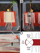
A Method to Analyze Local and Systemic Effects of Environmental Stimuli on Root Development in Plants

环境刺激对植物根部发育的局部和系统效应的分析方法

Neil E. Robbins II Neil E. Robbins II
José Dinneny José Dinneny
10239 Views
Sep 5, 2016
Root development in vascular plants is innately tied to the environment. However, relatively little attention has been paid toward understanding the spatial scales at which the root perceives and responds to external stimuli. While some environmental signals elicit global responses that affect root system architecture, others may have more localized effects. We have observed that various developmental processes can be induced or suppressed along the circumference of the main root depending on local contact with available water in a process termed hydropatterning (Bao et al., 2014). Our studies of hydropatterning indicate that the root can detect and respond to certain external stimuli at the resolution of the diameter of a single organ. In order to characterize developmental patterning at this spatial scale, we developed a procedure to vary environmental inputs across the circumferential axis of the root in vitro using agar media. Roots are grown between two blocks of agar media in a “sandwich”. Local environmental conditions can be varied depending on the composition of the media on either side. Stimuli that act locally can be distinguished from those that act systemically based on the developmental response of the root. Here we describe the overall method and provide an example of how it can be used to analyze lateral root patterning in Zea mays (maize) in response to an external water potential gradient. We also discuss how the method can be used more broadly for other plant species and environmental treatments.
洋麻的盐胁迫和干旱胁迫实验

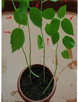
Salinity and Drought Treatment Assays in Kenaf (Hibiscus cannabinus L.)

洋麻的盐胁迫和干旱胁迫实验

Xiaoping Niu Xiaoping Niu
MC Meixia Chen
AT Aifen Tao
Jianmin Qi Jianmin Qi
9922 Views
Sep 5, 2016
Salinity and drought are the two main factors that cause fiber yield and quality losses in kenaf. It is reported that salinity and drought can affect more than 10% of arable land and cause a global decline in the average yields of major crops by more than 50%. Therefore, understanding plant tolerance of drought and salinity is of fundamental importance and has become the focus of intensive research. This protocol describes a simple and reproducible protocol to imitate natural salinity and drought stress under soil conditions. Even though the water-culture method is most frequently used for salinity and drought treatments, the soil-culture method in this study was more applicable to studying natural stress conditions.

干细胞

通过RNA染色显示单细胞的染色体转录区

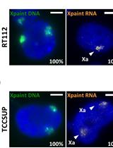
Single-cell Visualization of Chromosome Transcriptional Territories by RNA-paint

通过RNA染色显示单细胞的染色体转录区

CV Céline Vallot
CR Claire Rougeulle
8912 Views
Sep 5, 2016
We developed a FISH-based method to directly assess chromosome-wide transcriptional activity, thereby enabling the visualization of the actively transcribed fraction of a chromosome at the single-cell level. We applied this method to probe the activity of X-chromosomes and its instability in the context of human embryonic stem cells and cancer cells.