往期刊物2016

卷册: 6, 期号: 11

left

Dec

20

Dec

5

Nov

20

Nov

5

Oct

20

Oct

5

Sep

20

Sep

5

Aug

20

Aug

5

Jul

20

Jul

5

Jun

20

Jun

5

May

20

May

5

Apr

20

Apr

5

Mar

20

Mar

5

Feb

20

Feb

5

Jan

20

Jan

5

right

生物化学

全盲柔性肽-蛋白对接实验方法pepATTRACT的开发和应用

Development and Application of a Fully Blind Flexible Peptide-protein Docking Protocol, pepATTRACT

全盲柔性肽-蛋白对接实验方法pepATTRACT的开发和应用

Christina EM Schindler Christina EM Schindler
Sd Sjoerd J. de Vries
Martin Zacharias Martin Zacharias
7924 Views
Jun 5, 2016
Peptide-mediated interactions are involved in many signaling and regulatory pathways as well as the DNA replication machinery and are linked to many pathological disorders. Many research groups are currently working towards a more detailed understanding of these important interactions by characterizing the 3D complex structures with experimental methods like X-ray crystallography and NMR. However, for a large number of peptide-protein complexes such atomistic structural information is lacking to date. Computational peptide docking methods can yield information complementary to experimental information by predicting the protein-peptide complex structure from the 3D structure of the protein and the peptide sequence. This approach can also be used to study interactions between folded and disordered proteins/protein regions (e.g., the interactions of the disordered regions in tumor suppressor p53 with its different partners). Here, we describe the development and usage of the fully blind, flexible peptide-protein docking protocol pepATTRACT. The ATTRACT docking engine is implemented as a suite of command line tools and options that can be combined at will. Therefore, ATTRACT protocols like pepATTRACT are typically invoked via a custom, hand-written shell script. Although this approach is very flexible, it limits the accessibility of ATTRACT to expert users only. To make pepATTRACT easily accessible to non-expert users, we created a web-interface which helps the user set up a peptide docking protocol by editing parameters in a web browser (www.attract.ph.tum.de/peptide.html). pepATTRACT docking scripts can then executed on the user's local machine, once the ATTRACT software has been installed. Here, we describe all the steps necessary for setting up a pepATTRACT docking run via the web-interface including installation of the ATTRACT software.

癌症生物学

使用ThinCertsTM (一种侵袭迁移培养皿)进行体外肿瘤细胞迁移试验

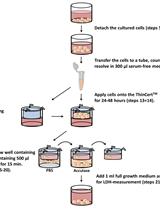
In vitro Tumor Cell Migration Assay Using ThinCertsTM (Transwells)

使用ThinCertsTM (一种侵袭迁移培养皿)进行体外肿瘤细胞迁移试验

Marc A. Schneider Marc A. Schneider
18169 Views
Jun 5, 2016
The high migration rate of tumor cells often results in poor prognosis for the survival of the patients. Here, we describe a protocol to measure the migration of cells using a quantitative assay. The relative tumor cell migration was measured using ThinCertsTM cell culture inserts and a lactate dehydrogenase (LDH) assay to quantify the relative cell number. The quantification of the migration with the LDH kit is much more precise than other methods using i.e. crystal blue to count the cells.
哺乳动物细胞的体外mTORC1激酶酶活测试法

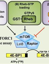
In vitro mTORC1 Kinase Assay for Mammalian Cells Protocol

哺乳动物细胞的体外mTORC1激酶酶活测试法

Kayleigh Margaret Dodd Kayleigh Margaret Dodd
AT Andrew Robert Tee
12320 Views
Jun 5, 2016
Historically, mechanistic target of rapamycin (mTOR) was purified from mammalian cells using mild nonionic detergents such as NP-40 and or Triton-X100 that resulted in dissociation of core regulatory components essential for its native kinase activity. Consequently, these older kinase assays required MnCl2 to artificially enhance the weak phosphotransfer activity observed (Bai et al., 2007; Kim et al., 2002). With the use of the zwitterionic detergent 3-[(3-Cholamidopropyl) dimethylammonio]-1-propanesulfonate (CHAPS), the mTOR complex 1 (mTORC1) containing Regulatory-associated protein of mTOR (Raptor) and Lst8 (also known as GbetaL) can be successfully purified as a complex. This in vitro kinase assay allows for purification of mTORC1 that resembles its physiological state and retains kinase activity under physiological MgCl2 concentrations (Sancak et al., 2007). The activity of mTORC1 can be further enhanced through the use of hyperactive mutations within the kinase domain of mTOR or inclusion of GTP-bound RAS enriched in brain (Rheb) that is supplemented into the in vitro kinase assays. Rheb is a small-G-protein that binds to and activates mTORC1 to phosphorylate downstream substrates, such as eukaryotic initiation factor 4E-BP1 (4E-BP1) (Burnett et al., 1998), ribosomal protein S6 kinase 1 (S6K1) (Kim et al., 2002), Signal transducer and activator of transcription 3 (STAT3) (Dodd et al., 2015), and proline-rich Akt substrate of 40 kDa (PRAS40) (Dunlop et al., 2009).

细胞生物学

高血脂小鼠中的主动脉粥样硬化病变分析

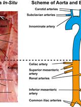
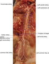
Aorta Atherosclerosis Lesion Analysis in Hyperlipidemic Mice

高血脂小鼠中的主动脉粥样硬化病变分析

Sarajo Mohanta Sarajo Mohanta
CY Changjun Yin
Christian Weber Christian Weber
DH Desheng Hu
AH Andreas J. R. Habenicht
21267 Views
Jun 5, 2016
Atherosclerosis is a chronic inflammatory disease of large and medium-sized arteries. Apolipoprotein E-deficient (ApoE-/-) mice are used as experimental models to study human atherosclerosis. ApoE-/- mice are constitutively hyperlipidemic and develop intima plaques that resemble human plaques. Various issues including experimental design for lesion analysis, dietary conditions, isolation of the aorta, staining methods, morphometry, group size, age, the location within the arterial tree, and statistical analyses are important parameters that need to be addressed to obtain robust data. Here, we provide detailed methods to quantify aorta atherosclerosis.
从C57BL/6J小鼠轮廓乳头中分离味蕾和味觉细胞以检测递质分泌

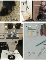
Isolating Taste Buds and Taste Cells from Vallate Papillae of C57BL/6J Mice for Detecting Transmitter Secretion

从C57BL/6J小鼠轮廓乳头中分离味蕾和味觉细胞以检测递质分泌

Anthony Y. Huang Anthony Y. Huang
SW Sandy Y. Wu
8945 Views
Jun 5, 2016
Mouse is a well-accepted model for studying taste bud function. Mice readily detect and respond to taste substances that humans consider to have sweet, bitter, salty, sour and umami taste qualities. A great deal of recent research on taste receptors is based on this species. Live mice are needed for these experiments because no alternative in vitro model incorporates all elements of taste transduction and peripheral signaling. The C57BL/6J strain was selected because these mice respond robustly to many taste stimuli and because of variety of transgenic animals, such as PLCβ2-GFP and GAD67-GFP, were derived from that strain. Prior analyses on behavior, nerve responses, cellular electrophysiology and molecular biology, all conducted on C57BL/6J mice will form a solid foundation for the proposed studies (Finger et al., 2005; Huang and Wu, 2015; Huang et al., 2007). Thus, freshly euthanized animals must be used as a source of taste buds from which we will isolate taste buds and taste cells.

免疫学

单克隆抗体介导的流感病毒血细胞凝集抑制(HI)试验

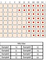
Hemagglutination Inhibition (HI) Assay of Influenza Viruses with Monoclonal Antibodies

单克隆抗体介导的流感病毒血细胞凝集抑制(HI)试验

YW Ying Wu
MC MyungSam Cho
DS David Shore
MS Manki Song
JC JungAh Choi
TJ Tao Jiang
YD Yong-Qiang Deng
MB Melissa Bourgeois
LA Lynn Almli
HY Hua Yang
LC Li-Mei Chen
YS Yi Shi
JQ Jianxu Qi
AL An Li
KY Kye Sook Yi
MC MinSeok Chang
JB Jin Soo Bae
HL HyunJoo Lee
JS JiYoung Shin
JS James Stevens
SH SeoungSuh Hong
CQ Cheng-Feng Qin
GG George F. Gao
SC Shin Jae Chang
RD Ruben O. Donis
33775 Views
Jun 5, 2016
Heamagglutination is inhibited when antibodies are present because antibodies to the influenza virus will prevent attachment of the virus to red blood cells. The highest dilution of antibody that prevents hemagglutination is called the HI titer. Human monoclonal antibodies generated from single human B cells were tested to characterize their ability to inhibit hemagglutination against virus A/California/07/2009 (H1N1) and A/Brisbane/10/2007 (H3N2).
从小鼠主动脉制备单细胞悬浮液

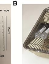
Preparation of Single Cell Suspensions from Mouse Aorta

从小鼠主动脉制备单细胞悬浮液

DH Desheng Hu
CY Changjun Yin
Sarajo Mohanta Sarajo Mohanta
Christian Weber Christian Weber
AH Andreas J. R. Habenicht
17455 Views
Jun 5, 2016
Atherosclerosis is a chronic inflammatory disease of the arterial wall characterized by lipid deposition, plaque formation, and immune cell infiltration. Innate and adaptive immune cells infiltrate the artery during development of the disease. Moreover, advanced disease leads to formation of artery tertiary lymphoid organs in the adventitia (Grabner et al., 2009; Hu et al., 2015). Various and diverse types of immune cells have been identified in the aorta adventitia vs atherosclerotic plaques (Elewa et al., 2016; Galkina et al., 2006; Lotzer et al., 2010; Mohanta et al., 2016; Mohanta et al., 2014; Moos et al., 2005; Srikakulapu et al., 2016; Zhao et al., 2004). There are conflicting reports on the number and subtypes of immune cells in the aorta depending on the age of the animals, the protocol that is used to obtain single cell suspensions, and the dietary conditions of the mice (Campbell et al., 2012; Clement et al., 2015; Galkina et al., 2006; Kyaw et al., 2012). The number of immune cells in the aorta differs as much as tenfold using different protocols (Butcher et al., 2012; Galkina et al., 2006; Gjurich et al., 2015; Grabner et al., 2009; Hu et al., 2015). These discrepant results call for a protocol that robustly documents bona fide aorta cells rather than those in the surrounding tissues or blood. Critical methodological hurdles include the removal of adjacent adipose tissue and small paraaortic lymph nodes lining the entire aortic tree that are not visible by the naked eye. A dissection microscope is therefore recommended. Moreover protocols of aorta preparations should ascertain that lymphocyte aggregates referred to as fat associated lymphoid clusters (FALCs) (Benezech et al., 2015; Elewa et al., 2015) that are often present at the border between the adipose tissue and the adventitia are removed before enzyme digestion. We propose - besides other approaches (Hu et al., 2015; Mohanta et al., 2014) - a combination of immunohistochemical staining and fluorescence activated cell sorter (FACS) analyses from single cell suspensions to quantify the cells of interest. This protocol describes isolation of single cells from mouse aorta for FACS and other analysis.
单克隆抗体介导的流感病毒微中和(MN)测定试验

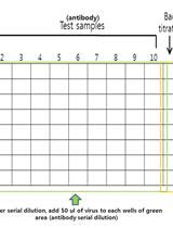
Micro Neutralization (MN) Assay of Influenza Viruses with Monoclonal Antibodies

单克隆抗体介导的流感病毒微中和(MN)测定试验

YW Ying Wu
MC MyungSam Cho
DS David Shore
MS Manki Song
JC JungAh Choi
TJ Tao Jiang
YD Yong-Qiang Deng
MB Melissa Bourgeois
LA Lynn Almli
HY Hua Yang
LC Li-Mei Chen
YS Yi Shi
JQ Jianxu Qi
AL An Li
KY Kye Sook Yi
MC MinSeok Chang
JB Jin Soo Bae
HL HyunJoo Lee
JS JiYoung Shin
JS James Stevens
SH SeoungSuh Hong
CQ Cheng-Feng Qin
GG George F. Gao
SC Shin Jae Chang
RD Ruben O. Donis
15130 Views
Jun 5, 2016
The human monoclonal antibodies generated from single human B cells were tested to characterize their ability to neutralize virus infectivity. The microneutralization assay is a highly sensitive and specific assay for detecting virus-specific neutralizing antibodies to influenza viruses. This protocol is to measure the ability of human monoclonal antibody to neutralize influenza virus by microneutralization assay.

微生物学

微球菌核酸酶消化法分析脉孢菌中核小体图谱

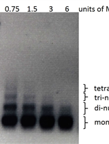
MNase Digestion for Nucleosome Mapping in Neurospora

微球菌核酸酶消化法分析脉孢菌中核小体图谱

CS Cigdem Sancar
GS Gencer Sancar
MB Michael Brunner
9682 Views
Jun 5, 2016
Digestion of chromatin by micrococcal nuclease MNase followed by high throughput sequencing allows us to determine the location and occupancy of nucleosomes on the genome. Here in this protocol we have described optimized conditions of MNase digestion of filamentous fungus Neurospora crassa chromatin without a requirement of a nuclear fractionation step.

神经科学

蔗糖偏好实验测量小鼠的应激诱导快感缺乏

Sucrose Preference Test to Measure Stress-induced Anhedonia

蔗糖偏好实验测量小鼠的应激诱导快感缺乏

Andrew L. Eagle Andrew L. Eagle
Michelle Mazei-Robison Michelle Mazei-Robison
AR Alfred J. Robison
32593 Views
Jun 5, 2016
The 2-bottle choice procedure for assessing sucrose preference is a useful test to investigate anhedonia (i.e., inability to feel pleasure) in laboratory rodents, particularly in stress-based models of depression. The 2-bottle choice procedure allows for a comparison between behavioral preference for sucrose solution in drinking water compared to water only. Preference is measured by volume and/or weight of liquid consumed daily, which is then converted to a percent preference compared to a water only baseline period. Sucrose preference is attenuated by a diversity of chronic stressors, including chronic mild and unpredictable stress (Willner et al., 1992; Willner, 1997; Pothion et al., 2004) and social defeat stress (Krishnan et al., 2007). It may also be susceptible to perturbation in mouse models of drug addiction because sucrose preference is altered in drug-dependent individuals (Kampov-Polevoy et al., 1997; Bogucka-Bonikowska et al., 2002; Janowsky et al., 2003). Both stress- and drug-induced alterations in sucrose preference may stem from maladaptations in the reward pathway, which consists of the dopaminergic neurons extending from the ventral tegmental area to the nucleus accumbens (NAc). Indeed, alterations in cyclic-AMP response element binding protein (CREB) activity in NAc underlie preference for sucrose (Barrot et al., 2002). Additionally, the transcription factor ΔFosB in NAc (Wallace et al., 2008), but not dorsal hippocampus (Eagle et al., 2015), regulates natural rewards, such as sucrose consumption. Therefore, the sucrose preference test described below provides a well-validated model to assess anhedonia and the function of specific brain regions and circuits.
子宫内电穿孔法检测小鼠小脑蒲肯野细胞

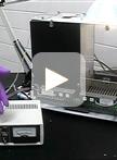
In utero Electroporation of Mouse Cerebellar Purkinje Cells

子宫内电穿孔法检测小鼠小脑蒲肯野细胞

Yukari H. Takeo Yukari H. Takeo
15616 Views
Jun 5, 2016
In utero electroporation (IUE) of mouse cerebellar Purkinje cells allows high expression levels of transgenes without toxicity (Nishiyama et al., 2012). This technique is suitable for co-transfection of multiple plasmid genes. Therefore, it is useful to express various sets of genes such as drug-inducible Cre/loxP constructs and CRISPR/Cas9 genome editing constructs (Takeo et al., 2015). Murine Purkinje cells arise from subventricular zone of fourth ventricle at embryonic day (E) 10-12. IUE at E11.5 into fourth ventricle results the most efficient transfection into Purkinje cells.
用时间解离性被动回避任务对海马依赖型学习敏感性的评估

Sensitive Assessment of Hippocampal Learning Using Temporally Dissociated Passive Avoidance Task

用时间解离性被动回避任务对海马依赖型学习敏感性的评估

Andrew L. Eagle Andrew L. Eagle
Hongbing Wang Hongbing Wang
AR Alfred J. Robison
8353 Views
Jun 5, 2016
The temporally dissociated passive avoidance (TDPA) paradigm is a variant of passive avoidance testing, and allows for more sensitive investigation of mild impairments in avoidance learning. Passive avoidance learning measures the latency to enter a “dark” context in which an aversive stimulus (foot shock) has been previously experienced using a light-dark box paradigm. Briefly, the animal is placed into the light side of the box and the time spent to cross into the dark side is measured. After entry into the dark chamber, the animal receives a mild (0.4-1.6 mA) footshock and is removed from the box. After a period of time, typically 24 h (note that this is entirely dependent on whether various levels of memory retention, e.g., short or long, are being measured), the animal is placed back into the box and cross-over latency is measured. Passive avoidance is learned after one trial and results in a robust increase in crossover latency. This behavior requires the association between a normally neutral environment and an aversive stimulus, and is dependent on hippocampal function (Stubley-Weatherly et al., 1996; Impey et al., 1998). TDPA extends this learning across multiple once-daily trials, producing a more graded and malleable latency score, and thus allows a more sensitive evaluation of changes in hippocampal function The task remains dependent on an intact hippocampus (Zhang et al., 2008), and subtle changes in hippocampal gene expression can result in robust alterations in TDPA latency scores (Eagle et al., 2015). We describe here a common method used to assess TDPA learning in mice.

植物科学

生物测定法研究昆虫产卵对植物抵御食草动物的影响

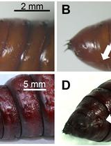
Bioassays to Investigate the Effects of Insect Oviposition on a Plant’s Resistance to Herbivores

生物测定法研究昆虫产卵对植物抵御食草动物的影响

Michele Bandoly Michele Bandoly
Anke Steppuhn Anke Steppuhn
12601 Views
Jun 5, 2016
Plants respond to herbivory with diverse defence responses (Schoonhoven et al., 2005). Many herbivorous insects deposit their eggs on their host plants before their larvae start to feed. Thus, plants could use insect eggs as a signal to increase their resistance to herbivores. Here, we report experimental procedures to explore whether and how insect oviposition impacts on plant resistance against the feeding larvae. The described approach revealed that Nicotiana attenuata (N. attenuata) plants that were previously exposed to oviposition by lepidopteran moths respond to herbivory by generalist Spodoptera exigua (S. exigua) and specialist Manduca sexta (M. sexta) larvae with an increased induction of defence responses, which results in a decreased performance or immune state of the feeding larvae (Bandoly et al., 2015; Bandoly et al., 2016). Consequently, insect oviposition can prime feeding-induced plant defence (priming: an enhanced plant response to stress upon the experience of a prior stimulus; Hilker et al., 2015). Full-factorial experiments with standardised procedures for insect oviposition and larval herbivory allow to decipher the effect of the plant exposure to insect eggs on the larval performance, feeding damage and immune state as well as to discriminate egg-induced plant responses from egg-primed responses to larval feeding.
采用飞秒激光技术定量测定过氧化物酶体和叶绿体之间的粘附强度

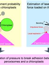
Quantification of the Adhesion Strength between Peroxisomes and Chloroplasts by Femtosecond Laser Technology

采用飞秒激光技术定量测定过氧化物酶体和叶绿体之间的粘附强度

YH Yoichiroh Hosokawa
TI Takanori Iino
Kazusato Oikawa Kazusato Oikawa
SM Shoji Mano
Kenji Yamada Kenji Yamada
Mikio Nishimura Mikio Nishimura
9708 Views
Jun 5, 2016
This is the detailed protocol to quantify adhesion strength between peroxisomes and chloroplasts in plant leaf palisade mesophyll cells described by Oikawa et al. (2015). The quantification was performed by utilizing local explosion induced by focusing femtosecond laser pulses into a mesophyll cell under a confocal microscope. When an impulsive force generated by an explosion is loaded on the interface between a peroxisome and a chloroplast, the peroxisome is frequently detached from the chloroplast. The probability of a peroxisome detaching from a chloroplast was estimated (left-top of Figure 1). Next, the magnitude of the impulsive force was quantified by an atomic force microscope (AFM) cantilever (right-top of Figure 1). On the basis of these results, the pressure to break adhesion between a peroxisome and a chloroplast was quantified as an index of the adhesion strength (bottom of Figure 1). In this protocol, these procedures are summarized. As the local explosion is induced not only in the medium of the mesophyll cells but also in aqueous medium generally, this method could be applied to various adhesions between organelles and between cells around 1 to 100 μm in diameter (e.g., adhesions between mitochondria and chloroplasts, between nucleus and cell membrane, and between two cells with weak physical interaction). Additionally, we have evaluated the interaction between peroxisomes and chloroplasts from the interaction length between two organelles. This protocol has been presented in Bio-protocol as “Measuring the interactions between peroxisomes and chloroplasts by in situ laser analysis” (Oikawa et al., 2015).Figure 1. Flow chart estimating adhesion strength between a peroxisome and a chloroplast by utilizing femtosecond laser and atomic force microscope
植物叶中细胞和气孔的微分和同步可视化

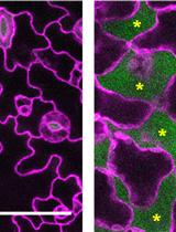
Differential and Simultaneous Visualization of Cells and Airspaces in Plant Leaves

植物叶中细胞和气孔的微分和同步可视化

TK Takashi Kawase
SS Shigeo S. Sugano
TS Tomoo Shimada
IH Ikuko Hara-Nishimura
8260 Views
Jun 5, 2016
This protocol is to differentially and simultaneously visualize both cells and airspaces in intact leaves and to create 3D structures of cells and airspaces from confocal images using the software DSLT_Demo (https://github.com/nslab2000/DSLT). Leaves stained with Nile Red in silicone-oil solution provide red color to cell membranes and green color to airspaces filled with silicone oil solution. This method is applicable to any tissues (except for dry seeds) of various plants including Arabidopsis, Nicotiana, Lemna and moss, and applicable even to hard leaves of plants such as switchgrass and Cinnamomum. Repeated use of this method enables time-lapse imaging of leaves over days and weeks because both Nile Red and silicone oil are harmless to plant tissues.