往期刊物2016

卷册: 6, 期号: 6

left

Dec

20

Dec

5

Nov

20

Nov

5

Oct

20

Oct

5

Sep

20

Sep

5

Aug

20

Aug

5

Jul

20

Jul

5

Jun

20

Jun

5

May

20

May

5

Apr

20

Apr

5

Mar

20

Mar

5

Feb

20

Feb

5

Jan

20

Jan

5

right

癌症生物学

荧光素酶标记癌细胞的体内生物发光成像

In vivo Bioluminescence Imaging of Luciferase-labeled Cancer Cells

荧光素酶标记癌细胞的体内生物发光成像

Marc Carceles-Cordon Marc Carceles-Cordon
Irene Rodriguez-Fernandez Irene Rodriguez-Fernandez
Veronica Rodriguez-Bravo Veronica Rodriguez-Bravo
CC Carlos Cordon-Cardo
Josep Domingo-Domenech Josep Domingo-Domenech
35058 Views
Mar 20, 2016
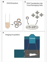
Over the past decade, in vivo bioluminescent imaging has emerged as a non-invasive and sensitive tool for studying ongoing biological processes within living organisms (Contag et al., 1997; Contag et al., 1998). Based on the detection and quantitation of the photons produced by the oxidation of luciferin by luciferase enzymes (Harvey, 1927), this technique has proved to be particularly useful in analyzing cancerous cells and monitoring tumor growth (Edinger et al., 1999; Sweeney et al., 1999; Vidal et al., 2015), providing a cost-effective insight into how the disease progresses in vivo, without the need of serial sacrifice of animals. This protocol describes in detail the procedure of obtaining luciferase-tagged tumors in immunocompromised mice that can be studied by bioluminescent imaging through the use of an IVIS Spectrum imager.

免疫学

流式细胞法体外评估免疫突触的形成

In vitro Assessment of Immunological Synapse Formation by Flow Cytometry

流式细胞法体外评估免疫突触的形成

Bo-Ra Na Bo-Ra Na
Chang-Duk Jun Chang-Duk Jun
16480 Views
Mar 20, 2016
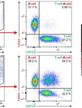
In adaptive immune system, formation of immunological synapse between T cells and antigen presenting cells (dendritic cells, B cells, and macrophages) or target cells (tumor cells and viral-infected cells) is critical for the execution of T cell immune responses via cytokine secretion or direct killing activity. Here, we describe the practical methods that directly measure the number of conjugates as a result of immunological synapse formation between T cells and superantigen-loaded B cells or between cytotoxic T cells and antigen-loaded target cells by dual-color flow cytometry.
基于鼠源淋巴祖细胞的体外分化获得先天免疫性淋巴细胞的方法

In vitro Differentiation of Murine Innate Lymphoid Cells from Common Lymphoid Progenitor Cells

基于鼠源淋巴祖细胞的体外分化获得先天免疫性淋巴细胞的方法

CS Corey Seehus
JK Jonathan Kaye
9668 Views
Mar 20, 2016
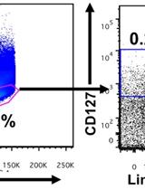
Subtypes of innate lymphoid cells (ILC), defined based on their cytokine secretion profiles and transcription factor expression, are important for host protection from pathogens and maintaining tissue homeostasis. ILCs develop from common lymphoid progenitors (CLP) in the bone marrow. Using the methods described here, we have previously shown that loss of the transcriptional regulator TOX (Thymocyte-selection associated HMG-box protein) leads to specific changes in ILC development and differentiation. Here, we describe how to obtain ILCs from in vivo isolated CLP grown in vitro.

微生物学

去NEDD化修饰的体外试验

In vitro Deneddylation Assay

去NEDD化修饰的体外试验

Anna M. Köhler Anna M. Köhler
Cindy Meister Cindy Meister
Gerhard H. Braus Gerhard H. Braus
9383 Views
Mar 20, 2016
Nedd8 is a small ubiquitin-like protein (9 kDa) covalently attached to a conserved lysine residue of a cullin protein which is part of cullin-RING ligases (CRLs). CRLs are major E3 ligases important for protein ubiquitination in the ubiquitin-proteasome pathway (UPP). The activity of CRLs is regulated by cycles of neddylation (CulA-N8, ~98 kDa) and deneddylation (CulA ~89 kDa). The COP9 signalosome (CSN) and Deneddylase A (DenA) are capable of cleaving the isopeptide bond between Nedd8 and CullinA. In contrast to the single protein DenA, CSN is an eight subunit multiprotein complex. Protein crude extracts of different Aspergillus nidulans csn deletion strains were mixed with recombinant CSN subunits expressed and purified from Escherichia coli (E. coli). Western hybridization experiments using anti-CulA or anti-Nedd8 antibodies could show the ratio of neddylated vs. deneddylated CulA. Using the deneddylation assay, we could show that CsnE is the last subunit joining a 7-subunit pre-assembled CSN in vitro and only then CSN can perform cullin deneddylation by the metalloprotease subunit CsnE. This assay is a fast and non-expensive method, which visualizes enzyme activity for deneddylating proteins. It might be also useful for testing the activity of other isopeptidases removing post-translational modifications from substrates in Aspergillus nidulans (A. nidulans) or other organisms.
痢疾变形虫的体外荧光基质降解试验

In vitro Fluorescent Matrix Degradation Assay for Entamoeba histolytica

痢疾变形虫的体外荧光基质降解试验

Merlyn Emmanuel Merlyn Emmanuel
SD Sunando Datta
8553 Views
Mar 20, 2016
Fluorescent matrix degradation assay is a popular and widely used assay in the field of invadopodium biology (Artym et al., 2009). Matrix remodeling and degradation can be observed under both physiological and pathological conditions. Cancer cells extensively remodel and degrade the underlying matrix by employing actin-rich protrusive structures called invadosomes. Similar structures are formed by the protozoan parasite Entamoeba histolytica (E. histolytica), upon coming in contact with fibronectin, a major component of the host (extracellular matrix) ECM. Here, we describe a similar assay to measure matrix degradation by Entamoeba histolytica.
使用还原性核糖核酸酶A检测蛋白质的氧化活性

Detection of Protein Oxidative Activity Using Reduced RNase A

使用还原性核糖核酸酶A检测蛋白质的氧化活性

MG Magdalena Grzeszczuk
KB Katarzyna Bocian-Ostrzycka
AL Anna Lasica
E. Katarzyna Jagusztyn-Krynicka E. Katarzyna Jagusztyn-Krynicka
6982 Views
Mar 20, 2016
This assay allows to determine whether proteins possess oxidative activity-the ability to introduce disulfide bond in vitro. The substrate for potential oxidases is a ribonuclease A which, for its activity, needs 4 properly formed disulfide bonds (Raines, 1998).RNase A activity can be detected by: Monitoring the digestion of RNA (Lambert and Freedman, 1983); Methylene Blue assay (Greiner-Stoeffele et al., 1996); Analyzing the cleavage of the cyclic CMP (Lyles and Gilbert, 1991; Lyles and Gilbert, 1991). We here describe method for measurements of oxidative activity, based on the cleavage of cCMP. Oxidative activity will be tested by measuring spectrophotometrically RNase A cleavage of cyclic-2’, 3’-cytidinemonophosphate (cCMP) to 3’-cytidinemonophosphate (3’ CMP), which results in an increase in absorption at 296 nm.The reaction equation: RNase A +2’ 3’-cCMP→RNase A + 3’ CMP.

神经科学

小鼠脑干听觉区反应测试

Mouse Auditory Brainstem Response Testing

小鼠脑干听觉区反应测试

Omar Akil Omar Akil
AO A. E. Oursler
Kevin Fan Kevin Fan
Lawrence R. Lustig Lawrence R. Lustig
17129 Views
Mar 20, 2016
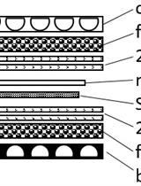
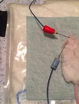
The auditory brainstem response (ABR) test provides information about the inner ear (cochlea) and the central pathways for hearing. The ABR reflects the electrical responses of both the cochlear ganglion neurons and the nuclei of the central auditory pathway to sound stimulation (Zhou et al., 2006; Burkard et al., 2007). The ABR contains 5 identifiable wave forms, labeled as I-V. Wave I represents the summated response from the spiral ganglion and auditory nerve while waves II-V represent responses from the ascending auditory pathway. The ABR is recorded via electrodes placed on the scalp of an anesthetized animal. ABR thresholds refer to the lowest sound pressure level (SPL) that can generate identifiable electrical response waves. This protocol describes the process of measuring the ABR of small rodents (mouse, rat, guinea pig, etc.), including anesthetizing the mouse, placing the electrodes on the scalp, recording click and tone burst stimuli and reading the obtained waveforms for ABR threshold values. As technology continues to evolve, ABR will likely provide more qualitative and quantitative information regarding the function of the auditory nerve and brainstem pathways involved in hearing.
小鼠脑室管膜细胞纤毛组织分析:脑室的解剖和染色

Dissection and Staining of Mouse Brain Ventricular Wall for the Analysis of Ependymal Cell Cilia Organization

小鼠脑室管膜细胞纤毛组织分析:脑室的解剖和染色

PL Paul Labedan
CM Cédric Matthews
LK Laurent Kodjabachian
HC Harold Cremer
FT Fadel Tissir
CB Camille Boutin
14628 Views
Mar 20, 2016
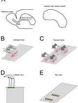
In the developing and mature central nervous system (CNS) the ventricular lumen is lined by the neuroepithelium and ependymal, respectively. These ventricular epithelia perform important functions related to the development, morphogenesis and physiology of the brain. In the mature CNS, ependyma constitutes a barrier between brain parenchyma and cerebro- spinal fluid (CSF). The most prominent feature of the apical surface of ependymal cells is the presence of multiple motile cilia that extend towards the ventricular lumen. The beating of cilia ensures the circulation of the CSF and its impairment leads to hydrocephalus. For an effective CSF flow, ciliary beating must be coordinated at the level of individual cells and at the tissue level. This coordination is achieved through the precise organization of cilia positioning within the plane of the ependyma. Two major features have been described regarding the planar organization of cilia in ependymal cells (Mirzadeh et al., 2010) and both have a cellular and tissular aspect (Boutin et al., 2014). The first one, rotational polarity, refers to the orientation of ciliary beating. At the cellular level, all cilia beat in the same direction (Figure 1B, black arrows). At the tissue level, each ependymal cell coordinates the direction of their beating with that of neighboring cells (Figure 1C, grey arrows). The second feature, translational polarity, is unique to ependymal cells and refers to the clustering of cilia in a tuft. At the cellular level, this tuft is displaced relative to the center of the ependymal cell (Figure 1B, red arrow). At the tissue level, the positioning of the ciliary tuft is coordinated between adjacent cells (Figure 1C). Alteration of any of these polarities at either level impairs CSF flow circulation (Mirzadeh et al., 2010; Boutin et al., 2014; Guirao et al., 2010; Hirota et al., 2010; Ohata et al., 2014). Cilia axonemes arise from basal bodies (BB) which are cylindrical structures anchored perpendicular to the sub-apical surface of the cells (Figure 1D). BBs are polarized by the presence of appendices such as basal foot or striated rootlets. The basal foot protrudes in a direction correlated with the direction of cilia beating, while the striated rootlet protrudes in the opposite direction of cilia beating (Marshall, 2008). The ‘en face view’ observation of BBs’ organization allows the visualization of ependymal polarities (Mirzadeh et al., 2010; Boutin et al., 2014). Here, we describe an immunofluorescence (IF) protocol for observation of ciliated cells in mouse brain ventricular lateral wall whole mounts (LWWM). This protocol can be used for classical confocal microscopy analysis. In addition, it is well suited for super-resolution STimulated Emission Depletion (STED) microscopy if observation of structures that have features which are smaller than the optical diffraction limit is needed. Finally, we describe a combination of antibodies that allow the concomitant observation, in a single sample, of ependymal polarities at the level of individual cilia, individual cells and at the tissue level.

植物科学

拟南芥和巴西固氮螺菌中一氧化氮的检测和硝酸盐浓度的测定

Detection of Nitric Oxide and Determination of Nitrite Concentrations in Arabidopsis thaliana and Azospirilum brasilense

拟南芥和巴西固氮螺菌中一氧化氮的检测和硝酸盐浓度的测定

Noelia Foresi Noelia Foresi
Natalia Correa-Aragunde Natalia Correa-Aragunde
Melina Amenta Melina Amenta
Andrés Arruebarrena Andrés Arruebarrena
Cecilia Creus Cecilia Creus
Lorenzo Lamattina Lorenzo Lamattina
20424 Views
Mar 20, 2016
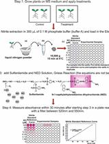
There is now general agreement that nitric oxide (NO) is an important and almost ubiquitous signal in plants. Nevertheless, there are still many controversial observations and differing opinions on the importance and functions of NO in plants. Partly, this may be due to the difficulties in detecting and quantifying NO. Here, we summarize protocols for detecting NO and quantifying nitrite concentration in Arabidopsis seedlings. We also present a method to measure NO in biofilms formed by the plant growth promoting rhizobacteria Azospirillum brasilense (A. brasilense). NO in oxygen-containing aqueous solutions has a short half-life that is often attributed to a rapid oxidation to nitrite. Here we detail the use of the fluorescent probe DAF-FM DA and the electrochemical method for directly detecting and quantifying NO, respectively, and the Griess reagent to indirectly detect NO through its oxidized nitrite form. These protocols could be useful in a variety of cell types and plant tissues, as well as for microorganisms.
体外微管结合实验和解离常数估算

In vitro Microtubule Binding Assay and Dissociation Constant Estimation

体外微管结合实验和解离常数估算

CK Christopher Kesten
RS René Schneider
SP Staffan Persson
14571 Views
Mar 20, 2016
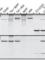
Microtubules (MTs) support an astonishing set of versatile cellular functions ranging from cell division, vesicle transport, and cell and tissue morphogenesis in various organisms. This versatility is in large mediated by MT-associated proteins (MAPs). The neuronal MAP Tau, for example, is stabilizing MTs in axons of the vertebrate nervous system and thus provides the basis for enduring axonal transport and the long life span of neurons (Mandelkow et al., 1994). Tau has been shown to bind to MTs directly in vitro and also to promote their nucleation from α-/β-tubulin subunits (Goode et al., 1994). Recently, we identified a plant-specific protein family called “companion of cellulose synthase” (CC), which was shown to bind MTs and enhance dynamics of the cortical MT array in plant cells under salt stress (Endler et al., 2015). The CCs were therefore hypothesized to help plant cells cope with stress conditions and thereby maintain biomass production under adverse growth conditions. Here, we provide detailed experimental information on in vitro MT binding assays, which allow assessing whether a protein of interest is binding to MTs. The assay utilizes the high molecular weight of MTs in a spin down approach and enables the determination of the dissociation constant Kd, a measure for the protein’s binding strength to MTs.
玉米中的玉蜀黎黑粉菌毒性试验

Ustilago maydis Virulence Assays in Maize

玉米中的玉蜀黎黑粉菌毒性试验

Amey Redkar Amey Redkar
Gunther Doehlemann Gunther Doehlemann
12630 Views
Mar 20, 2016
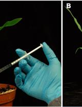
The basidiomycetous smut fungus Ustilago maydis (U. maydis) infects all aerial parts of its host plant maize (Zea mays L.). Infection symptoms are seen in the form of prominent tumors on all aerial parts of maize, after the establishment of a biotrophic interaction with the host usually around 5-6 days post infection (dpi). The fungus colonizes the various developmentally distinct aerial organs at different stages of development to form these prominent symptoms. Although being a biotrophic plant pathogen, U. maydis can easily be cultivated under axenic conditions to produce a standardized inoculum. The infections can be carried out under laboratory conditions by syringe inoculation on all the aerial organs of maize. This protocol has been successfully utilized to infect all the aerial organs of maize and formulate the virulence assays in U. maydis making it an excellent model system to study phyto-pathological investigations (Schilling et al., 2014; Redkar et al., 2015).
高粱根中独脚金内酯的提取和测定

Extraction and Measurement of Strigolactones in Sorghum Roots

高粱根中独脚金内酯的提取和测定

KY Kaori Yoneyama
XX Xiaonan Xie
TN Takahito Nomura
KY Koichi Yoneyama
10506 Views
Mar 20, 2016
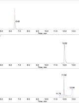
Strigolactones (SLs) are carotenoid-derived signaling chemicals containing two lactone moieties in their structures and induce seed germination of root parasitic plants, Striga and Orobanche spp. In the rhizosphere, SLs are essential host recognition signals not only for root parasitic plants but also for arbuscular mycorrhizal fungi. In plants, SLs play important roles as plant hormones regulating shoot and root architecture. Plants produce only trace amounts of chemically unstable SLs, which makes it difficult to determine SL contents in plant tissues. Here, we describe how to extract and quantify sorgomol and 5-deoxystrigol, major SLs produced in sorghum roots.
木本植物材料样本的制备用于X射线显微计算机断层扫描和木质部可视化技术

Sample Preparation for X-ray Micro-computed Tomography of Woody Plant Material and Associated Xylem Visualisation Techniques

木本植物材料样本的制备用于X射线显微计算机断层扫描和木质部可视化技术

NB Nicholas J. B. Brereton
9988 Views
Mar 20, 2016
Variation in the tissue structure of short rotation coppice (SRC) willow is a principle factor driving differences in lignocellulosic sugar yield yet much of the physiology and development of this tissue is unknown. Traditional sectioning can be both difficult and destructive in woody tissue; however, technology such as three dimensional X-ray micro-computational tomography (μCT) scanning can be used to move biological researchers beyond traditional two dimensional assessment of tissue variation without having to destructively cut cells. This technology does not replace classical microscopic techniques but rather can be carefully integrated with traditional methods to improve exploration of the world of plant biology in three dimensions. The procedures below outline preparation of willow for 3D X-ray μCT and associated xylem staining and visualisation techniques, in particular secondary xylem programmed-cell-death (PCD) delay during gelatinous fibre (g-fibre) development. Many of the staining techniques here are transferable to other woody species such as poplar and Eucalyptus.
在玉蜀黎黑粉菌感染玉米中进行基于EdU的DNA合成和细胞增殖实验

EdU Based DNA Synthesis and Cell Proliferation Assay in Maize Infected by the Smut Fungus Ustilago maydis

在玉蜀黎黑粉菌感染玉米中进行基于EdU的DNA合成和细胞增殖实验

Amey Redkar Amey Redkar
Gunther Doehlemann Gunther Doehlemann
9489 Views
Mar 20, 2016
The basidiomycetous smut fungus Ustilago maydis (U. maydis) infects all aerial parts of its host plant maize (Zea mays L.). Infection is seen in the form of prominent tumorous symptoms after the establishment of a biotrophic interaction with the host, usually around 5-6 days after infection. The fungus colonizes the various developmentally distinct aerial organs at different stages of development. Formation of tumors is coupled with the induction of host cell division. Activation of cell division can be understood as a measure of DNA synthesis which is triggered to induce rapid divisions in host cell. This developed protocol helps in tracking tumor induction in U. maydis by monitoring of DNA synthesis in planta. Infected leaves were treated with 5-ethynyl-2-deoxyuridine (EdU) at several stages of infection in the seedling leaves and labeled. EdU incorporation in the S phase cells, was visualized by attaching a fluorescent tag and non-dividing maize nuclei were stained with propidium iodide (PI). This protocol helped to understand the tumor development in U. maydis by confocal laser scanning microscopy (Kelliher and Walbot, 2011; Redkar et al., 2015)
玉米根中阿拉伯呋喃糖酶的活性测定

Assay of Arabinofuranosidase Activity in Maize Roots

玉米根中阿拉伯呋喃糖酶的活性测定

Liudmila V. Kozlova Liudmila V. Kozlova
PM Polina V. Mikshina
TG Tatyana A. Gorshkova
9076 Views
Mar 20, 2016
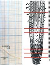
Root is a perfect model for studying the mechanisms of plant cell growth. Along the root length, several zones where cells are at different stages of development can be visualized (Figure 1). The dissection of the root on these zones allows the investigation of biochemical and genetic aspects of different growth steps. Maize primary root is much more massive than the root of other Monocots and thus more convenient for such type of research. Plant cell wall, mainly consisting of polysaccharides, plays an important role in plant life. Therefore, measurement of plant carbohydrate content and glycoside-modifying enzyme activity in plant cells has become an important aspect in plant physiology. One of the well-documented changes of hemicelluloses molecules during elongation growth of monocots cells is the decrease of arabinose substitution of glucuronoarabinoxylans. This might be caused by changes in synthesis of this polysaccharide or by the action of arabinofuranosidases. Here, we describe the protocol of spectrophotometric measuring of arabinofuranosidase activity in maize root by the rate of hydrolysis of chromogenic substrate (4-nitrophenyl α-L-arabinofuranoside).Figure 1. Scheme of plant material collection for further arabinofuranosidase assay. Four-day-old dark-grown maize seedling (left panel). Different zones of primary maize root and corresponding stages of cell development, according to Kozlova et al. (2012) (right panel).