往期刊物2016

卷册: 6, 期号: 3

left

Dec

20

Dec

5

Nov

20

Nov

5

Oct

20

Oct

5

Sep

20

Sep

5

Aug

20

Aug

5

Jul

20

Jul

5

Jun

20

Jun

5

May

20

May

5

Apr

20

Apr

5

Mar

20

Mar

5

Feb

20

Feb

5

Jan

20

Jan

5

right

癌症生物学

体外组织切片的稳定同位素分解代谢组学研究

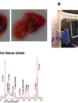
Stable Isotope Resolved Metabolomics Studies in ex vivo TIssue Slices

体外组织切片的稳定同位素分解代谢组学研究

TF Teresa W-M. Fan
AL Andrew N. Lane
RH Richard M. Higashi
10704 Views
Feb 5, 2016
An important component of this methodology is to assess the role of the tumor microenvironment on tumor growth and survival. To tackle this problem, we have adapted the original approach of Warburg (Warburg, 1923), by combining thin tissue slices with Stable Isotope Resolved Metabolomics (SIRM) to determine detailed metabolic activity of human tissues. SIRM enables the tracing of metabolic transformations of source molecules such as glucose or glutamine over defined time periods, and is a requirement for detailed pathway tracing and flux analysis. In our approach, we maintain freshly resected tissue slices (both cancerous and non- cancerous from the same organ of the same subject) in cell culture media, and treat with appropriate stable isotope-enriched nutrients, e.g., 13C6-glucose or 13C5, 15N2-glutamine. These slices are viable for at least 24 h, and make it possible to eliminate systemic influence on the target tissue metabolism while maintaining the original 3D cellular architecture. It is therefore an excellent pre-clinical platform for assessing the effect of therapeutic agents on target tissue metabolism and their therapeutic efficacy on individual patients (Xie et al., 2014; Sellers et al., 2015).

细胞生物学

小鼠卵母细胞分离、培养和RNA显微注射

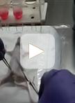
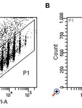
Mouse Oocyte Isolation, Cultivation and RNA Microinjection

小鼠卵母细胞分离、培养和RNA显微注射

Anna Tetkova Anna Tetkova
Marketa Hancova Marketa Hancova
16833 Views
Feb 5, 2016
Mammalian oocyte is a highly specialized cell, characterized by synthesis and storage of maternal proteins and RNAs that contributes to the meiotic cell cycle and early embryo development. The fully grown oocyte is transcriptionally quiescent and utilizes only transcripts synthesized and stored during the growing phase. Mouse oocytes are often used as a mammalian model for the study of molecular biology of the cell or biomedical research. Microinjection technique is a useful tool to deliver RNA coding for fluorescently tagged proteins to determine their subcellular localization or function, delivering biosensors for the study of various metabolic pathways or downregulation of specific targets by RNAi or oligo morpholinos to study gene function. Here, we describe a protocol for isolation, cultivation and microinjection of oocytes that might contribute to research or educational purposes.

免疫学

小鼠BMDC依赖性T淋巴细胞的极化试验

Mouse BMDC-dependent T Cell Polarization Assays

小鼠BMDC依赖性T淋巴细胞的极化试验

Vijaya Satchidanandam Vijaya Satchidanandam
14570 Views
Feb 5, 2016
In response to exposure to antigen, T cells whose T cell receptor (TCR) are capable of recognizing the self MHC-antigen derived peptide complex, respond to the antigen and differentiate into one of several subsets, namely TH1, TH2, TH17, Treg, etc. characterized by the signature cytokine they secrete, namely IFN-γ, IL-4, IL-17 or IL-10, respectively, referred to as syngeneic polarization as the MHC presenting the foreign antigen/epitope is self-derived. T cell responses following incubation for defined periods, usually 3 days for mouse splenocytes, are routinely measured by assessing the antigen-stimulated proliferation of T cells by measuring the radiolabeled precursor thymidine incorporated into the genomic DNA of the dividing T cell; the direction of polarization is assessed by measuring the cytokine produced by the proliferating or non-proliferating responding T cells using ELISA of culture supernatants or by intracellular cytokine staining followed by flow cytometry. In the protocols detailed below, we describe the use of syngeneic mouse bone marrow-derived primary dendritic cells (BMDC) as APC to stimulate spleen derived T cells. The proliferative response of the T cells is measured by incorporation of radiolabeled precursor thymidine into the genomic DNA and their direction of polarization is assessed by measuring the cytokines they secrete, namely IFN-γ, IL-4 and IL-17 over a 72 h period using ELISA. In addition, we used flow cytometry after intracellular cytokine staining to detect IL-17 positive T cells within the CD3+/CD4+/CD25low population. Prior live infection of BMDC with strains of Mycobacterium bovis- Bacille Calmette Guerin (BCG) was used as antigen to pre-condition the BMDC that presented antigens derived therefrom to T cells. We also measured cytokines secreted within 6 to 8 h of BCG infection by BMDC in order to correlate the BMDC cytokine profile with subsequent direction of T cell polarization.
基于细胞的监测AID活性的方法

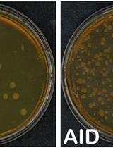
Cell-based Assays to Monitor AID Activity

基于细胞的监测AID活性的方法

Ludivine C. Litzler Ludivine C. Litzler
Stephen P. Methot Stephen P. Methot
Anne-Marie Patenaude Anne-Marie Patenaude
Astrid Zahn Astrid Zahn
Javier M. Di Noia Javier M. Di Noia
9166 Views
Feb 5, 2016
The enzyme Activation induced deaminase (AID) underpins antibody affinity maturation and isotype switching through its mutagenic activity of deaminating deoxycytidine to deoxyuridine in DNA. Subsequent processing of the deoxyuridine initiates the processes of somatic hypermutation (SHM) and class switch recombination (CSR) in B cells. Structure-function analysis of AID requires sensitive and biologically relevant methods to measure its various activities. Here we describe simple but effective methods to measure 1) the ability of AID to mutate the Escherichia coli genome, which provides an indication of its catalytic activity; 2) the capacity of AID to perform SHM by complementing a derivative of the DT40 chicken B cell line; 3) the ability of AID to perform CSR by complementing AID-deficient primary mouse B cells. The combination of the three methods, accompanied by the necessary analysis of AID subcellular localization and protein expression levels and stability, as controls, allows detailed structure-function interrogation of AID.

微生物学

酵母培养物中荧光硝酸盐和肽转运载体传感器的设计和功能分析

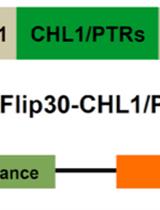
Design and Functional Analysis of Fluorescent Nitrate and Peptide Transporter Activity Sensors in Yeast Cultures

酵母培养物中荧光硝酸盐和肽转运载体传感器的设计和功能分析

CH Cheng-Hsun Ho
WF Wolf B. Frommer
10764 Views
Feb 5, 2016
This protocol describes the methods used to engineer and deploy genetically encoded fluorescence activity reporters for nitrate and peptide transporter activity in yeast cells. Fusion of the dual-affinity nitrate transceptor CHL1/AtNRT1.1/AtNPF6.3 or four different peptide transporters (AtPTR1, 2, 4, and 5) from Arabidopsis to a pair of fluorescent proteins with different spectral properties, enabled us to engineer the NiTracs (nitrate transporter activity tracking sensors) and the PepTracs (peptide transporter activity tracking sensors), ratiometric fluorescence activity sensors that monitor the activity of the plasma membrane nitrate transceptor or the peptide transporters in vivo (Ho et al., 2014). The NiTrac1 sensor responds specifically and reversibly to the addition of nitrate, while the PepTracs respond to addition of dipeptides, either by a reduction in donor and acceptor emission, while acceptor-excited emission remains unaltered, or by a change in ratio of the fluorophore emission. All sensors are suitable for ratiometric imaging. The similarity of the biphasic kinetics of the NiTrac1 sensor response [from µM to mM (Liu and Tsay, 2003)] and the nitrate transport kinetics of the native nitrate transceptor, intimates that NiTrac1 provides information on conformational rearrangements during the transport cycle, thereby reporting transporter activity over a wide range of external nitrate concentrations. Several variants of NiTrac have been engineered, which differ with respect to their affinity for nitrate (NiTrac1: CHL1; NiTracT101A: CHL1T101A). NiTrac also recognizes chlorate. Here we describe a simple method for the design, implementation, and detection of nitrate transceptor activity in yeast cells using a spectrofluorimeter.
采用微生物滞后时间计算衡量不同生长条件下的自适应能力

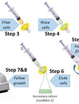
Calculation of Microorganism Lag Times as a Measure of Adaptative Capability between Different Growth Conditions

采用微生物滞后时间计算衡量不同生长条件下的自适应能力

Brice Enjalbert Brice Enjalbert
6540 Views
Feb 5, 2016
This protocol has been designed as a simple and efficient way to investigate microorganism adaptive capabilities (Enjalbert et al., 2015). It is performed using switch experiments in which cells are initially grown in the first condition (primary cultures), then rapidly switched to the second condition (secondary culture) without centrifugation or quenching. The measurement is based on the capacity of the secondary culture cells to resume growth. This protocol can be utilized for assessing metabolic or stress adaptation of microorganisms.

神经科学

开颅手术后利用电压敏感性染料进行小鼠脑皮层成像

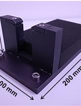
Craniotomy for Cortical Voltage-sensitive Dye Imaging in Mice

开颅手术后利用电压敏感性染料进行小鼠脑皮层成像

Takayuki Suzuki Takayuki Suzuki
MM Masanori Murayama
14176 Views
Feb 5, 2016
Cortico-cortical interactions play crucial roles in various brain functions. Here, we present a detailed surgical procedure for cortical voltage-sensitive dye (VSD) imaging that allows monitoring of spatiotemporal dynamics in cortical activity in living mice. Cortical neurons in the upper layers (layer 1-3) are stained with a VSD, and an image sensor with a fast sampling rate (500 Hz) detects fluorescent changes in corrective activity. The procedure includes fixing a mouse brain to a stereotaxic apparatus, craniotomy on a large cortical area, VSD staining, and wide-field imaging of cortical activity. The entire procedure can be completed in 5 h (from the administration of anesthesia to the start of cortical VSD imaging).

植物科学

使用结构照明显微镜(SIM)和光敏定位显微镜(PALM)分析RNA聚合酶II分子在流式分选的拟南芥细胞核中的丰度及分布

Structured Illumination Microscopy (SIM) and Photoactivated Localization Microscopy (PALM) to Analyze the Abundance and Distribution of RNA Polymerase II Molecules on Flow-sorted Arabidopsis Nuclei

使用结构照明显微镜(SIM)和光敏定位显微镜(PALM)分析RNA聚合酶II分子在流式分选的拟南芥细胞核中的丰度及分布

Klaus Weisshart Klaus Weisshart
JF Jörg Fuchs
VS Veit Schubert
12780 Views
Feb 5, 2016
RNA polymerase II (RNAPII) is the enzyme transcribing most of the eukaryotic protein-coding genes. Analysing the distribution and quantification of RNAPII can help understanding its function in interphase nuclei. Although several investigations in mammals indicate the organization of RNAPII in so-called ‘transcription factories’ (Jackson et al., 1993; Rieder et al., 2012; Papantonis and Cook, 2013), their existence is still controversially discussed (Zhao et al., 2014). Recently, based on super-resolution microscopy the presence of transcription factories was also suggested in plants. Applying structured illumination microscopy (SIM) and photoactivated localization microscopy (PALM) the distribution and number of RNAPII molecules in Arabidopsis nuclei were analysed and a positive correlation between RNAPII abundance and endopolyploidy was found (Schubert, 2014; Schubert and Weisshart, 2015). Here, we present a protocol describing the isolation of Arabidopsis thaliana interphase nuclei via flow-sorting according to their endopolyploidy level, followed by a double immunostaining using antibodies specific for different RNAPII modifications and the subsequent evaluation by spatial SIM and PALM to achieve results regarding the abundance, distribution and co-localization of single inactive and active RNAPII molecules.
黄单胞菌感染番茄叶片后乙烯产生的定量测定

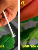
Quantification of Ethylene Production in Tomato Leaves Infected by Xanthomonas euvesicatoria

黄单胞菌感染番茄叶片后乙烯产生的定量测定

JK Jung-Gun Kim
WS William Stork
MM Mary Beth Mudgett
10897 Views
Feb 5, 2016
Ethylene is a gaseous plant hormone controlling fruit ripening, flower opening, leaf senescence as well as abscission, and disease symptom development. Ethylene plays a critical role in the bacterial pathogen Xanthomonas euvesicatoria (X. euvesicatoria)-elicited symptom development in tomato. This protocol describes the measurement of ethylene gas produced by tomato leaves infected with X. euvesicatoria. Infected leaflets are placed in a glass tube for 30 min without sealing. The glass tubes are then capped with a septa stopper, and incubated for an hour. A 1 ml gas sample is removed from the tube using a syringe and then injected into a gas chromatograph to quantify ethylene gas levels. This protocol will be applicable for other plants with other pathogens with modifications.
叶绿体膜脂的制备及脂质-蛋白质相互作用的分析

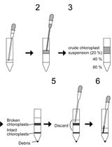
Preparation of Chloroplast Lipid Membrane and Lipid-protein Interaction Assay

叶绿体膜脂的制备及脂质-蛋白质相互作用的分析

KO Kumiko Okazaki
SM Shin-ya Miyagishima
HW Hajime Wada
10077 Views
Feb 5, 2016
Lipid-Protein interaction assay is a method to search lipids, which are bound with proteins in vitro. Since membranes that are spotted with chloroplast lipids such as monogalactosyldiacylglycerol (MGDG), digalactosyldiacylglycerol (DGDG), and sulfoquinovosyldiacylglycerol (SQDG) are not commercially available, we extracted these lipids from cyanobacterial cells and spotted them onto membranes. The prepared membranes could be used for lipid-protein interaction assay.
测量从拟南芥中分离的完整叶绿体中的PI4P含量

Measurement of PI4P Levels in Intact Chloroplasts Isolated from Arabidopsis thaliana

测量从拟南芥中分离的完整叶绿体中的PI4P含量

KO Kumiko Okazaki
SM Shin-ya Miyagishima
HW Hajime Wada
9757 Views
Feb 5, 2016
Phosphatidylinositol 4-phosphate (PI4P), a major species of phosphoinositides, modulates many fundamental cellular processes. We have recently revealed that PI4P plays an important role in chloroplast division as a negative regulator. Despite its importance in chloroplasts, the content of PI4P in chloroplasts is very low and it is difficult to measure PI4P levels. In this protocol, we describe a simple method that we have developed for measurement of low level of PI4P in chloroplasts. Intact chloroplasts were isolated by a basic method using Percoll gradient centrifugation and acidic lipids were extracted from the isolated chloroplasts. The extracted acidic lipids including PI4P were spotted onto the membrane strip, which had been pre-spotted with PI4P standards and other phosphoinositides as negative controls. PI4P in the spot of acidic lipids on the membrane was detected using a PI4P binding protein.

干细胞

用miRNA前体转染拟胚体来诱导心肌分化

Transfection of Embryoid Bodies with miRNA Precursors to Induce Cardiac Differentiation

用miRNA前体转染拟胚体来诱导心肌分化

Federica Pisano Federica Pisano
MG Massimiliano Gnecchi
8955 Views
Feb 5, 2016
In recent years, the utilization of stem cell therapy to regenerate cardiac tissue has been proposed as a possible strategy to treat cardiac damage (Gnecchi et al., 2012, Aguirre et al., 2013; Sanganalmath and Bolli, 2013). Although encouraging results have been obtained in experimental models, the efficiency of cardiac regeneration is very poor and one of the major barriers to progress in the area of cell therapy for damaged heart is represented by the limited capacity of cells to differentiate into mature cardiomyocytes (CMC) (Laflamme and Murry, 2011). Cell manipulation and transfection represent versatile tools in this context (Melo et al., 2005; Dzau et al., 2005). Murine P19 embryonal carcinoma cells are a well-established cell line capable of differentiating in vitro into spontaneously beating CMC. This cell system with its limited cell culture requirements, protocol reproducibility and ease in uptake and subsequent expression of ectopic genetic materials render it ideal for the study of the cardiac differentiation process. P19 cells have been successfully used to gain important insights into the early molecular processes of CMC differentiation (van der Heyden and Defize, 2003; van der Heyden et al., 2003). P19 cells can also be maintained in an undifferentiated state in a monolayer culture when grown in adherence; this condition allows the enrichment of large cell numbers useful for cardiac differentiation protocols (McBurney, 1993). On the other hand, when cultured in bacterial dishes, P19 cells will grow in suspension and generate embryoid bodies (EB). When exposed to dimethyl sulfoxide (DMSO), EB differentiate into spontaneously beating cells, which can be defined as CMC. This definition is based on their gene and protein expression and their electrophysiological properties (Wobus et al., 1994; van der Heyden et al., 2003). In our laboratory, we used this in vitro model to verify whether the over-expression of a defined combination of miRNA can synergistically induce effective cardiac differentiation (Pisano et al., 2015). We used miRNA1, miRNA133 and miRNA499 alone or in combination. Here, we describe how we transiently transfect P19 cells to over-express a single or a combination of miRNA precursors (pre-miRNA).