往期刊物2025

卷册: 15, 期号: 2

left

Sep

5

Aug

20

Aug

5

Jul

20

Jul

5

Jun

20

Jun

5

May

20

May

5

Apr

20

Apr

5

Mar

20

Mar

5

Feb

20

Feb

5

Jan

20

Jan

5

right

生物信息学与计算生物学

基于DIMet的标记代谢组学数据差异分析:以胶质母细胞瘤代谢为例的分步指南

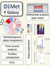
Using DIMet for Differential Analysis of Labeled Metabolomics Data: A Step-by-step Guide Showcasing the Glioblastoma Metabolism

基于DIMet的标记代谢组学数据差异分析:以胶质母细胞瘤代谢为例的分步指南

JG Johanna Galvis
JG Joris Guyon
TD Thomas Daubon
MN Macha Nikolski
1679 Views
Jan 20, 2025
Stable-isotope resolved metabolomics (SIRM) is a powerful approach for characterizing metabolic states in cells and organisms. By incorporating isotopes, such as 13C, into substrates, researchers can trace reaction rates across specific metabolic pathways. Integrating metabolomics data with gene expression profiles further enriches the analysis, as we demonstrated in our prior study on glioblastoma metabolic symbiosis. However, the bioinformatics tools for analyzing tracer metabolomics data have been limited. In this protocol, we encourage the researchers to use SIRM and transcriptomics data and to perform the downstream analysis using our software tool DIMet. Indeed, DIMet is the first comprehensive tool designed for the differential analysis of tracer metabolomics data, alongside its integration with transcriptomics data. DIMet facilitates the analysis of stable-isotope labeling and metabolic abundances, offering a streamlined approach to infer metabolic changes without requiring complex flux analysis. Its pathway-based "metabologram" visualizations effectively integrate metabolomics and transcriptomics data, offering a versatile platform capable of analyzing corrected tracer datasets across diverse systems, organisms, and isotopes. We provide detailed steps for sample preparation and data analysis using DIMet through its intuitive, web-based Galaxy interface. To showcase DIMet's capabilities, we analyzed LDHA/B knockout glioblastoma cell lines compared to controls. Accessible to all researchers through Galaxy, DIMet is free, user-friendly, and open source, making it a valuable resource for advancing metabolic research.

生物工程

在酿酒酵母和大肠杆菌中克隆叶绿体基因组

Cloning a Chloroplast Genome in Saccharomyces cerevisiae and Escherichia coli

在酿酒酵母和大肠杆菌中克隆叶绿体基因组

EW Emma Jane Lougheed Walker
BK Bogumil Jacek Karas
2894 Views
Jan 20, 2025
Chloroplast genomes present an alternative strategy for large-scale engineering of photosynthetic eukaryotes. Prior to our work, the chloroplast genomes of Chlamydomonas reinhardtii (204 kb) and Zea mays (140 kb) had been cloned using bacterial and yeast artificial chromosome (BAC/YAC) libraries, respectively. These methods lack design flexibility as they are reliant upon the random capture of genomic fragments during BAC/YAC library creation; additionally, both demonstrated a low efficiency (≤ 10%) for correct assembly of the genome in yeast. With this in mind, we sought to create a highly flexible and efficient approach for assembling the 117 kb chloroplast genome of Phaeodactylum tricornutum, a photosynthetic marine diatom. Our original article demonstrated a PCR-based approach for cloning the P. tricornutum chloroplast genome that had 90%–100% efficiency when screening as few as 10 yeast colonies following assembly. In this article, we will discuss this approach in greater depth as we believe this technique could be extrapolated to other species, particularly those with a similar chloroplast genome size and architecture.

生物科学

脂多糖(LPS)诱导牙髓炎的小鼠模型

Mouse Model of Lipopolysaccharide (LPS)-Induced Pulpitis

脂多糖(LPS)诱导牙髓炎的小鼠模型

LS Lanting Shao
BC Baian Chen
YZ Ying Zheng
2230 Views
Jan 20, 2025
Pulpitis is an important and prevalent disease within the oral cavity. Thus, animal models are necessary tools for basic research focused on pulpitis. Researchers worldwide often use dogs and miniature pigs to construct animal models of pulpitis. However, gene editing in miniature pigs is difficult, the surgical modeling process is complex, and tooth demineralization time is lengthy. Although some researchers have attempted to establish a mouse model of pulpitis, most models have involved direct exposure of dental pulp. However, the causes of pulpitis vary considerably among individuals, hindering effective research. In this study, we established a mouse model of pulpitis by accessing the pulp cavity, exposing the pulp to lipopolysaccharide (LPS), and then filling the tooth. One day after surgery, we observed many necrotic tissues and extensive inflammatory exudate, including neutrophils, around the coronal cavity preparation. Additionally, we noted many more neutrophils and a small amount of chronic inflammatory cell infiltrates at the junction between inflamed and normal tissue. These findings indicated that our model can be used to explore the early stage of pulpitis. Ten days after surgery, we observed vacuolar degeneration in some fibroblasts and proliferation in others at the distal end of the inflamed tissue. We also noted dilation and congestion of the pulp blood vessels. Therefore, our model can also be used to explore the middle and later stages of pulpitis. Thirty days after surgery, we observed necrosis in the coronal pulp cavity and upper half of the root pulp, indicating that our model can also be used to explore the end stage of pulpitis. This model is easy to establish, shows pulpitis progression in the dental pulp, exhibits a clear inflammatory phenotype, and can be readily combined with gene editing techniques. Accordingly, it is suitable for basic research focused on pulpitis and has substantial practical value.

癌症生物学

优化的患者来源类器官与匹配癌相关成纤维细胞同步培养方案

An Optimized Protocol for Simultaneous Propagation of Patient-derived Organoids and Matching CAFs

优化的患者来源类器官与匹配癌相关成纤维细胞同步培养方案

JH Jenny M. Högström
TM Taru Muranen
3043 Views
Jan 20, 2025
Recurrent hormone receptor-positive (HR+) breast cancer is a leading cause of cancer mortality in women. Recurrence and resistance to targeted therapies have been difficult to study due to the long clinical course of the disease, the complex nature of resistance, and the lack of clinically relevant model systems. Existing models are limited to a few HR+ cell lines, organoid models, and patient-derived xenograft models, all lacking components of the human tumor microenvironment. Furthermore, the low take rate and loss of estrogen receptor (ER) expression in patient-derived organoids (PDOs) has been challenging. Our protocol allows simultaneous isolation of PDOs and matching cancer-associated fibroblasts (CAFs) from primary and metastatic HR+ breast cancers. Importantly, our protocol has a higher take rate and enables long-term culturing of PDOs that retain ER expression. Our matching PDOs and CAFs will provide researchers with a new resource to study the influence of the tumor microenvironment on various aspects of cancer biology such as cell growth and drug resistance in HR+ breast cancer.

细胞生物学

小鼠耳蜗器官解剖、免疫染色及共聚焦成像

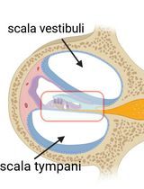
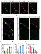
Cochlear Organ Dissection, Immunostaining, and Confocal Imaging in Mice

小鼠耳蜗器官解剖、免疫染色及共聚焦成像

CC Chenyu Chen
BC Binjun Chen
XQ Xiaoqing Qian
HS Haojie Sun
XF Xiao Fu
DR Dongdong Ren
2383 Views
Jan 20, 2025
The organ of Corti, located in the inner ear, is the primary organ responsible for animal hearing. Each hair cell has a V-shaped or U-shaped hair bundle composed of actin-filled stereocilia and a kinocilium supported by true transport microtubules. Damage to these structures due to noise exposure, drug toxicity, aging, or environmental factors can lead to hearing loss and other disorders. The challenge when examining auditory organs is their location within the bony labyrinth and their small and fragile nature. This protocol describes the dissection procedure for the cochlear organ, followed by confocal imaging of immunostained endogenous and fluorescent proteins. This approach can be used to understand hair cell physiology and the molecular mechanisms required for normal hearing.
基于流式的体内方法测定翻译核糖体数量及翻译延长速率

Flow-based In Vivo Method to Enumerate Translating Ribosomes and Translation Elongation Rate

基于流式的体内方法测定翻译核糖体数量及翻译延长速率

MS Mina O. Seedhom
DD Devin Dersh
JY Jonathan W. Yewdell
2025 Views
Jan 20, 2025
Protein synthesis is by far the most energetically costly cellular process in rapidly dividing cells. Quantifying translating ribosomes in individual cells and their average mRNA transit rate is arduous. Quantitating assembled ribosomes in individual cells requires electron microscopy and does not indicate ribosome translation status. Measurement of average transit rates entails in vitro pulse-chase radiolabeling of isolated cells or ribosome profiling after ribosome runoff, which is expensive and extremely demanding technically. Here, we detail protocols based on ribosome-mediated nascent chain puromycylation, harringtonine to stall initiating ribosomes while allowing ribosome elongation to continue normally, and cycloheximide to freeze translating ribosomes in place. Each compound is delivered intravenously to mice in the appropriate order, and after ex vivo cell fixation and permeabilization, translating ribosome numbers and transit rates are measured by flow cytometry using a directly conjugated puromycin-specific antibody.

发育生物学

通过CRISPR介导的Tp53基因敲除高效永生化小鼠胚胎成纤维细胞

An Efficient Method for Immortalizing Mouse Embryonic Fibroblasts by CRISPR-mediated Deletion of the Tp53 Gene

通过CRISPR介导的Tp53基因敲除高效永生化小鼠胚胎成纤维细胞

SS Srisathya Srinivasan
HH Hsin-Yi Henry Ho
1680 Views
Jan 20, 2025
Mouse embryonic fibroblasts (MEFs) derived from genetically modified mice are a valuable resource for studying gene function and regulation. The MEF system can also be combined with rescue studies to characterize the function of mutant genes/proteins, such as disease-causing variants. However, primary MEFs undergo senescence soon after isolation and passaging, making long-term genetic manipulations difficult. Previously described methods for MEF immortalization are often inconsistent or alter the physiological properties of the cells. Here, we describe an optimized method that overcomes these limitations. By using electroporation to deliver CRISPR constructs that target the Tp53 gene, the method reliably generates immortalized MEFs (iMEFs) within three weeks. Importantly, iMEFs closely resemble the parent cell populations, and individual iMEFs can be cloned and expanded for subsequent genetic manipulation and characterization. We envision that this protocol can be adopted broadly to immortalize other mouse primary cell types.

分子生物学

利用叉式PCR进行基因组步移以扩增未知侧翼DNA的实验方案

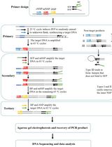
Protocol to Retrieve Unknown Flanking DNA Using Fork PCR for Genome Walking

利用叉式PCR进行基因组步移以扩增未知侧翼DNA的实验方案

HW Hongjing Wu
HP Hao Pan
HL Haixing Li
1636 Views
Jan 20, 2025
PCR-based genome walking is one of the prevalent techniques implemented to acquire unknown flanking genomic DNAs. The worth of genome walking includes but is not limited to cloning full-length genes, mining new genes, and discovering regulatory regions of genes. Therefore, this technique has advanced molecular biology and related fields. However, the PCR amplification specificity of this technique needs to be further improved. Here, a practical protocol based on fork PCR is proposed for genome walking. This PCR uses a fork primer set of three arbitrary primers to execute walking amplification task, where the primary fork primer mediates walking by partially annealing to an unknown flank, and the fork-like structure formed between the three primers participates in inhibiting non-target amplification. In primary fork PCR, the low-annealing temperature (25 °C) cycle allows the primary fork primer to anneal to many sites of the genome, synthesizing a cluster of single-stranded DNAs; the subsequent 65 °C cycle processes the target single-strand into double-strand via the site-specific primer; then, the remaining 65 °C cycles selectively enrich this target DNA. However, any non-target single-stranded DNA formed in the 25 °C cycle cannot be further processed in the following 65 °C cycles because it lacks an exact binding site for any primer. Secondary, or even tertiary nested fork PCR further selectively enriches the target DNA. The practicability of fork PCR was validated by walking three genes in Levilactobacillus brevis CD0817 and one gene in Oryza sativa. The results indicated that the proposed protocol can serve as a supplement to the existing genome walking protocols.

神经科学

基于FlashTag的胚胎脑室内巨噬细胞标记方法

FlashTag-mediated Labeling for Intraventricular Macrophages in the Embryonic Brain

基于FlashTag的胚胎脑室内巨噬细胞标记方法

HA Hisa Asai
MO Mizuki Ono
TM Takaki Miyata
YH Yuki Hattori
1201 Views
Jan 20, 2025
The fate mapping technique is essential for understanding how cells differentiate and organize into complex structures. Various methods are used in fate mapping, including dye injections, genetic labeling (e.g., Cre-lox recombination systems), and molecular markers to label cells and track their progeny. One such method, the FlashTag system, was originally developed to label neural progenitors. This technique involves injecting carboxyfluorescein diacetate succinimidyl ester (CFSE) into the lateral ventricles of mouse embryos, relying on the direct uptake of dye by cells. The injection of CFSE into the lateral ventricle allows for the pulse labeling of mitotic (M-phase) neural progenitors in the ventricular zone and their progeny throughout the brain. This approach enables us to trace the future locations and differentiation paths of neural progenitors. In our previous study, we adapted this method to selectively label central nervous system–associated macrophages (CAMs) in the lateral ventricle by using a lower concentration of CFSE compared to the original protocol. Microglia, the brain's immune cells, which play pivotal roles in both physiological and pathological contexts, begin colonizing the brain around embryonic day (E) 9.5 in mice, with their population expanding as development progresses. The modified FlashTag technique allowed us to trace the fate of intraventricular CAMs, revealing that certain populations of microglia are derived from these cells. The optimized approach offers deeper insights into the developmental trajectories of microglia. This protocol outlines the modified FlashTag method for labeling intraventricular CAMs, detailing the CFSE injection procedure, evaluation of CFSE dilution, and preparation of tissue for immunohistochemistry.
原代神经元培养与瞬时转染

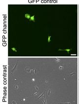
Primary Neuronal Culture and Transient Transfection

原代神经元培养与瞬时转染

ST Shun-Cheng Tseng
PC Peng-Tzu Chen
EH Eric Hwang
1132 Views
Jan 20, 2025
Primary neuronal culture and transient transfection offer a pair of crucial tools for neuroscience research, providing a controlled environment to study the behavior, function, and interactions of neurons in vitro. These cultures can be used to investigate fundamental aspects of neuronal development and plasticity, as well as disease mechanisms. There are numerous methods of transient transfection, such as electroporation, calcium phosphate precipitation, or cationic lipid transfection. In this protocol, we used electroporation for neurons immediately before plating and cationic lipid transfection for neurons that have been cultured for a few days in vitro. In our experience, the transfection efficiency of electroporation can be as high as 30%, and cationic lipid transfection has an efficiency of 1%–2%. While cationic lipid transfection has much lower efficiency than electroporation, it does offer the advantage of a higher expression level. Therefore, these transfection methods are suitable for different stages of neurons and different expression requirements.
利用蛋白涂层质谱技术解析乙酰胆碱结合蛋白的配体受体相互作用位点

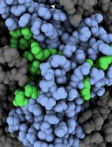
Using Protein Painting Mass Spectrometry to Define Ligand Receptor Interaction Sites for Acetylcholine Binding Protein

利用蛋白涂层质谱技术解析乙酰胆碱结合蛋白的配体受体相互作用位点

AG Alexandru Graur
NE Natalie Erickson
NK Nadine Kabbani
889 Views
Jan 20, 2025
Nicotinic acetylcholine receptors (nAChRs) are a family of ligand-gated ion channels expressed in nervous and non-nervous system tissue important for memory, movement, and sensory processes. The pharmacological targeting of nAChRs, using small molecules or peptides, is a promising approach for the development of compounds for the treatment of various human diseases including inflammatory and neurogenerative disorders such as Alzheimer’s disease. Using the Aplysia californica acetylcholine binding protein (Ac-AChBP) as an established structural surrogate for human homopentameric α7 nAChRs, we describe an innovative protein painting mass spectrometry (MS) method that can be used to identify interaction sites for various ligands at the extracellular nAChR site. We describe how the use of small molecule dyes can be optimized to uncover contact sites for ligand–protein interactions based on MS detection. Protein painting MS has been recently shown to be an effective tool for the identification of residues within Ac-AChBP involved in the binding of know ligands such as α-bungarotoxin. This strategy can be used with computational structural modeling to identify binding regions involved in drug targeting at the nAChR.
基于小鼠突触体的胰蛋白酶切割实验解析突触蛋白亚细胞定位

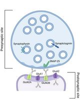
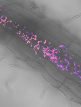
Mouse-derived Synaptosomes Trypsin Cleavage Assay to Characterize Synaptic Protein Sub-localization

基于小鼠突触体的胰蛋白酶切割实验解析突触蛋白亚细胞定位

JS Jasmeet Kaur Shergill
DA Domenico Azarnia Tehran
853 Views
Jan 20, 2025
Neurons communicate through neurotransmission at highly specialized junctions called synapses. Each neuron forms numerous synaptic connections, consisting of presynaptic and postsynaptic terminals. Upon the arrival of an action potential, neurotransmitters are released from the presynaptic site and diffuse across the synaptic cleft to bind specialized receptors at the postsynaptic terminal. This process is tightly regulated by several proteins at both presynaptic and postsynaptic sites. The localization, abundance, and function of these proteins are essential for productive neurotransmission and are often affected in neurological and neurodegenerative disorders. Here, we outline a method for purifying mouse synaptosomes and using limited tryptic digestion to assess the subcellular localization of synaptic proteins. During synaptosomes purification, presynaptic terminals reseal and are protected from proteolysis, while postsynaptic proteins remain susceptible to tryptic cleavage. These changes can easily be evaluated by western blot analysis. This approach offers a straightforward and reliable method to evaluate the subcellular localization of synaptic proteins based on their proteolytic sensitivity, providing valuable insights into synaptic physiology and pathology.

植物科学

利用基于FRET的生物传感器测量丛枝菌根共生黑麦草根中的细胞磷水平

Utilizing FRET-based Biosensors to Measure Cellular Phosphate Levels in Mycorrhizal Roots of Brachypodium distachyon

利用基于FRET的生物传感器测量丛枝菌根共生黑麦草根中的细胞磷水平

SZ Shiqi Zhang
LJ Lucas Jurgensen
MH Maria J. Harrison
1175 Views
Jan 20, 2025
Arbuscular mycorrhizal (AM) fungi engage in symbiotic relationships with plants, influencing their phosphate (Pi) uptake pathways, metabolism, and root cell physiology. Despite the significant role of Pi, its distribution and response dynamics in mycorrhizal roots remain largely unexplored. While traditional techniques for Pi measurement have shed some light on this, real-time cellular-level monitoring has been a challenge. With the evolution of quantitative imaging with confocal microscopy, particularly the use of genetically encoded fluorescent sensors, live imaging of intracellular Pi concentrations is now achievable. Among these sensors, fluorescence resonance energy transfer (FRET)-based biosensors stand out for their accuracy. In this study, we employ the Pi-specific biosensor (cpFLIPPi-5.3m) targeted to the cytosol or plastids of Brachypodium distachyon plants, enabling us to monitor intracellular Pi dynamics during AM symbiosis. A complementary control sensor, cpFLIPPi-Null, is introduced to monitor non-Pi-specific changes. Leveraging a semi-automated ImageJ macro for sensitized FRET analysis, this method provides a precise and efficient way to determine relative intracellular Pi levels at the level of individual cells or organelles.

更正

校正:基于微孔板的拟南芥硝酸盐转运蛋白2.1表达监测系统——利用荧光素酶报告基因

Correction Notice: A Microplate-Based Expression Monitoring System for Arabidopsis NITRATE TRANSPORTER2.1 Using the Luciferase Reporter

校正:基于微孔板的拟南芥硝酸盐转运蛋白2.1表达监测系统——利用荧光素酶报告基因

YU Yoshiaki Ueda
SY Shuichi Yanagisawa
287 Views
Jan 20, 2025