往期刊物2024
卷册: 14, 期号: 22
生物化学
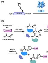
Preparation of Protein Lysates Using Biorthogonal Chemical Reporters for Click Reaction and in-Gel Fluorescence Analysis
利用生物正交化学标记制备蛋白裂解物用于点击反应及凝胶内荧光分析
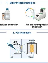
Utilizing the Planar Lipid Bilayer Technique to Investigate Drosophila melanogaster dMpv17 Channel Activity
利用平面脂质双层技术研究果蝇dMpv17通道活性
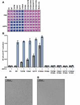
Measurement of the Activity of Wildtype and Disease-Causing ALPK1 Mutants in Transfected Cells With a 96-Well Format NF-κB/AP-1 Reporter Assay
96孔板NF-κB/AP-1报告基因检测转染细胞中野生型及致病性ALPK1突变体活性
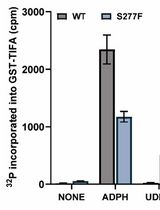
Quantitative Measurement of the Kinase Activity of Wildtype ALPK1 and Disease-Causing ALPK1 Mutants Using Cell-Free Radiometric Phosphorylation Assays
野生型和致病突变体ALPK1激酶活性的无细胞放射性磷酸化定量测定
生物工程
A Simple Staining Method Using Pyronin Y for Laser Scanning Confocal Microscopy to Evaluate Gelatin Cryogels
利用Pyronin Y的简单染色方法评估明胶冷冻凝胶的激光扫描共聚焦显微镜技术
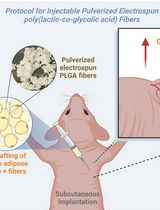
A Protocol for co-Injecting Cells with Pulverized Fibers for Improved Cell Survival and Engraftment
细胞与粉碎纤维共注射提高细胞存活率和移植效果的实验方案
生物物理学
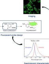
Small-Molecule Probe for Imaging Oxidative Stress–Induced Carbonylation in Live Cells
小分子探针用于活细胞氧化应激诱导羰基化成像
细胞生物学
Preparation of Polarity-Marked Microtubules Using a Plus-End Capping DARPin
利用正端封闭DARPin制备极性标记微管
免疫学
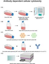
Display of Native SARS-CoV-2 Spike on Mammalian Cells to Measure Antibody Affinity and ADCC
天然SARS-CoV-2刺突蛋白在哺乳动物细胞上的展示及其抗体亲和力和ADCC测定
植物科学
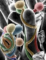
Fast and High-Resolution Imaging of Pollinated Stigmatic Cells by Tabletop Scanning Electron Microscopy
利用台式扫描电子显微镜快速高分辨成像授粉柱头细胞
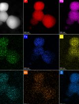
Optimized Isolation of Lysosome-Related Organelles from Stationary Phase and Iron-Overloaded Chlamydomonas reinhardtii Cells
静止期及铁超载莱茵衣藻溶酶体相关细胞器的优化分离
干细胞
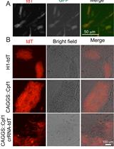
Multiplex Genome Editing of Human Pluripotent Stem Cells Using Cpf1
利用Cpf1实现人类多能干细胞的多重基因组编辑
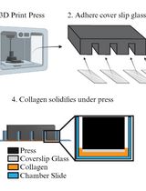
Preparing Chamber Slides With Pressed Collagen for Live Imaging Monolayers of Primary Human Intestinal Stem Cells
制备带压制胶原的腔室载玻片用于原代人肠道干细胞单层活细胞成像