往期刊物2024

卷册: 14, 期号: 18

left

Dec

20

Dec

5

Nov

20

Nov

5

Oct

20

Oct

5

Sep

20

Sep

5

Aug

20

Aug

5

Jul

20

Jul

5

Jun

20

Jun

5

May

20

May

5

Apr

20

Apr

5

Mar

20

Mar

5

Feb

20

Feb

5

Jan

20

Jan

5

right

生物化学

体外 GT 阵列 (i-GT-ray):筛选糖基转移酶活性和蛋白质相互作用的平台

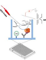
In Vitro GT-array (i-GT-ray), a Platform for Screening of Glycosyltransferase Activities and Protein–Protein Interactions

体外 GT 阵列 (i-GT-ray):筛选糖基转移酶活性和蛋白质相互作用的平台

MB Matrika Bhattarai
Tasleem Javaid Tasleem Javaid
AV Akshayaa Venkataraghavan
AF Ahmed Faik
1118 Views
Sep 20, 2024
Progress in bioinformatics has facilitated the identification of a large number of putative glycosyltransferases (GTs) associated with many physiological processes. However, many of these GTs remain with unknown biochemical function due to numerous technical limitations. One of these limitations is the lack of innovative tools for large-scale screening of enzyme activity in vitro and testing protein–protein interactions (PPIs) between GT partners. Currently, testing the enzyme activity of a protein requires its production in a heterologous expression system and purification before enzyme assays, a process that is time-consuming and not amenable to high-throughput screening. To overcome this, we developed a platform called in vitro GT-array (i-GT-ray). In this platform, 96-well microplates are coated with plasmid DNA encoding for tagged GTs and a capture antibody. Tagged GTs are produced from plasmid DNA via a cell-free in vitro transcription/translation (IVTT) system and captured through the anti-tag capture antibody directly on microplates. After washing to remove IVTT components, the captured enzymes can be considered purified, and their activity can be tested directly on microplates. The whole process can be performed in less than two days, compared to several weeks for currently available screening methods. The i-GT-ray platform has also been adapted to investigate PPIs between GTs. Here, we provide a practical user guide for the preparation of GT-arrays coated with plasmid DNA and a capture antibody that can be used for monitoring enzyme activity and PPIs of GTs in a high-throughput manner.

生物工程

人源化小鼠切割性伤口愈合模型中植入支架的方法

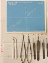
Protocol for the Implantation of Scaffolds in a Humanized Mouse Cutaneous Excisional Wound Healing Model

人源化小鼠切割性伤口愈合模型中植入支架的方法

DG Dina Gadalla
MK Maeve M. Kennedy
DL David G. Lott
724 Views
Sep 20, 2024
Tissue-engineered constructs combine the mechanical properties of biomaterials with biological agents to serve as scaffolds that direct the wound-healing process and promote tissue regeneration. A limitation to studying wound healing in vivo is that mouse skin contracts to heal rather than exhibiting granulation tissue formation and epithelialization like human skin. Therefore, it became necessary to develop a mouse model to better recapitulate human wound healing. The first splinted excisional wound healing model in mice, described in 2004, utilized silicone splints to prevent skin contracture. This model has been used to test a variety of wound healing strategies; however, to our knowledge, this model has not been adapted to test the effect of implants on wound healing. In our established protocol, circular bilateral excisional wounds are made on the mouse’s dorsum. A circular implant made of porous polyethylene is sutured to the skin within the wound. A thin, donut-shaped silicone splint is secured to the skin surrounding the wound, and a thick, donut-shaped splint is placed on top to tent the wound dressing. Finally, the mouse’s abdomen is wrapped in a bandage and tape to protect the implants. Our protocol offers a significant enhancement to the existing model by enabling the testing of implants for wound healing, as well as using an additional splint that prevents direct contact between the wound dressing and the wound bed. This model can be used to study tissue-engineered implant designs in a relatively low-cost, simple, and high-throughput manner before advancing to larger animal studies.

生物物理学

通过迭代免疫染色与NEDD去噪提升ExM-LSCM的信噪比

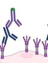
Iterative Immunostaining and NEDD Denoising for Improved Signal-To-Noise Ratio in ExM-LSCM

通过迭代免疫染色与NEDD去噪提升ExM-LSCM的信噪比

LA Lucio Azzari
MV Minnamari Vippola
SN Soile Nymark
TI Teemu O. Ihalainen
EM Elina Mäntylä
1491 Views
Sep 20, 2024
Expansion microscopy (ExM) has significantly reformed the field of super-resolution imaging, emerging as a powerful tool for visualizing complex cellular structures with nanoscale precision. Despite its capabilities, the epitope accessibility, labeling density, and precision of individual molecule detection pose challenges. We recently developed an iterative indirect immunofluorescence (IT-IF) method to improve the epitope labeling density, improving the signal and total intensity. In our protocol, we iteratively apply immunostaining steps before the expansion and exploit signal processing through noise estimation, denoising, and deblurring (NEDD) to aid in quantitative image analyses. Herein, we describe the steps of the iterative staining procedure and provide instructions on how to perform NEDD-based signal processing. Overall, IT-IF in ExM–laser scanning confocal microscopy (LSCM) represents a significant advancement in the field of cellular imaging, offering researchers a versatile tool for unraveling the structural complexity of biological systems at the molecular level with an increased signal-to-noise ratio and fluorescence intensity.

医学

用于包封或游离形式药物局部递送的小鼠肾动脉插管

Mouse Renal Artery Catheterization for Local Delivery of Drugs in Capsulated or Free Forms

用于包封或游离形式药物局部递送的小鼠肾动脉插管

OS Olga A. Sindeeva
OG Olga I. Gusliakova
EP Ekaterina S. Prikhozhdenko
NS Natalia A. Shushunova
GS Gleb B. Sukhorukov
707 Views
Sep 20, 2024
Arterial delivery to the kidney offers significant potential for targeted accumulation and retention of cells, genetic material, and drugs, both in free and encapsulated forms, because the entire dose passes through the vessels feeding this organ during the first circulation of blood. At the same time, a detailed study on the safety and effectiveness of developed therapies in a large number of experimental animals is required. Small laboratory animals, especially mice, are the most sought-after in experimental and preclinical testing due to their cost-effectiveness. Most of the described manipulations in mice involve puncturing the walls of the abdominal aorta or renal artery for direct administration of solutions and suspensions. Such manipulations are temporary and, in some cases, result in long-term occlusion of the aorta. Ultimately, this can lead to disruption of blood flow as well as functional and morphological changes to the kidneys. In addition, few of these protocols describe targeted delivery to the kidney. The presented protocol involves the injection of test substances or suspensions through the renal artery into one of the kidneys. The catheter is implanted into the femoral artery and then advanced into the abdominal aorta and renal artery within the vessels. In this case, the integrity violation of the renal artery or abdominal aorta is absent. Occlusion of the renal artery is necessary only immediately at the time of injection to minimize the entry of the injected substance into the aorta. This protocol is similar to the clinical procedure for delivering a catheter into the renal artery and is designed for real-world operating conditions.

神经科学

秀丽隐杆线虫进食行为的咽喉抽动实验

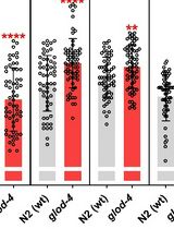
Pharyngeal Pumping Assay for Quantifying Feeding Behavior in Caenorhabditis elegans

秀丽隐杆线虫进食行为的咽喉抽动实验

MM Muniesh Muthaiyan Shanmugam
Pankaj Kapahi Pankaj Kapahi
948 Views
Sep 20, 2024
C. elegans is a well-established nematode model organism, with 83% of its genes conserved in humans with translation potential. C. elegans is translucent, with clearly defined cellular organization, and robustly identifiable under a microscope, being an excellent model for studying feeding behavior. Its neuromuscular pharyngeal pump undergoes a pumping motion that can be quantified to study feeding behavior at specific treatment conditions and in genetically modified worms. Understanding the evolutionarily conserved feeding behaviors and regulatory signals is vital, as unhealthy eating habits increase the risk of associated diseases. The current protocol was developed to identify and study evolutionary conserved signals regulating feeding behavior. The protocol described here is very robust in calculating the pumping rate (pumping per minute) as it directly counts the pharyngeal pumping for 30 s. This protocol uses basic laboratory instrumentation, such as a stereomicroscope with an attached camera and a computer with a video program that can be used to count manually. The advantages of studying C. elegans feeding include understanding the genetic basis of feeding regulation, dysregulation of feeding behavior in a disease model, the influence of toxic or environmental substances in feeding behavior, and modulation of feeding behavior by pharmacological agents.
幼体斑马鱼毛特讷氏细胞突触连接的扩展显微镜研究

Expansion Microscopy of Synaptic Contacts on the Mauthner Cells of Larval Zebrafish

幼体斑马鱼毛特讷氏细胞突触连接的扩展显微镜研究

SI Sundas Ijaz
SC Sandra P. Cárdenas-García
AP Alberto E. Pereda
859 Views
Sep 20, 2024
Because of its genetic tractability and amenability for live imaging, larval zebrafish (Danio rerio) have emerged as a model to study the cellular and synaptic properties underlying behavior. The accessibility of Mauthner cells, a pair of escape-organizing neurons located in the brainstem of teleost fish, along with their associated sensory inputs, enables exploration of the correlation between structural and functional synaptic features. This is the case of the endings of auditory afferents on the lateral dendrite of this cell, known as large myelinated club endings, which provide the excitatory drive for the initiation of auditory-evoked escape responses mediated by the Mauthner cell and its spinal network. Here, we describe the procedures that make it possible to expose the molecular composition of these synapses using protein-retention expansion microscopy (proExM). This method allowed us to generate a map of the distribution of synaptic proteins at these identifiable synapses, which could also be applied to examine the organization of other synaptic contacts in this cell.
日本鹌鹑胚胎原代脑细胞培养的开发与特性分析

Development and Characterization of Primary Brain Cultures from Japanese Quail Embryos

日本鹌鹑胚胎原代脑细胞培养的开发与特性分析

SZ Shaden Zoabi
AB Achinoam Blau
Shai Berlin Shai Berlin
810 Views
Sep 20, 2024
Cell cultures play a crucial role in neuroscience research, facilitating the elucidation of the complexities of cellular physiology and pathology. The relative simplicity in producing cultures and the accessibility to cells that the cultures provide, in contrast to in vivo settings, allow users to manipulate and monitor cells more easily at higher throughputs and lower costs. These are ideal for screening purposes and electrophysiological characterizations. Despite the prevalence of methodologies for producing brain cultures from various animal models, rodents in particular, approaches for culturing neurons (and glia) from birds are less established or completely absent as in the case of the Japanese quail model. Here, we present a unique culturing protocol for brain cells (e.g., neurons at different maturation levels, such as progenitor cells, excitatory and inhibitory neurons, microglia, and endothelial cells) from entire forebrains of Japanese quail embryos for high-throughput screening of viral vectors in vitro and other various purposes. Following dissection and digestion methods uniquely suited for avian brains, we tailored the growth media and culturing surface to allow the survival of quail brain cultures for more than three weeks in vitro.

植物科学

亚洲和非洲水稻品种杂交与标记辅助育种逐步指南

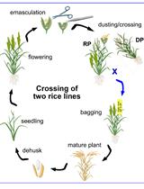
A Step-by-step Protocol for Crossing and Marker-Assisted Breeding of Asian and African Rice Varieties

亚洲和非洲水稻品种杂交与标记辅助育种逐步指南

Yugander Arra Yugander Arra
EL Eliza P.-I. Loo
BD B.N. Devanna
MS Melissa Stiebner
WF Wolf B. Frommer
1118 Views
Sep 20, 2024
Improving desirable traits of popular rice varieties is of particular importance for small-scale food producers. Breeding is considered the most ecological and economic approach to improve yield, especially in the context of pest and pathogen-resistant varieties development. Being able to cross rice lines is also a critical step when using current transgene-based genome editing technologies, e.g., to remove transgenes. Moreover, rice breeders have developed accelerated breeding methods, including marker-assisted backcross breeding (MABB) to develop novel rice varieties with in-built resistance to biotic and abiotic stressors, grain, and nutritional quality. MABB is a highly efficient and cost-effective approach in accelerating the improvement of recipient variety by introgressing desirable traits, especially from landrace cultivars and wild rice accessions. Here, we provide a detailed protocol including video instructions for rice crossing and MABB to introgress target trait(s) of interest into the elite rice line. Further, we also highlight tips and tricks to be considered for a successful crossing and MABB.

干细胞

符合GMP标准的角膜基质干细胞培养全流程

A Full Good Manufacturing Practice–Compliant Protocol for Corneal Stromal Stem Cell Cultivation

符合GMP标准的角膜基质干细胞培养全流程

MS Mithun Santra
YH Yen-Michael S. Hsu
SK Shaheen Khadem
SR Sydney Radencic
MF Martha L. Funderburgh
OS Onkar B. Sawant
DD Deepinder K. Dhaliwal
VJ Vishal Jhanji
GY Gary H.F. Yam
1010 Views
Sep 20, 2024
Corneal scarring, a significant cause of global blindness, results from various insults, including trauma, infections, and genetic disorders. The conventional treatment to replace scarred corneal tissues includes partial or full-thickness corneal transplantation using healthy donor corneas. However, only 1 in 70 individuals with treatable corneal scarring can undergo surgery, due to the limited supply of transplantable donor tissue. Our research focuses on cell-based strategies, specifically ex vivo–expanded corneal stromal stem cells (CSSCs), to address corneal scarring. Preclinical studies have demonstrated the efficacy of CSSC treatment in reducing corneal inflammation and fibrosis, inhibiting scar formation, and regenerating native stromal tissue. Mechanisms include CSSC differentiation into stromal keratocytes and the expression of regenerative cytokines. Here, we present a good manufacturing practice (GMP)-compliant protocol to isolate and expand human CSSCs. This method paves the way to produce clinical-grade CSSCs for transplantation and clinical trials.