往期刊物2024

卷册: 14, 期号: 16

left

Dec

20

Dec

5

Nov

20

Nov

5

Oct

20

Oct

5

Sep

20

Sep

5

Aug

20

Aug

5

Jul

20

Jul

5

Jun

20

Jun

5

May

20

May

5

Apr

20

Apr

5

Mar

20

Mar

5

Feb

20

Feb

5

Jan

20

Jan

5

right

生物工程

快速且完全的蛋白质生物共轭的四嗪氨基酸编码技术

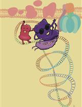
Tetrazine Amino Acid Encoding for Rapid and Complete Protein Bioconjugation

快速且完全的蛋白质生物共轭的四嗪氨基酸编码技术

AE Alex J. Eddins
AP Abigail H. Pung
RC Richard B. Cooley
RM Ryan A. Mehl
1639 Views
Aug 20, 2024
Generating protein conjugates using the bioorthogonal ligation between tetrazines and trans-cyclooctene groups avoids the need to manipulate cysteine amino acids; this ligation is rapid, site-specific, and stoichiometric and allows for labeling of proteins in complex biological environments. Here, we provide a protocol for the expression of conjugation-ready proteins at high yields in Escherichia coli with greater than 95% encoding and labeling fidelity. This protocol focuses on installing the Tet2 tetrazine amino acid using an optimized genetic code expansion (GCE) machinery system, Tet2 pAJE-E7, to direct Tet2 encoding at TAG stop codons in BL21 E. coli strains, enabling reproducible expression of Tet2-proteins that quantitatively react with trans-cyclooctene (TCO) groups within 5 min at room temperature and physiological pH. The use of the BL21 derivative B95(DE3) minimizes premature truncation byproducts caused by incomplete suppression of TAG stop codons, which makes it possible to use more diverse protein construct designs. Here, using a superfolder green fluorescent protein construct as an example protein, we describe in detail a four-day process for encoding Tet2 with yields of ~200 mg per liter of culture. Additionally, a simple and fast diagnostic gel electrophoretic mobility shift assay is described to confirm Tet2-Et encoding and reactivity. Finally, strategies are discussed to adapt the protocol to alternative proteins of interest and optimize expression yields and reactivity for that protein.

细胞生物学

使用3D-Speckler校准荧光显微镜(3D荧光散斑分析仪)

Calibrating Fluorescence Microscopy With 3D-Speckler (3D Fluorescence Speckle Analyzer)

使用3D-Speckler校准荧光显微镜(3D荧光散斑分析仪)

CL Chieh-Chang Lin
AS Aussie Suzuki
883 Views
Aug 20, 2024
Fluorescence microscopy has been widely accessible and indispensable in cell biology research. This technique enables researchers to label targets, ranging from individual entities to multiple groups, with fluorescent markers. It offers precise determinations of localization, size, and shape, along with accurate quantifications of fluorescence signal intensities. Furthermore, an ideal fluorescence microscope can achieve approximately 250 nm in lateral and 600 nm in axial resolution. Despite its integral role in these measurements, the calibration of fluorescence microscopes is often overlooked. This protocol introduces the use of 3D-Speckler (3D fluorescence speckle analyzer), a semi-automated software tool we have recently developed, for calibrating fluorescence microscopy. Calibration of fluorescence microscopy includes determining resolution limits, validating accuracy in size measurements, evaluating illumination flatness, and determining chromatic aberrations. 3D-Speckler is user-friendly and enables precise quantification of fluorescence puncta, including nanoscale 2D/3D particle size, precise locations, and intensity information. By utilizing multispectral fluorescence beads of known sizes alongside 3D-Speckler, the software can effectively calibrate imaging systems. We emphasize the importance of routine calibration for imaging systems to maintain their integrity and reproducibility, ensuring accurate quantification. This protocol provides a detailed step-by-step guide on using 3D-Speckler to calibrate imaging systems.

发育生物学

在不同发育阶段成像同一类IV型神经元的操作步骤

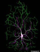
Protocol for Imaging the Same Class IV Neurons at Different Stages of Development

在不同发育阶段成像同一类IV型神经元的操作步骤

SS Sonal Shree
JH Jonathon Howard
769 Views
Aug 20, 2024
In this protocol, we focused on analyzing internal branches of Drosophila class IV neurons. These neurons are characterized by their highly branched axons and dendrites and intricately tile the larval body. As Drosophila larvae progress through developmental stages, the dendritic arbors of Class IV neurons undergo notable transformations. As Drosophila larvae develop, their Class IV dendritic arbors grow. In the initial 24 h after egg laying (AEL), the dendrites are smaller than segments. During the subsequent 24 h of the first instar larval stage, dendritic arbors outpace segment growth, achieving tiling. After 48 h, arbors and segments grow concurrently. Epidermal cells near Class IV dendrites expand in proportion to segment growth. This observation suggested that Class IV cells might grow via branch dilation—uniformly elongating branches, akin to Class I cells [1,2]. To understand whether the class IV complex arbor structure is formed by dilation or simply from growing tips, we developed this protocol to introduce a systematic approach for quantitatively assessing the growth dynamics of internal branches.

微生物学

通过密度梯度超速离心法提取细菌膜泡和噬菌体复合物

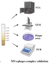
Extraction of Bacterial Membrane Vesicle and Phage Complex by Density Gradient Ultracentrifugation

通过密度梯度超速离心法提取细菌膜泡和噬菌体复合物

SL Shangru Li
AR Anmin Ren
ML Menglu Li
GL Guobao Li
LY Liang Yang
TJ Tianyuan Jia
1065 Views
Aug 20, 2024
The bacterial membrane vesicles (MVs) are non-replicative, nanoscale structures that carry specific cargos and play multiple roles in microbe–host interactions. An appropriate MV isolation method that mimics complex pathogen infections in vivo is needed. After bacterial MVs extraction, flagella or pili can be frequently observed along with MVs by transmission electron microscope (TEM). Recently, MVs from Pseudomonas aeruginosa were found to coexist with Pf4 phages, and this MV–phages complex exhibited a different impact on host cell innate immunity compared with MVs or phages solely. The presence of this MVs–phages complex simulates the real condition of complex pathogen infections within the host. This protocol outlines the extraction of the MVs and Pf4 phages complex of P. aeruginosa PAO1, including the respective isolation and qualification approaches. Our step-by-step bacterial MVs–phages complex extraction protocol provides valuable insights for further studying microbe–host cell interactions and the development of novel phage therapies.

分子生物学

使用IOCBIO Gel软件简单分析胶图像

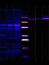
Simple Analysis of Gel Images With IOCBIO Gel Software

使用IOCBIO Gel软件简单分析胶图像

LJ Lucia Jaska
RB Rikke Birkedal
ML Martin Laasmaa
MV Marko Vendelin
877 Views
Aug 20, 2024
Gel image analyses are often difficult to reproduce, as the most commonly used software, the ImageJ Gels plugin, does not automatically record any steps in the analysis process. This protocol provides detailed steps for image analysis using IOCBIO Gel software with western blot as an example; however, the protocol is applicable to all images obtained by electrophoresis, such as Southern blotting, northern blotting, and isoelectric focusing. IOCBIO Gel allows multiple sample analyses, linking the original image to all the operations performed on it, which can be stored in a central database or on a PC, ensuring ease of access and the possibility to perform corrections at each analysis stage. In addition, IOCBIO Gel is lightweight, with only minimal computer requirements.

神经科学

使用定位显微技术量化突触前终扣的钙通道

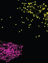
Using Localization Microscopy to Quantify Calcium Channels at Presynaptic Boutons

使用定位显微技术量化突触前终扣的钙通道

BM Brian D. Mueller
SM Sean A. Merrill
LV Lexy Von Diezmann
EJ Erik M. Jorgensen
1259 Views
Aug 20, 2024
Calcium channels at synaptic boutons are critical for synaptic function, but their number and distribution are poorly understood. This gap in knowledge is primarily due to the resolution limits of fluorescence microscopy. In the last decade, the diffraction limit of light was surpassed, and fluorescent molecules can now be localized with nanometer precision. Concurrently, new gene editing strategies allowed direct tagging of the endogenous calcium channel genes—expressed in the correct cells and at physiological levels. Further, the repurposing of self-labeling enzymes to attach fluorescent dyes to proteins improved photon yields enabling efficient localization of single molecules. Here, we describe tagging strategies, localization microscopy, and data analysis for calcium channel localization. In this case, we are imaging calcium channels fused with SNAP or HALO tags in live anesthetized C. elegans nematodes, but the analysis is relevant for any super-resolution preparations. We describe how to process images into localizations and protein clusters into confined nanodomains. Finally, we discuss strategies for estimating the number of calcium channels present at synaptic boutons.

植物科学

利用Rhizophagus irregularis进行体外菌丝分枝实验

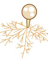
In Vitro Hyphal Branching Assay Using Rhizophagus irregularis

利用Rhizophagus irregularis进行体外菌丝分枝实验

TT Takaya Tominaga
HK Hironori Kaminaka
1306 Views
Aug 20, 2024
Most terrestrial plants are associated with symbiotic Glomeromycotina fungi, commonly known as arbuscular mycorrhizal (AM) fungi. AM fungi increase plant biomass in phosphate-depleted conditions by allocating mineral nutrients to the host; therefore, host roots actively exude various specialized metabolites and orchestrate symbiotic partners. The hyphal branching activity induced by strigolactones (SLs), a category of plant hormones, was previously discovered using an in vitro assay system. For this bioassay, AM fungi of the Gigaspora genus (Gigasporaeae) are commonly used due to their linear hyphal elongation and because the simple branching pattern is convenient for microscopic observation. However, many researchers have also used Glomeraceae fungi, such as Rhizophagus species, as the symbiotic partner of host plants, although they often exhibit a complex hyphal branching pattern. Here, we describe a method to produce and quantify the hyphal branches of the popular model AM fungus Rhizophagus irregularis. In this system, R. irregularis spores are sandwiched between gels, and chemicals of interest are diffused from the surface of the gel to the germinating spores. This method enables the positive effect of a synthetic SL on R. irregularis hyphal branching to be reproduced. This method could thus be useful to quantify the physiological effects of synthesized chemicals or plant-derived specialized metabolites on R. irregularis.

系统生物学

用于自下而上蛋白质组学分析的氯仿/甲醇蛋白提取与溶液内胰蛋白酶消化方法

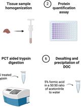
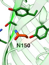
Chloroform/Methanol Protein Extraction and In-solution Trypsin Digestion Protocol for Bottom-up Proteomics Analysis

用于自下而上蛋白质组学分析的氯仿/甲醇蛋白提取与溶液内胰蛋白酶消化方法

TP Tess Puopolo
NS Navindra P. Seeram
CL Chang Liu
1530 Views
Aug 20, 2024
Bottom-up proteomics utilizes sample preparation techniques to enzymatically digest proteins, thereby generating identifiable and quantifiable peptides. Proteomics integrates with other omics methodologies, such as genomics and transcriptomics, to elucidate biomarkers associated with diseases and responses to drug or biologics treatment. The methodologies employed for preparing proteomic samples for mass spectrometry analysis exhibit variability across several factors, including the composition of lysis buffer detergents, homogenization techniques, protein extraction and precipitation methodologies, alkylation strategies, and the selection of digestion enzymes. The general workflow for bottom-up proteomics consists of sample preparation, mass spectrometric data acquisition (LC-MS/MS analysis), and subsequent downstream data analysis including protein quantification and differential expression analysis. Sample preparation poses a persistent challenge due to issues such as low reproducibility and inherent procedure complexities. Herein, we have developed a validated chloroform/methanol sample preparation protocol to obtain reproducible peptide mixtures from both rodent tissue and human cell line samples for bottom-up proteomics analysis. The protocol we established may facilitate the standardization of bottom-up proteomics workflows, thereby enhancing the acquisition of reliable biologically and/or clinically relevant proteomic data.

更正

更正:重组人线粒体RNA聚合酶(POLRMT)及启动因子TFAM和TFB2M的表达和纯化

Correction Notice: Expression and Purification of Recombinant Human Mitochondrial RNA Polymerase (POLRMT) and the Initiation Factors TFAM and TFB2M

更正:重组人线粒体RNA聚合酶(POLRMT)及启动因子TFAM和TFB2M的表达和纯化

AH An H. Hsieh
SR Sean D. Reardon
JM Jubilee H. Munozvilla-Cabellon
JS Jiayu Shen
SP Smita S. Patel
TM Tatiana V. Mishanina
439 Views
Aug 20, 2024