往期刊物2015

卷册: 5, 期号: 24

left

Dec

20

Dec

5

Nov

20

Nov

5

Oct

20

Oct

5

Sep

20

Sep

5

Aug

20

Aug

5

Jul

20

Jul

5

Jun

20

Jun

5

May

20

May

5

Apr

20

Apr

5

Mar

20

Mar

5

Feb

20

Feb

5

Jan

20

Jan

5

right

癌症生物学

微型PET-CT成像分析鼠肺部肿瘤

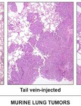
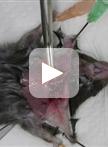
Analysis of Murine Lung Tumors by Micro PET-CT Imaging

微型PET-CT成像分析鼠肺部肿瘤

Chiara Ambrogio Chiara Ambrogio
Juan Antonio Cámara Juan Antonio Cámara
Patricia Nieto Patricia Nieto
David Santamaría David Santamaría
Francisca Mulero Francisca Mulero
14584 Views
Dec 20, 2015
Accurate live tumor imaging in mice is now possible by means of high-resolution positron emission tomography (micro-PET) and X-ray computed tomography (micro-CT). By providing a powerful tool to examine biological samples with complex structure in vivo, this technology generated a significant advance in the cancer research field, particularly regarding the ability to perform longitudinal studies in combination with a therapeutic intervention. Here, we describe methods to optimize visualization of murine lung tumors by micro-PET, micro-CT and combined micro-PET-CT.
从原代鼠肿瘤中生成鼠甲状腺产降血钙素的肿瘤细胞

Generation of Mouse Thyroid Calcitonin-producing Cell Tumors from Primary Mouse Tumors

从原代鼠肿瘤中生成鼠甲状腺产降血钙素的肿瘤细胞

SK Shunsuke Kitajima
FL Fengkai Li
Chiaki Takahashi Chiaki Takahashi
8289 Views
Dec 20, 2015
Medullary thyroid cancers (MTCs) are derived from calcitonin-producing cells (C cells) of neuroendocrine origin. Rb heterozygous mice develop low-grade C cell adenocarcinoma following biallelic inactivation of the Rb tumor suppressor gene loci. Additional inactivation of another tumor suppressor gene such as Trp53, Arf or Cdkn1a allows Rb-deficient mice to generate more aggressive C cell adenocarcinoma (Takahashi et al., 2006; Shamma et al., 2009; Kitajima et al., 2015). To characterize C cell adenocarcinoma cells derived from Rb-deficient mice of different genetic backgrounds, we attempted to extract C cell adenocarcinoma cells from primary thyroid tumor tissue. Since primary mouse small cell lung cancer (SCLC) cells those originate in neuroendocrine cells that also stems C cells, can be established both as non-adhesive and adhesive cells (Calbo et al., 2011), we applied their method to MTCs. Here we describe our isolation technique for non-adhesive and adhesive cell cultures from primary medullary thyroid tumor tissue. We found that the molecular markers of C cell such as Calcitonin and Ascl1 are predominantly enriched in the non-adhesive population (Kitajima et al., 2015). This is in line with the fact that one of most commonly distributed human MTC cell line TT is non-adhesive.

微生物学

大肠杆菌脂多糖中酮基-脱氧-d-甘露-8辛酸(KDO)的测定

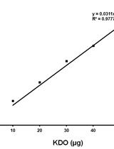
Determination of Keto-deoxy-d-manno-8-octanoic acid (KDO) from Lipopolysaccharide of Escherichia coli

大肠杆菌脂多糖中酮基-脱氧-d-甘露-8辛酸(KDO)的测定

M. R. Sunayana M. R. Sunayana
Manjula Reddy Manjula Reddy
17062 Views
Dec 20, 2015
2-Keto-3-deoxy-octonate (KDO) is an essential constituent of lipopolysaccharide (LPS) that forms the outermost leaflet of Gram-negative bacterial outer membrane. LPS is mainly composed of lipid A, O-antigen and a core oligosaccharide. Two molecules of KDO are present per one molecule of LPS. A proper level of LPS is required to maintain the outer membrane integrity and either high or low levels of LPS are toxic to the cell. Various methods are available for quantification of LPS; of these, determination of KDO is a simple and accurate method and it can be estimated either directly from crude bacterial cell lysates or from purified LPS by a simple colorimetric assay. Although this procedure can be theoretically used for any Gram-negative bacterium, we used it routinely to measure KDO from cell lysates of Escherichia coli (E. coli) K12 strains.Method: The protocol is taken from Karkhanis et al. (1978). It is a simple, sensitive and reliable method to measure KDO. The assay is performed after complete acid hydrolysis of cell lysates or LPS to release the various components of LPS. Further, reaction with periodate, arsenite and thiobarbituric acid gives a pink to red color chromophore, which is measured at 548 nm after stabilizing with DMSO.
制备并分析葡萄球菌属的天然自溶酶提取物

Preparation and Analysis of Crude Autolytic Enzyme Extracts from Staphylococcus aureus

制备并分析葡萄球菌属的天然自溶酶提取物

Filipa Vaz Filipa Vaz
Sérgio R. Filipe Sérgio R. Filipe
13101 Views
Dec 20, 2015
The metabolism of the cell surface during bacterial cell division involves synthesis and degradation of peptidoglycan (PGN), the major component of the bacterial cell wall. Bacteria have to ensure that their surface remains capable of withstanding high turgor pressures and, simultaneously, that the PGN at their surface is concealed from receptors produced by the host innate immune system. For cell separation to occur, and for PGN to be kept concealed, “old” PGN is degraded by specific PGN hydrolases, also known as autolysins, that are found at the bacterial cell surface or that are secreted into the growth medium. Bacterial PGN hydrolases are cell wall lytic enzymes that comprise a broad and diverse group of proteins. It is often difficult to assign a specific function to a PGN hydrolase mainly because an organism can have a large number of hydrolases with redundant activities and one hydrolase can have more than one enzymatic activity and participate in various cell processes (Vollmer et al. 2008). Bacillus subtilis has ca. 35 known or hypothetical PGN hydrolases, whereas Escherichia coli (E. coli) and Staphylococcus aureus (S. aureus) have, respectively, ca.16 and 19 PGN hydrolases (Vollmer, 2012; Heidrich et al., 2001; Singh et al., 2012). PGN hydrolases can be classified in three main classes: glycosidases, amidases and peptidases. Glycosidases cleave the glycan backbone and are divided into N-acetylglucosaminidases and N-acetylmuramidases. Amidases cleave the linkage between the peptide chain and the N-acetylmuramic residue of the glycan chain. Peptidases, such as endopeptidases and carboxypeptidases, are able to cleave peptide bonds between different amino acids of the PGN stem peptide. Here we describe a method to extract PGN hydrolases, which are non-covalently linked to the S. aureus cell wall (Vollmer, 2008). Analysis of extracts containing denatured PGN hydrolytic enzymes is performed by running a zymogram gel (a SDS-PAGE gel containing crude bacterial cell walls or substrate cells), which is then incubated in a non-denaturing buffer to allow renaturation of the PGN hydrolases. These renatured enzymes can then be identified through the production of clear bands that are observed where cell wall digestion has occurred. The protocol is divided into three steps: A) Preparation of the crude autolytic extracts from S. aureus cells; B) Preparation of substrate cells for gel zymograms; C) Analysis of crude autolytic extracts by gel zymography. We also show that this method can be used to determine the absence or altered activity of PGN hydrolases produced by different S. aureus mutant strains.
电击法转化蓝藻鞘丝藻

Transformation of the Cyanobacterium Leptolyngbya boryana by Electroporation

电击法转化蓝藻鞘丝藻

RT Ryoma Tsujimoto
HK Hiroya Kotani
AN Aoi Nonaka
YM Yuri Miyahara
YH Yuto Hiraide
YF Yuichi Fujita
11735 Views
Dec 20, 2015
Leptolyngbya boryana (L. boryana) (formerly Plectonema boryanum) is a versatile, filamentous cyanobacterium that has the ability to fix nitrogen under microoxic conditions and to grow heterotrophically with glucose in the dark, providing an excellent system to investigate photosynthesis, nitrogen fixation, and their regulatory mechanisms. While L. boryana is not naturally transformable different from the unicellular cyanobacterium Synechocystis sp. PCC 6803, it can be transformed by electroporation. Here we describe the transformation of L. boryana by electroporation to isolate mutants in which a targeted gene is disrupted.
病人血清样本和克隆中HBV分离体的特性描述

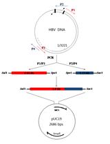
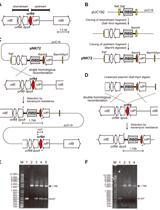
Characterization of HBV Isolates from Patient Serum Samples and Cloning

病人血清样本和克隆中HBV分离体的特性描述

LC Liang Cao
CW Chunchen Wu
KZ Kaitao Zhao
XC Xinwen Chen
10028 Views
Dec 20, 2015
Hepatitis B virus (HBV) mutants can lead to vaccine failure, diagnostic failure of HBV detection, increase viral replication and resistance to antiviral agents. To study the biological characteristics of these mutations may contribute to our knowledge on viral pathogenesis. Therefore, it is essential to isolate and characterize HBV strains from patients. Here we describe the experimental methods to isolate and clone HBV DNA from patient serum. The method will facilitate isolation and functional analysis of new HBV variants.
蓝藻细菌中烷类的提取和量化

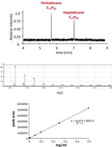
Extraction and Quantification of Alkanes in Cyanobacteria

蓝藻细菌中烷类的提取和量化

HK Hakuto Kageyama
RW Rungaroon Waditee-Sirisattha
SS Sophon Sirisattha
YT Yoshito Tanaka
AM Aparat Mahakhant
TT Teruhiro Takabe
8788 Views
Dec 20, 2015
Many species of cyanobacteria accumulate alkanes in their cells. It has been reported that these cyanobacteria accumulate mainly 15 long carbon chain alkane, pentadecane (C15H32), or/and 17 long chain alkane, heptadecane (C17H36). Here we describe a protocol of our laboratory for extraction and quantification of cyanobacterial intracellular pentadecane and heptadecane. We have confirmed this protocol was applicable to at least three kinds of cyanobacteria, nitrogen-fixing filamentous cyanobacterium Anabaena sp. PCC7120, non-diazotrophic unicellular cyanobacterium Synechococcus elongatus PCC7942 and halotolerant unicellular cyanobacterium Aphanothece halophytica.
霍乱弧菌中[14C] 亚油酸的摄取和分级分离试验

[14C] Linoleic Acid Uptake and Fractionation Assay in Vibrio cholerae

霍乱弧菌中[14C] 亚油酸的摄取和分级分离试验

SP Sarah C. Plecha
JW Jeffrey H. Withey
6943 Views
Dec 20, 2015
The gram-negative curved bacillus Vibrio cholerae (V. cholerae) causes the severe diarrheal illness cholera. The work presented here is to assess whether unsaturated fatty acids (UFAs), such as linoleic acid, have the potential to directly affect proteins involved in DNA binding because they are able to enter the cell. In this protocol, we show how to measure linoleic acid entering V. cholerae when added exogenously and determine whether it is able to enter the cytoplasm. This protocol will quantify how much linoleic acid is able to enter the cell and then identify the amount of linoleic acid that stays in the membrane or ultimately enters the cytoplasm.

植物科学

鲁米诺反应检测拟南芥叶片中免疫激发产生的过氧化氢

Luminol-based Assay for Detection of Immunity Elicitor-induced Hydrogen Peroxide Production in Arabidopsis thaliana Leaves

鲁米诺反应检测拟南芥叶片中免疫激发产生的过氧化氢

NB Nora Gigli Bisceglia
MG Matteo Gravino
Daniel V. Savatin Daniel V. Savatin
21148 Views
Dec 20, 2015
In Arabidopsis thaliana, one of the very early immune-related responses induced after elicitor perception is the oxidative burst, i.e., reactive oxygen species (ROS) generation including superoxide anion and hydrogen peroxide (H2O2). ROS production plays different roles in a wide range of biotic and abiotic stress responses, including the closure of stomata and the regulation of cell expansion. In particular, elicitor-induced H2O2 is produced mainly by the membrane localized NAD(P)H oxidases RESPIRATORY BURST OXIDASE HOMOLOGUE D and F. In this protocol, we describe a simple and reproducible luminol/peroxidase-based assay to detect and evaluate immunity-related accumulation of H2O2 produced in Arabidopsis leaf discs treated with immunity elicitors, such as oligogalacturonides (OGs), flagellin (flg22) or the elongation factor-thermo-unstable (EF-Tu - elf18). This method is based on the detection of the luminescence released by excited-luminol molecules generated after the horseradish peroxidase (HRP)-catalyzed oxidation of luminol molecules in the presence of H2O2. Levels as well as duration of the luminescence are proportional to the amount of H2O2 produced by elicited leaf discs.
从拟南芥叶提取质外体洗涤液和叶柄渗出液

Extraction of Apoplastic Wash Fluids and Leaf Petiole Exudates from Leaves of Arabidopsis thaliana

从拟南芥叶提取质外体洗涤液和叶柄渗出液

TA Takao Araya
AB Anne Bohner
Nicolaus von Wirén Nicolaus von Wirén
14329 Views
Dec 20, 2015
The long-distance translocation of metabolites and mineral elements is crucial for plant growth and reproduction. In most cases, source-to-sink translocation of metabolites and minerals requires their passage through the apoplast, irrespective whether they are transported via the xylem or the phloem. This apoplast-mediated pathway is of particular importance during plant senescence, when photoassimilates as well as organic, inorganic or chelated forms of nutrients are translocated from leaves to fruits or seeds. Recent genetic and physiological studies revealed the involvement of numerous membrane transporters mediating phloem loading of amino acids, sugars, urea or mineral elements. To evaluate the contribution of individual transporters to xylem unloading or phloem loading, the collection of apoplastic fluids and of phloem sap is essential. Here, we describe a method for the extraction of apoplastic fluids and the collection of leaf petiole exudates from Arabidopsis leaves, the latter representing an approximation to the real composition of the phloem sap.
采用GC-MS分析氯仿提取的拟南芥和其它植物物种中的木栓质根蜡

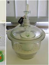
GC-MS-Based Analysis of Chloroform Extracted Suberin-Associated Root Waxes from Arabidopsis and Other Plant Species

采用GC-MS分析氯仿提取的拟南芥和其它植物物种中的木栓质根蜡

DK Dylan K. Kosma
Isabel Molina Isabel Molina
OR Owen Rowland
11552 Views
Dec 20, 2015
The periderm and exodermis of taproots and tuberous taproots contain an extracellular lipid polymer, suberin, deposited in their cell walls. This polymer is intractable in organic solvents, and is co-deposited with chloroform-extractable waxes. These suberin-associated root waxes are typically composed of alkanes, primary alcohols, fatty acids, alkyl ferulates, alkyl caffeates, and alkyl coumarates (Espelie et al., 1980; Li et al., 2007; Kosma et al., 2015). They are believed to contribute to the diffusion barrier properties of suberized cell walls (Soliday et al., 1979), and possibly have other roles yet to be discovered. Here we describe a protocol to extract and analyze waxes associated with root suberin. This fraction of aliphatic components is extracted by whole root immersion in chloroform, and is then chemically modified to prepare samples that are more suitable to gas-chromatography analysis. This protocol is optimized for Arabidopsis thaliana, but can be used with roots of other plants as described herein.
植物根部CLE肽的生物活性的体外实验

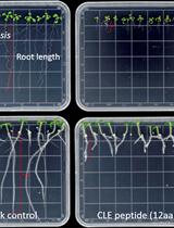
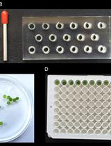
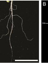
In vitro CLE Peptide Bioactivity Assay on Plant Roots

植物根部CLE肽的生物活性的体外实验

Shiyan  Chen Shiyan Chen
Xiaohong  Wang Xiaohong Wang
9839 Views
Dec 20, 2015
Plant CLAVATA3/ESR (CLE)-related proteins play diverse roles in plant growth and development including regulating the development of root meristem. The mature functional forms of CLE peptides are typically 12-13 amino acids (aa) in length that are derived from the conserved C-termini of their precursor proteins. Genes encoding small secreted peptides sharing similarity to plant CLE proteins have recently been cloned from plant-parasitic nematodes, pests that infect many important crops. It is demonstrated that exogenous application of synthetic 12-14 aa CLE peptides corresponding to the CLE domain of their precursor proteins can suppress plant root growth. This protocol is to evaluate the bioactivity of CLE peptides originated from plant-parasitic nematodes by measuring the growth of plant roots or the size of root apical meristem (RAM) after CLE peptide treatment. Plants used in the study included Arabidopsis and potato.
从番茄果皮中分离液泡膜微囊

Isolation of Tonoplast Vesicles from Tomato Fruit Pericarp

从番茄果皮中分离液泡膜微囊

CS Christopher J. Snowden
BT Benjamin Thomas
CB Charles J. Baxter
J. Andrew C. Smith J. Andrew C. Smith
Lee J. Sweetlove Lee J. Sweetlove
9809 Views
Dec 20, 2015
This protocol describes the isolation of tonoplast vesicles from tomato fruit. The vesicles isolated using this procedure are of sufficiently high purity for downstream proteomic analysis whilst remaining transport competent for functional assays. The methodology was used to study the transport of amino acids during tomato fruit ripening (Snowden et al., 2015) and based on the procedure used by Betty and Smith (Bettey and Smith, 1993). Such vesicles may be useful in further studies into the dynamic transfer of metabolites across the tonoplast for storage and metabolism during tomato fruit development.
莱茵衣藻的插入突变

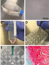
Insertional Mutagenesis of Chlamydomonas reinhardtii

莱茵衣藻的插入突变

CT Chia-Hong Tsai
CB Christoph Benning
8506 Views
Dec 20, 2015
The unicellular microalga Chlamydomonas reinhardtii (C. reinhardtii) has been used as a reference model for numerous fields of research. Principle research areas are eukaryotic flagellar structure and function, basal bodies (centrioles), cell-cell recognition, cell cycle control, chloroplast biogenesis, phototaxis, nonphotochemical quenching, and especially photosynthesis for C. reinhardtii can grow in the dark on an organic carbon (e.g. acetate), and thus provides advantages over land plants (Harris, 2001; Peers et al., 2009). C. reinhardtii has a short life cycle, a sequenced genome (Merchant et al., 2007), and a growing molecular toolbox for forward and reverse genetic studies, including transformation protocols, gene silencing (Kim and Cerutti, 2009; Molnar et al., 2009), and fluorescent protein-tag (Rasala et al., 2013). There are two commonly used methods for C. reinhardtii transformation – electroporation and glass bead agitation. Electroporation is normally restricted to strains with cell wall, as it kills cell-wall-deficient strains effectively if without careful handling of osmosis. Electroporation also requires special instruments such as electroporator and cuvettes. In contrast, glass bead agitation uses simple lab equipment. The mild shear created by agitation in the presence of glass bead allows cell-wall-deficient strains to take up DNA. If glass bead method is to be applied to cell-wall strains, cells need to be treated with autolysin (http://www.chlamy.org/methods/autolysin.html) to partially lyse the wall components. A pitfall of both methods is that the DNAs are often shortened by nuclease once entering the cells, making the downstream PCR-based genotyping of insertion site rather difficult. Here I describe an improved design of insertional mutagenesis used in (Tsai et al., 2014), and the transformation protocol using glass bead as previously described in (Kindle, 1990) with minor modification. The putative mutants can be selected by autotrophic or antibiotic resistance markers, and the disrupted loci can be mapped by methods such as plasmid rescue (Peers et al., 2009) and SiteFinding PCR (Tan et al., 2005).

干细胞

BMSC结籽支架的多孔支架播种和软骨分化

Porous Scaffold Seeding and Chondrogenic Differentiation of BMSC-seeded Scaffolds

BMSC结籽支架的多孔支架播种和软骨分化

Troy D. Bornes Troy D. Bornes
Nadr M. Jomha Nadr M. Jomha
Aillette Mulet-Sierra Aillette Mulet-Sierra
Adetola B. Adesida Adetola B. Adesida
9056 Views
Dec 20, 2015
Bone marrow-derived mesenchymal stromal stem cells (BMSCs) are a promising cell source for treating articular cartilage defects (Bornes et al., 2014). BMSCs can be seeded within porous biomaterial scaffolds that support three-dimensional cell organization, chondrogenic differentiation and extracellular matrix deposition for the creation of engineered cartilage. This protocol describes our defined methods for isolation and expansion of human and ovine BMSCs, seeding of BMSCs within porous scaffolds and in vitro chondrogenic differentiation (Adesida et al., 2012; Bornes et al., 2015).