往期刊物2024
卷册: 14, 期号: 12
免疫学
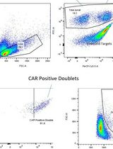
A Flow Cytometry–Based Method for Assessing CAR Cell Binding Kinetics Using Stable CAR Jurkat Cells
基于流式细胞术的稳定CAR-Jurkat细胞结合动力学评估方法

微生物学
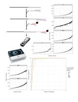
Direct RNA Sequencing of Foot-and-mouth Disease Virus Genome Using a Flongle on MinION
使用MinION的Flongle直接测序口蹄疫病毒RNA基因组
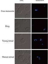
Transfection of Babesia duncani: A Genetic Toolbox of This Pathogen to Advance Babesia Biology
邓氏巴贝斯虫基因转染:推进巴贝斯虫生物学的遗传工具箱
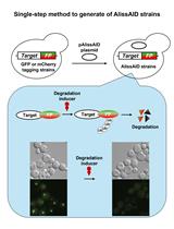
A Single-step Generation of AlissAID-based Conditional Knockdown Strains Using Nanobody that Targets GFP or mCherry in Budding Yeast
利用靶向GFP或mCherry的纳米抗体在出芽酵母中一步生成AlissAID条件性敲降菌株
神经科学

Flow Cytometry Analysis of Microglial Phenotypes in the Murine Brain During Aging and Disease
衰老和疾病过程中小鼠大脑小胶质细胞表型的流式细胞术分析
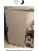
Measuring Sleep and Activity Patterns in Adult Zebrafish
测量成体斑马鱼的睡眠和活动模式
植物科学
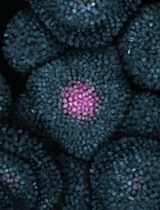
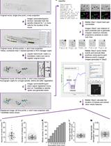
Live Imaging of the Shoot Apical Meristem of Intact, Soil-Grown, Flowering Arabidopsis Plants
完整土培开花拟南芥植物茎尖分生组织的实时成像
干细胞
Isolation of Human Bone Marrow Non-hematopoietic Cells for Single-cell RNA Sequencing
用于单细胞RNA测序的人骨髓非造血细胞的分离