

往期刊物2023
卷册: 13, 期号: 14
生物化学
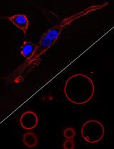
Visualizing Loss of Plasma Membrane Lipid Asymmetry Using Annexin V Staining
使用Annexin V染色可视化质膜脂质不对称性的丧失
生物工程
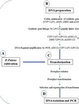
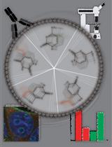
Heterologous Production of Artemisinin in Physcomitrium patens by Direct in vivo Assembly of Multiple DNA Fragments
通过在体内直接组装多个DNA片段在青蒿中异源生产青蒿素
生物物理学
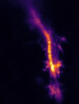
Recoil Measurements in Drosophila Embryos: from Mounting to Image Analysis
果蝇胚胎的反冲测量:从安装到图像分析
癌症生物学
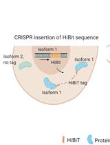
Isoform-specific, Semi-quantitative Determination of Highly Homologous Protein Levels via CRISPR-Cas9-mediated HiBiT Tagging
通过CRISPR-Cas9介导的HiBiT标签对高度同源蛋白水平进行异构体特异性半定量测定
细胞生物学
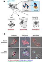
Optogenetic Induction of Pyroptosis, Necroptosis, and Apoptosis in Mammalian Cell Lines
光遗传学诱导哺乳动物细胞株的细胞焦亡、细胞坏死性凋亡和细胞凋亡
Biophysical Analysis of Mechanical Signals in Immotile Cilia of Mouse Embryonic Nodes Using Advanced Microscopic Techniques
利用先进显微技术对小鼠胚胎结节中不动纤毛的机械信号进行生物物理分析
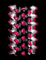
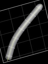
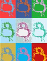
Characterization of Microtubule Lattice Heterogeneity by Segmented Subtomogram Averaging
分段亚谱平均表征微管晶格异质性
发育生物学
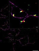
Three-color dSTORM Imaging and Analysis of Recombination Foci in Mouse Spread Meiotic Nuclei
小鼠扩散型减数分裂细胞核中重组病灶的三色dSTORM成像及分析
免疫学
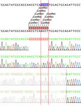
HDR-based CRISPR/Cas9-mediated Knockout of PD-L1 in C57BL/6 Mice
基于HDR的CRISPR/Cas9介导的C57BL/6小鼠PD-L1敲除
LiverQuant: An Improved Method for Quantitative Analysis of Liver Pathology
LiverQuant:肝脏病理定量分析的改进方法
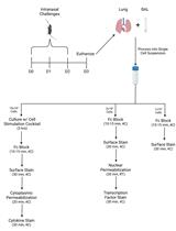
Monitoring Group 2 Innate Lymphoid Cell Biology in Models of Lung Inflammation
监测组2肺部炎症模型中的先天淋巴细胞生物学
Intravital Imaging of Intestinal Intraepithelial Lymphocytes
肠上皮内淋巴细胞的活体成像
微生物学
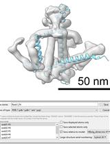
Protein Structure Predictions, Atomic Model Building, and Validation Using a Cryo-EM Density Map from Hepatitis B Virus Spherical Subviral Particle
利用乙型肝炎病毒球形亚病毒颗粒的低温电子显微镜密度图进行蛋白质结构预测、原子模型构建和验证
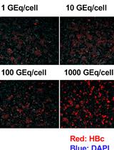
Production and Purification of Cell Culture–generated Hepatitis B Virus by Transient Transfection and Density Gradient
通过瞬时转染和密度梯度法生产和纯化细胞培养产生的乙型肝炎病毒
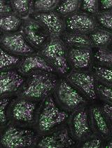
Simplifying Barley Leaf Rust Research: An Easy and Reproducible Infection Protocol for Puccinia hordei on a Small Laboratory Scale
简化大麦叶锈病研究:一种简单且可重复的小型实验室规模的大麦锈病感染方案
分子生物学
In situ Quantification of Cytosine Modification Levels in Heterochromatic Domains of Cultured Mammalian Cells
培养哺乳动物细胞异染色质中胞嘧啶修饰水平的原位定量分析
神经科学
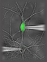
An ex vivo Model of Paired Cultured Hippocampal Neurons for Bi-directionally Studying Synaptic Transmission and Plasticity
成对培养的海马神经元的离体模型,用于双向研究突触传递和可塑性
植物科学
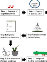
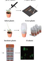
Inoculation of Maize with Sugarcane Mosaic Virus Constructs and Application for RNA Interference in Fall Armyworms
用甘蔗花叶病毒构建体接种玉米并应用于秋季棉铃虫的RNA干扰
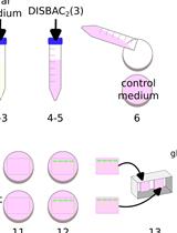
Relative Membrane Potential Measurements Using DISBAC2(3) Fluorescence in Arabidopsis thaliana Primary Roots
使用DISBAC2(3)荧光测量拟南芥主根中的相对膜电位
干细胞
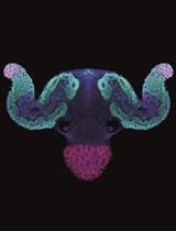
A New Approach to Generate Gastruloids to Develop Anterior Neural Tissues
一种产生原肠胚样细胞以发育前神经组织的新方法
系统生物学

Chromatin-RNA in situ Reverse Transcription Sequencing (CRIST-seq) Approach to Profile the Non-coding RNA Interaction Network
染色质-RNA原位逆转录测序(CRIST-seq)方法分析非编码RNA相互作用网络