往期刊物2023

卷册: 13, 期号: 10

left

Dec

20

Dec

5

Nov

20

Nov

5

Oct

20

Oct

5

Sep

20

Sep

5

Aug

20

Aug

5

Jul

20

Jul

5

Jun

20

Jun

5

May

20

May

5

Apr

20

Apr

5

Mar

20

Mar

5

Feb

20

Feb

5

Jan

20

Jan

5

right

生物工程

小鼠脂肪组织腺相关病毒直接注射

Direct Adeno-associated Viruses Injection of Murine Adipose Tissue

小鼠脂肪组织腺相关病毒直接注射

SW Shao-Chin Wu
CL Chi-Hung Lin
1202 Views
May 20, 2023
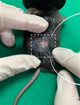
The adipose tissue is a central metabolic organ that regulates whole-body energy homeostasis. The abnormal expansion of adipose tissue leads to the progression of obesity. The adipose tissue microenvironment is affected by pathological hypertrophy of adipocytes, highly correlated with systemic metabolic disorders. In vivo genetic modification is a great tool for understanding the role of genes involved in such processes. However, obtaining new conventional engineered mice is time consuming and costly. Here, we provide a simple and speedy method to efficiently transduce genes into adipose tissue by injecting the adeno-associated virus vector serotypes 8 (AAV8) into the fat pads of adult mice.

生物物理学

用放射性标记的ATP高灵敏度测定体外ATP酶活性

Measuring in vitro ATPase Activity with High Sensitivity Using Radiolabeled ATP

用放射性标记的ATP高灵敏度测定体外ATP酶活性

SV Sarina Veit
TP Thomas Günther Pomorski
1360 Views
May 20, 2023
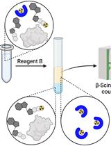
ATPase assays are a common tool for the characterization of purified ATPases. Here, we describe a radioactive [γ-32P]-ATP-based approach, utilizing complex formation with molybdate for phase separation of the free phosphate from non-hydrolyzed, intact ATP. The high sensitivity of this assay, compared to common assays such as the Malachite green or NADH-coupled assay, enables the examination of proteins with low ATPase activity or low purification yields. This assay can be used on purified proteins for several applications including the identification of substrates, determination of the effect of mutations on ATPase activity, and testing specific ATPase inhibitors. Furthermore, the protocol outlined here can be adapted to measure the activity of reconstituted ATPases.Graphical overview

癌症生物学

线粒体复制测定(MIRA)可有效原位定量新生线粒体DNA和蛋白质与新生线粒体DNA(mitoSIRF)的相互作用

Mitochondrial Replication Assay (MIRA) for Efficient in situ Quantification of Nascent mtDNA and Protein Interactions with Nascent mtDNA (mitoSIRF)

线粒体复制测定(MIRA)可有效原位定量新生线粒体DNA和蛋白质与新生线粒体DNA(mitoSIRF)的相互作用

ML Macy Lozen
YC Yue Chen
RB Rebecca A. Boisvert
KS Katharina Schlacher
1269 Views
May 20, 2023
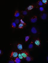
Mitochondria play decisive roles in bioenergetics and intracellular communication. These organelles contain a circular mitochondrial DNA (mtDNA) genome that is duplicated within one to two hours by a mitochondrial replisome, independently from the nuclear replisome. mtDNA stability is regulated in part at the level of mtDNA replication. Consequently, mutations in mitochondrial replisome components result in mtDNA instability and are associated with diverse disease phenotypes, including premature aging, aberrant cellular energetics, and developmental defects. The mechanisms ensuring mtDNA replication stability are not completely understood. Thus, there remains a need to develop tools to specifically and quantifiably examine mtDNA replication. To date, methods for labeling mtDNA have relied on prolonged exposures of 5′-bromo-2′-deoxyuridine (BrdU) or 5′-ethynyl-2′-deoxyuridine (EdU). However, labeling with these nucleoside analogs for a sufficiently short time in order to monitor nascent mtDNA replication, such as under two hours, does not produce signals suited for efficient or accurate quantitative analysis. The assay system described here, termed Mitochondrial Replication Assay (MIRA), utilizes proximity ligation assay (PLA) combined with EdU-coupled Click-IT chemistry to address this limitation, thereby enabling sensitive and quantitative analysis of nascent in situ mtDNA replication with single-cell resolution. This method can be further paired with conventional immunofluorescence (IF) for multi-parameter cell analysis. By enabling monitoring nascent mtDNA prior to the complete replication of the entire mtDNA genome, this new assay system allowed the discovery of a new mitochondrial stability pathway, mtDNA fork protection. Moreover, a modification in primary antibodies application allows the adaptation of our previously described in situ protein Interactions with nascent DNA Replication Forks (SIRF) for the detection of proteins of interest to nascent mtDNA replication forks on a single molecule level (mitoSIRF).Graphical overviewSchematic overview of Mitochondrial Replication Assay (MIRA). 5′-ethynyl-2′-deoxyuridine (EdU; green) incorporated in DNA is tagged with biotin (blue) using Click-IT chemistry. Subsequent proximity ligation assay (PLA, pink circles) using antibodies against biotin allows the fluorescent tagging of the nascent EdU and amplification of the signal sufficient for visualization by standard immunofluorescence. PLA signals outside the nucleus denote mitochondrial DNA (mtDNA) signals. Ab, antibody. In in situ protein interactions with nascent DNA replication forks (mitoSIRF), one of the primary antibodies is directed against a protein of interest, while the other detects nascent biotinylated EdU, thus enabling in situ protein interactions with nascent mtDNA.

药物发现

在Twist1a-ERT2转基因斑马鱼中进行体内药物筛选以确定抗转移药物

In vivo Drug Screening to Identify Anti-metastatic Drugs in Twist1a-ERT2 Transgenic Zebrafish

Twist1a-ERT2转基因斑马鱼中进行体内药物筛选以确定抗转移药物

JN Joji Nakayama
HM Hideki Makinoshima
ZG Zhiyuan Gong
1076 Views
May 20, 2023
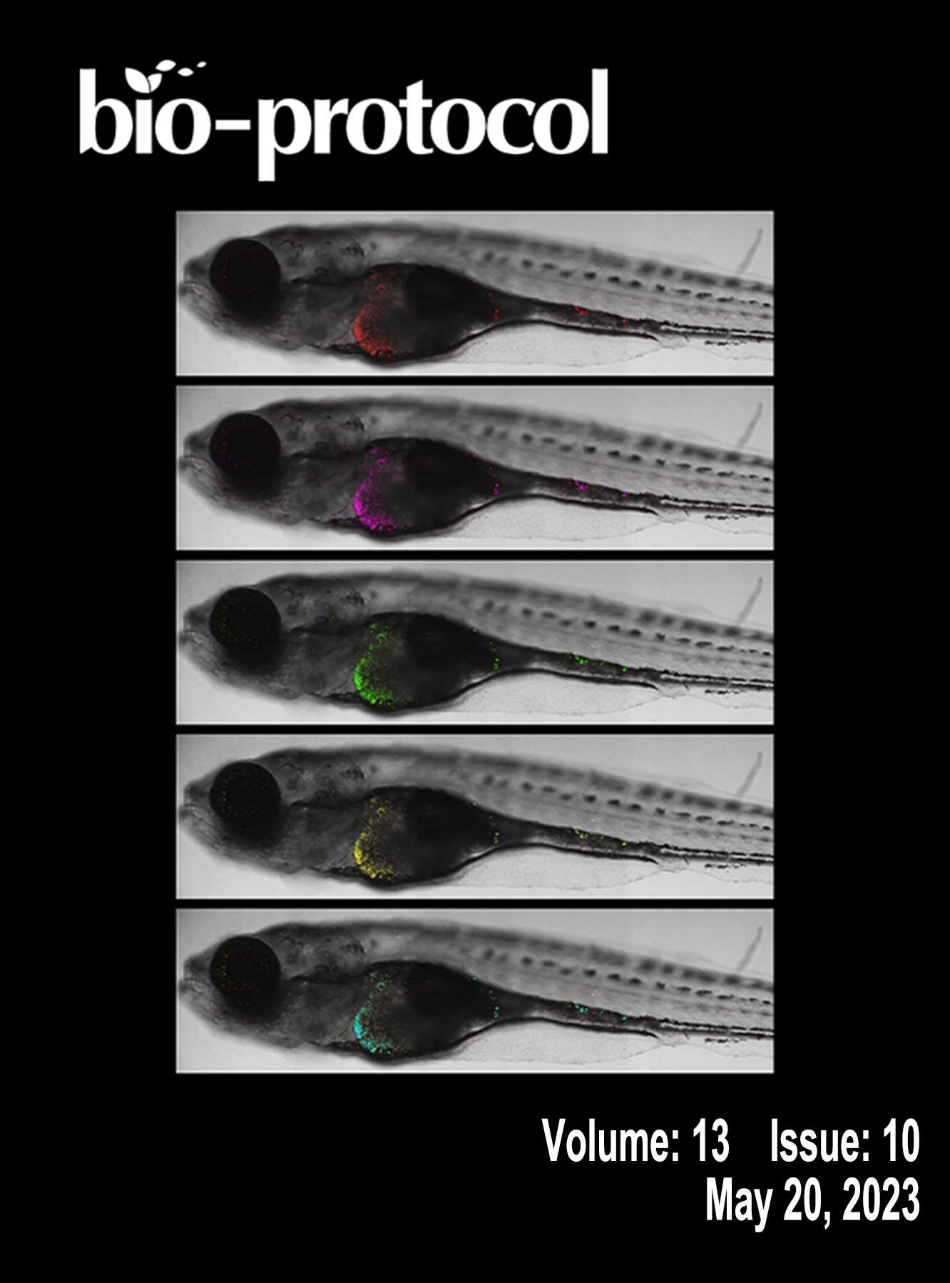
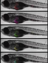
Here, we present an in vivo drug screening protocol using a zebrafish model of metastasis for the identification of anti-metastatic drugs. A tamoxifen-controllable Twist1a-ERT2 transgenic zebrafish line was established to serve as a platform for the identification. By crossing Twist1a-ERT2 with xmrk (a homolog of hyperactive form of the epidermal growth factor receptor) transgenic zebrafish, which develop hepatocellular carcinoma, approximately 80% of the double transgenic zebrafish show spontaneous cell dissemination of mCherry-labeled hepatocytes from the liver to the entire abdomen and tail regions in five days, through induction of epithelial to mesenchymal transition (EMT). This rapid and high-frequency induction of cell dissemination makes it possible to perform an in vivo drug screen for the identification of anti-metastatic drugs targeting metastatic dissemination of cancer cells. The protocol evaluates the suppressor effect of a test drug on metastasis in five days, by comparing the frequencies of the fish showing abdominal and distant dissemination patterns in the test drug–treated group with those in the vehicle-treated group. Our study previously identified that adrenosterone, an inhibitor for hydroxysteroid (11-beta) dehydrogenase 1 (HSD11β1), has a suppressor effect on cell dissemination in the model. Furthermore, we validated that a pharmacologic and genetic inhibition of HSD11β1 suppressed metastatic dissemination of highly metastatic human cell lines in a zebrafish xenotransplantation model. Taken together, this protocol opens new routes for the identification of anti-metastatic drugs. Graphical overviewTimingDay 0: Zebrafish spawningDay 8: Primary tumor inductionDay 11: Chemical treatmentDay 11.5: Metastatic dissemination induction in the presence of a test chemicalDay 16: Data analysis

免疫学

从小鼠淋巴器官、血管周围脂肪组织、肾脏和肺分离的γδ、CD4+和CD8+T细胞中的白细胞介素-17A和干扰素-γ产生的测定

Determination of Interleukin-17A and Interferon-γ Production in γδ, CD4+, and CD8+ T Cells Isolated from Murine Lymphoid Organs, Perivascular Adipose Tissue, Kidney, and Lung

从小鼠淋巴器官、血管周围脂肪组织、肾脏和肺分离的γδ、CD4+和CD8+T细胞中的白细胞介素-17A和干扰素-γ产生的测定

KC Kevin Comeau
AC Antoine Caillon
Pierre  Paradis Pierre Paradis
ES Ernesto L. Schiffrin
2636 Views
May 20, 2023
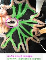
T cells localized to the kidneys and vasculature/perivascular adipose tissue (PVAT) play an important role in hypertension and vascular injury. CD4+, CD8+, and γδ T-cell subtypes are programmed to produce interleukin (IL)-17 or interferon-γ (IFNγ), and naïve T cells can be induced to produce IL-17 via the IL-23 receptor. Importantly, both IL-17 and IFNγ have been demonstrated to contribute to hypertension. Therefore, profiling cytokine-producing T-cell subtypes in tissues relevant to hypertension provides useful information regarding immune activation. Here, we describe a protocol to obtain single-cell suspensions from the spleen, mesenteric lymph nodes, mesenteric vessels and PVAT, lungs, and kidneys, and profile IL-17A- and IFNγ-producing T cells using flow cytometry. This protocol is different from cytokine assays such as ELISA or ELISpot in that no prior cell sorting is required, and various T-cell subsets can be identified and individually assessed for cytokine production simultaneously within an individual sample. This is advantageous as sample processing is kept to a minimum, yet many tissues and T-cell subsets can be screened for cytokine production in a single experiment. In brief, single-cell suspensions are activated in vitro with phorbol 12-myristate 13-acetate (PMA) and ionomycin, and Golgi cytokine export is inhibited with monensin. Cells are then stained for viability and extracellular marker expression. They are then fixed and permeabilized with paraformaldehyde and saponin. Finally, antibodies against IL-17 and IFNγ are incubated with the cell suspensions to report cytokine production. T-cell cytokine production and marker expression is then determined by running samples on a flow cytometer. While other groups have published methods to perform T-cell intracellular cytokine staining for flow cytometry, this protocol is the first to describe a highly reproducible method to activate, phenotype, and determine cytokine production by CD4, CD8, and γδ T cells isolated from PVAT. Additionally, this protocol can be easily modified to investigate other intracellular and extracellular markers of interest, allowing for efficient T-cell phenotyping.
靶向人CD20的双模块融合蛋白scFv2H7-P18F3的表达和纯化

Expression and Purification of scFv2H7-P18F3, a Bi-Modular Fusion Protein (BMFP) Targeting Human CD20

靶向人CD20的双模块融合蛋白scFv2H7-P18F3的表达和纯化

CB Carine Brousse
NR Nathan E. Rainey
AD Alexandra Desrames
JT Jean-Luc Teillaud
BG Benoît Gamain
AC Arnaud Chêne
1005 Views
May 20, 2023
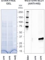
P18F3-based bi-modular fusion proteins (BMFPs), designed to re-direct pre-existing anti-Epstein-Barr virus (EBV) endogenous polyclonal antibodies towards defined target cells, demonstrated efficient biological activity in a mouse tumor model and could potentially represent a universal and versatile platform to develop novel therapeutics against a broad range of diseases. This protocol provides step-by-step instructions for expressing scFv2H7-P18F3, a BMFP targeting human CD20, in Escherichia coli (SHuffle®), and for purifying soluble proteins using a two-step process, namely immobilized metal affinity chromatography (IMAC) followed by size exclusion chromatography. This protocol can also be used for expression and purification of other BMFPs with alternative binding specificities.

微生物学

用物种特异性细菌检测器(SSBD)快速鉴定重症肺炎病原体

Rapid Identification of Pathogens in Severe Pneumonia by Species-specific Bacterial Detector (SSBD)

用物种特异性细菌检测器(SSBD)快速鉴定重症肺炎病原体

CZ Cong Zhang
XL Xiaohui Liang
YQ Yali Qin
WY Wenkui Yu
QC Qihan Chen
917 Views
May 20, 2023
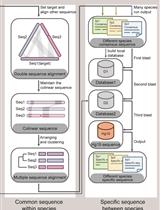
Fast and accurate detection of pathogenic bacterial infection in patients with severe pneumonia is significant to its treatment. The traditional culture method currently used by most medical institutions relies on a time-consuming culture process (over two days) that is unable to meet clinical needs. Rapid, accurate, and convenient species-specific bacterial detector (SSBD) has been developed to provide timely information on pathogenic bacteria. The SSBD was designed based on the fact that Cas12a indiscriminately cleaves any DNA following the binding of the crRNA-Cas12a complex to the target DNA molecule. SSBD involves two processes, starting with PCR of the target DNA using primers specific for the pathogen, followed by detection of the existence of pathogen target DNA in the PCR product using the corresponding crRNA and Cas12a protein. Compared to the culture test, the SSBD can obtain accurate pathogenic information in only a few hours, dramatically shortening the detection time and allowing more patients to benefit from timely clinical treatment.

分子生物学

THRIFTY-一种基于免疫荧光检测的高通量单肌纤维分型方法

THRIFTY—A High-throughput Single Muscle Fiber Typing Method Based on Immunofluorescence Detection

THRIFTY-一种基于免疫荧光检测的高通量单肌纤维分型方法

SE Sebastian Edman
OH Oscar Horwath
WA William Apró
926 Views
May 20, 2023
Skeletal muscle consists of a mixture of fiber types with different functional and metabolic characteristics. The relative composition of these muscle fiber types has implications for muscle performance, whole-body metabolism, and health. However, analyses of muscle samples in a fiber type–dependent manner are very time consuming. Therefore, these are often neglected in favor of more time-efficient analyses on mixed muscle samples. Methods such as western blot and myosin heavy chain separation by SDS-PAGE have previously been utilized to fiber type–isolated muscle fibers. More recently, the introduction of the dot blot method significantly increased the speed of fiber typing. However, despite recent advancements, none of the current methodologies are feasible for large-scale investigations because of their time requirements. Here, we present the protocol for a new method, which we have named THRIFTY (high-THRoughput Immunofluorescence Fiber TYping), that enables rapid fiber type identification using antibodies towards the different myosin heavy chain (MyHC) isoforms of fast and slow twitch muscle fibers. First, a short segment (<1 mm) is cut off from isolated muscle fibers and mounted on a customized gridded microscope slide holding up to 200 fiber segments. Second, the fiber segments attached to the microscope slide are stained with MyHC-specific antibodies and then visualized using a fluorescence microscope. Lastly, the remaining pieces of the fibers can either be collected individually or pooled together with fibers of the same type for subsequent analyses. The THRIFTY protocol is approximately three times as fast as the dot blot method, which enables not only time-sensitive assays to be performed but also increases the feasibility to conduct large-scale investigations into fiber type specific physiology.Graphical OverviewGraphical overview of the THRIFTY workflow. Cut off a small segment (0.5 mm) of an individually dissected muscle fiber and mount it onto the customized microscope slide containing a printed grid system. Using a Hamilton syringe, fixate the fiber segment by applying a small droplet of distilled water on the segment and let it fully dry (1A). The remaining large segment of the fiber should be placed in the corresponding square on a black A4 paper (1B). Once the microscope slide has been fully mounted with fiber segments, submerge the slide in a polypropylene slide mailer (illustrated as a Coplin jar in the figure) containing acetone to permeabilize the fiber segments. Thereafter, incubate the slide with primary antibodies targeting MyHC-I and MyHC-II. Following washes in PBS solution, incubate the slides with fluorescently labeled secondary antibodies, wash again, and mount with a cover glass and antifade reagent (2). Identification of fiber type can be performed using a digital fluorescence microscope (3), whereafter the remaining pieces of the fiber segments (large) are pooled together according to their fiber type or individually collected for experiments on single fibers (4). Image modified from Horwath et al. (2022).

神经科学

量化单通道和双通道实时成像数据: 神经元中细胞器运动的Kymograph分析

Quantifying Single and Dual Channel Live Imaging Data: Kymograph Analysis of Organelle Motility in Neurons

量化单通道和双通道实时成像数据: 神经元中细胞器运动的Kymograph分析

DL Laura Digilio
LM Lloyd P. McMahon
AD Alois Duston
CY Chan Choo Yap
BW Bettina Winckler
1624 Views
May 20, 2023
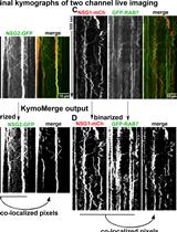
Live imaging is commonly used to study dynamic processes in cells. Many labs carrying out live imaging in neurons use kymographs as a tool. Kymographs display time-dependent microscope data (time-lapsed images) in two-dimensional representations showing position vs. time. Extraction of quantitative data from kymographs, often done manually, is time-consuming and not standardized across labs. We describe here our recent methodology for quantitatively analyzing single color kymographs. We discuss the challenges and solutions of reliably extracting quantifiable data from single-channel kymographs. When acquiring in two fluorescent channels, the challenge becomes analyzing two objects that may co-traffic together. One must carefully examine the kymographs from both channels and decide which tracks are the same or try to identify the coincident tracks from an overlay of the two channels. This process is laborious and time consuming. The difficulty in finding an available tool for such analysis has led us to create a program to do so, called KymoMerge. KymoMerge semi-automates the process of identifying co-located tracks in multi-channel kymographs and produces a co-localized output kymograph that can be analyzed further. We describe our analysis, caveats, and challenges of two-color imaging using KymoMerge.

植物科学

农杆菌介导的棉花遗传转化和通过体细胞胚胎发生的再生

Agrobacterium-mediated Genetic Transformation of Cotton and Regeneration via Somatic Embryogenesis

农杆菌介导的棉花遗传转化和通过体细胞胚胎发生的再生

Alka  Srivastava Alka Srivastava
AS Anoop K. Shukla
Subhi   Srivastava Subhi Srivastava
RD Rama S. Dubey
PS Pradyumna K. Singh
PV Praveen C. Verma
2749 Views
May 20, 2023
Cotton is a significant industrial crop, playing an essential role in the global economy that suffers several setbacks due to biotic and abiotic adversities. Despite such problems, biotechnological advances in cotton are limited because of genetic transformation and regeneration limitations. Here, we present a detailed protocol optimized based on previously published papers, along with our modifications. These involve changes in Agrobacterium concentration, co-cultivation time and temperature, hormones used for regeneration, media manipulation for embryogenic callus production, and efficient rescue of deformed embryos. Further, this protocol has been used in genetic studies on biotic and abiotic stress in cotton. This protocol assures a reproducible stable transgenic cotton development procedure via somatic embryogenesis that can be used by researchers worldwide.

更新

更新:利用Gibson组装技术高效生成蛋白质配体筛选全基因组文库

Update Notice: Efficient Generation of Genome-wide Libraries for Protein–ligand Screens Using Gibson Assembly

更新:利用Gibson组装技术高效生成蛋白质配体筛选全基因组文库

TS Tamara Sternlieb
ML Mira Loock
MG Mengjin Gao
IC Igor Cestari
500 Views
May 20, 2023