往期刊物2023

卷册: 13, 期号: 3

left

Dec

20

Dec

5

Nov

20

Nov

5

Oct

20

Oct

5

Sep

20

Sep

5

Aug

20

Aug

5

Jul

20

Jul

5

Jun

20

Jun

5

May

20

May

5

Apr

20

Apr

5

Mar

20

Mar

5

Feb

20

Feb

5

Jan

20

Jan

5

right

生物化学

分泌性胚胎碱性磷酸酶的测定

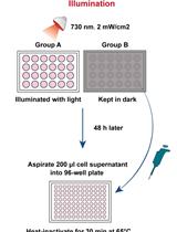
Measurement of Secreted Embryonic Alkaline Phosphatase

分泌性胚胎碱性磷酸酶的测定

MW Meiyan Wang
XW Xinyi Wang
HY Haifeng Ye
1255 Views
Feb 5, 2023
Secreted reporters have been demonstrated to be simple and useful tools for analyzing transcriptional regulation in mammalian cells. The distinctive feature of these assays is the ability to detect reporter gene expression in the culture supernatant without affecting the cell physiology or leading to cell lysis, which allows repeated experimentation and sampling of the culture medium using the same cell cultures. Secreted embryonic alkaline phosphatase (SEAP) is one of the most widely used reporter, which can be easily detected using colorimetry following incubation with a substrate, such as p-nitrophenol phosphate. In this report, we present detailed procedures for detection and quantification of the SEAP reporter. We believe that this step-by-step protocol can be easily used by researchers to monitor and measure molecular genetic events in a variety of mammalian cells due to its simplicity and ease of handling.Graphical abstractSchematic overview of the workflow described in this protocol

细胞生物学

小鼠仿人切除伤口夹板模型的实验方案

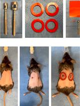
Protocol for the Splinted, Human-like Excisional Wound Model in Mice

小鼠仿人切除伤口夹板模型的实验方案

KF Katharina S. Fischer
BL Ben Litmanovich
DS Dharshan Sivaraj
HK Hudson C. Kussie
WH William W. Hahn
AH Andrew C. Hostler
KC Kellen Chen
GG Geoffrey C. Gurtner
2198 Views
Feb 5, 2023
While wound healing in humans occurs primarily through re-epithelization, in rodents it also occurs through contraction of the panniculus carnosus, an underlying muscle layer that humans do not possess. Murine experimental models are by far the most convenient and inexpensive research model to study wound healing, as they offer great variability in genetic alterations and disease models. To overcome the obstacle of contraction biasing wound healing kinetics, our group invented the splinted excisional wound model. While other rodent wound healing models have been used in the past, the splinted excisional wound model has persisted as the most used model in the field of wound healing. Here, we present a detailed protocol of updated and refined techniques necessary to utilize this model, generate results with high validity, and accurately analyze the collected data. This model is simple to conduct and provides an easy, standardizable, and replicable model of human-like wound healing.
基于 CRISPR 的 RNA 修饰的时间控制位点特异性编辑策略

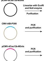
A CRISPR-based Strategy for Temporally Controlled Site-Specific Editing of RNA Modifications

基于 CRISPR 的 RNA 修饰的时间控制位点特异性编辑策略

YX Ying Xu
YW Yufan Wang
FL Fu-Sen Liang
980 Views
Feb 5, 2023
Chemical modifications on RNA play important roles in regulating its fate and various biological activities. However, the impact of RNA modifications varies depending on their locations on different transcripts and cells/tissues contexts; available tools to dissect context-specific RNA modifications are still limited. Herein, we report the detailed protocol for using a chemically inducible and reversible platform to achieve site-specific editing of the chosen RNA modification in a temporally controlled manner by integrating the clustered regularly interspaced short palindromic repeats (CRISPR) technology and the abscisic acid (ABA)-based chemically induced proximity (CIP) system. The procedures were demonstrated using the example of inducible and reversible N6-methyladenosine (m6A) editing and the evaluation of its impact on RNA properties with ABA addition and reversal with the control of ABA or light.

发育生物学

果蝇胚胎组织成体肌肉干细胞的双色实时成像

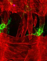
Dual-Color Live Imaging of Adult Muscle Stem Cells in the Embryonic Tissues of Drosophila melanogaster

果蝇胚胎组织成体肌肉干细胞的双色实时成像

MZ Monika Zmojdzian
BD Binoj Dhanarajan
KJ Krzysztof Jagla
RA Rajaguru Aradhya
984 Views
Feb 5, 2023
Adult muscle stem cells (MuSCs) show remarkable capability in repairing injured tissues. Studying MuSCs in suitable model organisms, which show strong homology with vertebrate counterparts, helps in dissecting the mechanisms regulating their behavior. Additionally, ease of handling and access to technological tools make model organisms well suited for studying biological processes that are conserved across species. MuSCs quiescence, proliferation, and migration are regulated by various input of signals from the surrounding tissues that constitute the MuSCs niche. Observing MuSCs along with their niche in vivo through live imaging provides key information on how MuSCs behave in quiescent and activated states. Drosophila melanogaster is well known for its genetic tool arsenal and the similarity of its different biological processes with vertebrates. Hence, it is widely used to study different types of stem cells. Gained knowledge could then be extrapolated to the vertebrate/mammalian homologous systems to enhance our knowledge in stem cell fields. In this protocol, we discuss how to perform live cell imaging of Drosophila MuSCs, called adult muscle precursors (AMPs) at embryonic stages, using dual-color labelling to visualize both AMPs and the surrounding tissues. This dual-color fluorescent labelling enables the observation of in vivo behavior of two types of cells simultaneously and provides key information on their interactions. The originality of this protocol resides in its biological application to MuSCs and their niche.

免疫学

使用荧光酶标仪连续测量受细菌感染的骨髓源性巨噬细胞中活性氧的生成

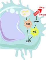
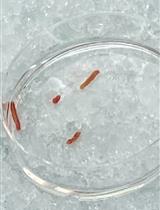
Continuous Measurement of Reactive Oxygen Species Formation in Bacteria-infected Bone Marrow–derived Macrophages Using a Fluorescence Plate Reader

使用荧光酶标仪连续测量受细菌感染的骨髓源性巨噬细胞中活性氧的生成

NB Natascha Brigo
PG Philipp Grubwieser
IT Igor Theurl
MN Manfred Nairz
GW Günter Weiss
CP Christa Pfeifhofer-Obermair
1602 Views
Feb 5, 2023
Macrophages are at the center of innate immunity and are the main target cells of the intracellular pathogen Salmonella enterica serovar Typhi. The production of reactive oxygen and nitrogen species (ROS/RNS) is the host’s early response to invading microbes, as oxidative stress is highly toxic for bacteria. Adequate ROS/RNS production in infected macrophages is critical for the clearance of intracellular pathogens; this is achieved by several enzymes, including inducible NADPH phagocyte oxidase (NOX) and nitric oxide synthase (iNOS), respectively. The pro-inflammatory cytokine interferon gamma (IFNγ), primarily produced by activated natural killer cells and T-helper cells type 1, is a potent inducer of iNOS. Therefore, it is crucial for infection control through oxidative microbicidal activity.To characterize the early oxidative stress response via ROS formation, which is critical for the reduction of Salmonella proliferation within macrophages, we established an in vitro model of murine macrophages infected with Salmonella enterica serovar Typhimurium (S.tm). This serovar induces a systemic infection in mice that is frequently used as a model for typhoid fever, which, in human subjects, is caused by Salmonella Typhi. We generated bone marrow–derived macrophages (BMDM) from C57BL/6N wildtype mice using macrophage colony-stimulating factor (M-CSF) stimulation for six days. Thereafter, we infected BMDM with S.tm for one hour. Shortly before infection, cells were stained with CellROXTM Deep Red reagent. In its reduced form, CellROXTM is non-fluorescent. As a result of oxidation by ROS, this reagent exhibits strong fluorescence and persists within the cells. Subsequently, changes as a result of the oxidative stress response can be measured with a TECAN Spark microplate reader over time.We designed this protocol to measure oxidative stress in macrophages through the course of an infection with an intracellular bacterium. The protocol has several advantages over established techniques. First, it allows to continuously monitor and quantify ROS production in living cells from the very start of the infection to the final clearance of the intracellular pathogen. Second, this protocol enables efficient ROS detection without stressing the cells by detaching or staining procedures.Graphical abstract

分子生物学

从人速冻肝组织中分离细胞核用于单核 RNA 测序

Isolation of Nuclei from Human Snap-frozen Liver Tissue for Single-nucleus RNA Sequencing

从人速冻肝组织中分离细胞核用于单核 RNA 测序

MA Marcus Alvarez
JB Jihane N. Benhammou
SR Shuyun Rao
LM Lopa Mishra
JP Joseph R. Pisegna
PP Päivi Pajukanta
1603 Views
Feb 5, 2023
Single-nucleus RNA sequencing (snRNA-seq) provides a powerful tool for studying cell type composition in heterogenous tissues. The liver is a vital organ composed of a diverse set of cell types; thus, single-cell technologies could greatly facilitate the deconvolution of liver tissue composition and various downstream omics analyses at the cell-type level. Applying single-cell technologies to fresh liver biopsies can, however, be very challenging, and snRNA-seq of snap-frozen liver biopsies requires some optimization given the high nucleic acid content of the solid liver tissue. Therefore, an optimized protocol for snRNA-seq specifically targeted for the use of frozen liver samples is needed to improve our understanding of human liver gene expression at the cell-type resolution. We present a protocol for performing nuclei isolation from snap-frozen liver tissues, as well as guidance on the application of snRNA-seq. We also provide guidance on optimizing the protocol to different tissue and sample types.
在局部紫外线诱导的 DNA 损伤位点进行程序外DNA合成,以量化人类细胞中的全基因组核苷酸切除修复活性

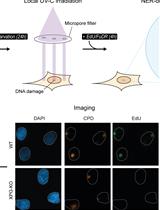
Unscheduled DNA Synthesis at Sites of Local UV-induced DNA Damage to Quantify Global Genome Nucleotide Excision Repair Activity in Human Cells

在局部紫外线诱导的 DNA 损伤位点进行程序外DNA合成,以量化人类细胞中的全基因组核苷酸切除修复活性

Pv Paula J. van der Meer
Dv Diana van den Heuvel
ML Martijn S. Luijsterburg
1323 Views
Feb 5, 2023
Nucleotide excision repair (NER) removes a wide variety of structurally unrelated lesions from the genome, including UV-induced photolesions such as 6–4 pyrimidine–pyrimidone photoproducts (6-4PPs) and cyclobutane pyrimidine dimers (CPDs). NER removes lesions by excising a short stretch of single-stranded DNA containing the damaged DNA, leaving a single-stranded gap that is resynthesized in a process called unscheduled DNA synthesis (UDS). Measuring UDS after UV irradiation in non-dividing cells provides a measure of the overall NER activity, of which approximately 90% is carried out by the global genome repair (GGR) sub pathway. Here, we present a protocol for the microscopy-based analysis and quantification of UDS as a measurement for GGR activity. Following local UV-C irradiation, serum-starved human cells are supplemented with the thymidine analogue 5-ethynyl-2'-deoxyuridine (EdU), which is incorporated into repair patches following NER-dependent dual incision. The incorporated nucleotide analogue is coupled to a fluorophore using Click-iT chemistry, followed by immunodetection of CPD photolesions to simultaneously visualize both signals by fluorescence microscopy. Accompanying this protocol is a custom-built ImageJ plug-in to analyze and quantify UDS signals at sites of CPD-marked local damage. The local UDS assay enables an effective and sensitive fluorescence-based quantification of GGR activity in single cells with application in basic research to better understand the regulatory mechanism in NER, as well as in diagnostics to characterize fibroblasts from individuals with NER-deficiency disorder.Graphical abstract
使用 Mito-FUNCAT FACS 对哺乳动物细胞中线粒体蛋白合成进行高通量评估

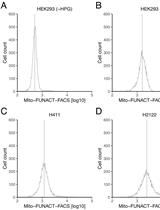
High-throughput Assessment of Mitochondrial Protein Synthesis in Mammalian Cells Using Mito-FUNCAT FACS

使用 Mito-FUNCAT FACS 对哺乳动物细胞中线粒体蛋白合成进行高通量评估

HS Hironori Saito
TO Tatsuya Osaki
YI Yoshiho Ikeuchi
SI Shintaro Iwasaki
1146 Views
Feb 5, 2023
In addition to cytosolic protein synthesis, mitochondria also utilize another translation system that is tailored for mRNAs encoded in the mitochondrial genome. The importance of mitochondrial protein synthesis has been exemplified by the diverse diseases associated with in organello translation deficiencies. Various methods have been developed to monitor mitochondrial translation, such as the classic method of labeling newly synthesized proteins with radioisotopes and the more recent ribosome profiling. However, since these methods always assess the average cell population, measuring the mitochondrial translation capacity in individual cells has been challenging. To overcome this issue, we recently developed mito-fluorescent noncanonical amino acid tagging (FUNCAT) fluorescence-activated cell sorting (FACS), which labels nascent peptides generated by mitochondrial ribosomes with a methionine analog, L-homopropargylglycine (HPG), conjugates the peptides with fluorophores by an in situ click reaction, and detects the signal in individual cells by FACS equipment. With this methodology, the hidden heterogeneity of mitochondrial translation in cell populations can be addressed.

植物科学

生成可重复的缺氧条件用于植物表型分析

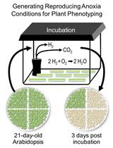
Generating Reproducing Anoxia Conditions for Plant Phenotyping

生成可重复的缺氧条件用于植物表型分析

IM Iny E. Mathew
HR Hormat Shadgou Rhein
AG Ardawna J. Green
KH Kendal D. Hirschi
1333 Views
Feb 5, 2023
Based on the availability of oxygen, plant growth environment can be normoxic (normal environment), hypoxic (reduced oxygen, <21%), or anoxic (complete depletion of oxygen). Hypoxic/anoxic environment is created when a plant is exposed to stresses such as submergence, flooding, or pathogen attack. Survival of the plants following stress conditions is in part dependent on their ability to overcome the stress induced by anoxia/hypoxia conditions. This shows the need for the development of strategies for understanding the mechanisms involved in plant tolerance to anoxia. Previous studies have employed different methods for establishing an anerobic environment. Here, we describe a simple method for creating anoxic environment using an anaerobic atmosphere generation bag. Anoxic conditions can be maintained in a cylindrical jar, a rectangular box, or a vacuum sealer bag, enabling the screening of a large number of samples. This protocol is particularly useful to screen plant mutants that are tolerant to anoxia. The method is simple, easy, cost-efficient, reproducible, and does not require any sophisticated instruments.Graphic abstract
使用合成寡肽底物测定植物蛋白酶介导的肽前体裂解

Assay for Phytaspase-mediated Peptide Precursor Cleavage Using Synthetic Oligopeptide Substrates

使用合成寡肽底物测定植物蛋白酶介导的肽前体裂解

SR Sven Reichardt
AS Annick Stintzi
AS Andreas Schaller
917 Views
Feb 5, 2023
Proteases control plant growth and development by limited proteolysis of regulatory proteins at highly specific sites. This includes the processing of peptide hormone precursors to release the bioactive peptides as signaling molecules. The proteases involved in this process have long remained elusive. Confirmation of a candidate protease as a peptide precursor–processing enzyme requires the demonstration of protease-mediated precursor cleavage in vitro. In vitro cleavage assays rely on the availability of suitable substrates and the candidate protease with high purity. Here, we provide a protocol for the expression, purification, and characterization of tomato (Solanum lycopersicum) phytaspases as candidate proteases for the processing of the phytosulfokine precursor. We also show how synthetic oligopeptide substrates can be used to demonstrate site-specific precursor cleavage.Graphical abstract