往期刊物2015

卷册: 5, 期号: 19

left

Dec

20

Dec

5

Nov

20

Nov

5

Oct

20

Oct

5

Sep

20

Sep

5

Aug

20

Aug

5

Jul

20

Jul

5

Jun

20

Jun

5

May

20

May

5

Apr

20

Apr

5

Mar

20

Mar

5

Feb

20

Feb

5

Jan

20

Jan

5

right

细胞生物学

用于细胞微生物学中关联光学和电子显微镜技术(CLEM)分析的样本制备

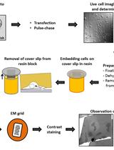
Sample Preparation for Correlative Light and Electron Microscopy (CLEM) Analyses in Cellular Microbiology

用于细胞微生物学中关联光学和电子显微镜技术(CLEM)分析的样本制备

VL Viktoria Liss
MH Michael Hensel
16934 Views
Oct 5, 2015
Dynamic processes in cells are usually monitored by live cell fluorescence microscopy. Unfortunately, this method lacks the ultrastructural information about the structure of interest (SOI). Currently, electron microscopy (EM) is the best tool to achieve highest spatial resolution. In addition, correlative light and electron microscopy (CLEM) analysis of the same structure allows combining authentic live cell imaging with the resolution power of EM. Additionally the reference space of the SOI is revealed. Our CLEM analyses of HeLa cells allow tracing the morphology and dynamic behavior of intracellular micro-compartments in living cells and their ultrastructure and subcellular organization in a highly resolved manner.

免疫学

对EBV 和CMV特异性人CD8+T淋巴细胞的TCRβ克隆型分析

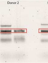
TCRβ Clonotype Analysis of EBV and CMV-specific Human CD8+ T Cells

对EBV 和CMV特异性人CD8+T淋巴细胞的TCRβ克隆型分析

NN Nening M. Nanlohy
DK Dan Koning
EQ Esther D. Quakkelaar
DB Debbie van Baarle
8034 Views
Oct 5, 2015
This protocol describes the quantification of all expressed T-cell antigen receptor (TCR) gene products within sorted (by flow cytometry) EBV and CMV-specific memory CD8+ T-cell populations using a template-switch anchored reverse transcription polymerase chain reaction (RT-PCR).

微生物学

密度梯度离心法富集和鉴定丝状真菌中GFP标记的壳聚糖小囊泡

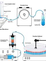
Density Gradient Centrifugation for Enrichment and Identification of GFP-tagged Chitosomal Microvesicles of Filamentous Fungi

密度梯度离心法富集和鉴定丝状真菌中GFP标记的壳聚糖小囊泡

Jorge Verdín Jorge Verdín
Eddy Sánchez-León Eddy Sánchez-León
RF Rosa Fajardo-Somera
CM Carlos A. Leal Morales
SB Salomón Bartnicki-García
MR Meritxell Riquelme
15301 Views
Oct 5, 2015
Density gradient centrifugation has been utilized to characterize the subcellular distribution of physiologically relevant enzymes in yeasts and filamentous fungi (Leal-Morales et al., 1988; Martínez et al., 1989; Kamada et al., 1991). This approach is now potentiated by protein tagging and live imaging techniques, which make possible to relate a single protein with, for example, a discrete population of intracellular vesicles and their in vivo dynamics (Verdín et al., 2009; Fajardo-Somera et al., 2013; Sánchez-León et al., 2015). Here, we describe the density gradient centrifugation and fractionation analysis of cell-free homogenates of a Neurospora crassa (N. crassa) strain that expresses CHS-6 chitin synthase fused to the green fluorescent protein (Riquelme et al., 2007).
采用 LC/MS/MS 分析法提取和量化细菌卟啉

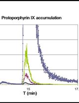
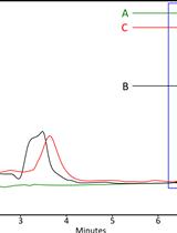
Bacterial Porphyrin Extraction and Quantification by LC/MS/MS Analysis

采用 LC/MS/MS 分析法提取和量化细菌卟啉

SM Stefano Mancini
JI James A. Imlay
9189 Views
Oct 5, 2015
Heme is an iron-containing porphyrin which acts as a prosthetic group in several enzymes involved in disparate functions, such as respiration and H2O2-scavenging. Escherichia coli is able to produce heme endogenously since it contains all the enzymes involved in the nine-step biosynthesis pathway, which in absence of stress and in iron-replete media proceeds unabated. However, we recently showed that two steps are affected by H2O2 stress (Mancini and Imlay, 2015). To compensate, two enzymes, namely the ferrochelatase (HemH) and an isozyme of coproporphyrinogen III oxidase (HemF), are activated by the H2O2-responsive regulator OxyR. Genetic mutations that block either adaptation cause the intracellular accumulation of protoporphyrin IX and coproporphyrinogen III, the substrates of HemH and HemF, respectively. We here describe a method used to extract and quantify protoporphyrin IX and coproporphyrin III, the product of the spontaneous oxidation of coproporphyrinogen III.
体外研究:肝脏微粒体中去甲替林对奈韦拉平新陈代谢的抑制

In vitro Studies: Inhibition of Nevirapine Metabolism by Nortriptyline in Hepatic Microsomes

体外研究:肝脏微粒体中去甲替林对奈韦拉平新陈代谢的抑制

Iris Usach Iris Usach
José-Esteban Peris José-Esteban Peris
8008 Views
Oct 5, 2015
One of the most prevalent and interfering psychosocial comorbidities of HIV infection is clinical depression (22 to 45%). For this reason, a study of a possible interaction between the nonnucleoside reverse transcriptase inhibitor nevirapine (NVP) and the tricyclic antidepressant nortriptyline (NT) was carried out. In vitro studies with rat and human hepatic microsomes showed a marked inhibition of NVP metabolism by NT being more intense in rat than in human. The extrapolation of these results to humans suggests increased NVP side effects when both drugs are coadministered, but additional in vivo human studies are required to evaluate the clinical implication of this interaction.This protocol describes a technique for detecting and measuring the inhibition of the nevirapine metabolism by nortriptyline in hepatic microsomes.

神经科学

鸡胚胎神经管外植体培养

Chick Neural Tube Explant Culture

鸡胚胎神经管外植体培养

Zhanna Alekseenko Zhanna Alekseenko
Elisabet Andersson Elisabet Andersson
José M. Dias José M. Dias
10303 Views
Oct 5, 2015
The neural tube explant culture technique allows in vitro culturing of small pieces of neural tissue isolated from e.g., chick or mouse embryonic tissue in a matrix of collagen for defined periods of time. This method can be used to study the effects of defined molecules on developmental processes such as neural progenitor proliferation and neuronal differentiation and/or survival. Since the explant material can also be prepared from embryonic tissue electroporated with expression vectors, this technique can be adapted to study gene function in the presence of specific environmental signals. Different regions of the neural tube can also be isolated during the dissection step, allowing specific regions of the neural tube to be studied separately. Here, we present a neural tube explant culture method that we have used in several studies (Dias et al., 2014; Lek et al., 2010; Vallstedt et al., 2005).

植物科学

对拟南芥样本的cDNA 3'末端进行快速扩增(3' RACE)

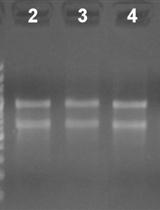
3’ Rapid Amplification of cDNA Ends (3’ RACE) Using Arabidopsis Samples

对拟南芥样本的cDNA 3'末端进行快速扩增(3' RACE)

ER Encarnación Rodríguez-Cazorla
Alfonso Andújar Alfonso Andújar
Juan José Ripoll Juan José Ripoll
LB Lindsay J. Bailey
Antonio Martínez-Laborda Antonio Martínez-Laborda
Martin F. Yanofsky Martin F. Yanofsky
Antonio Vera Antonio Vera
20293 Views
Oct 5, 2015
Production of functional eukaryotic RNA is a very elaborate process that involves a complex interplay between transcription and various RNA processing activities, including splicing, 5’ capping, and 3’ cleavage and polyadenylation (Bentley, 2014). Accurate mapping of RNA ends provides a valuable tool to assess transcriptional and post-transcriptional events giving rise to different gene transcripts. The abundance of such transcripts most likely depends on exogenous and developmental cues, or mutations. In the reference plant Arabidopsis, perturbation of the HUA-PEP post-transcriptional regulatory factors (Rodríguez-Cazorla et al., 2015) leads to the accumulation of aberrant transcripts of the key floral homeotic gene AGAMOUS (AG) (Yanofsky et al., 1990) that retain intronic sequence. It was determined by 3’ RACE reactions that such erroneous transcripts correspond to premature processing and polyadenylation events taking place at the AG intron region. Here we describe a protocol that is suitable for analysis of relatively abundant transcripts and also for detecting aberrant RNA species that are likely prone to rapid turnover. Likewise, the method, here adapted to Arabidopsis reproductive tissues, can be applied to characterize RNA species from other organs (leaf, root) and/or other plant species. We provide a detailed protocol of our 3’ RACE procedure comprising four major parts: Total RNA extraction, RNA amount determination and quality control, the RACE procedure itself, and isolation of the resulting RACE products for cloning and sequencing.
植物叶中胼胝质沉积的定量测定

Quantification of Callose Deposition in Plant Leaves

植物叶中胼胝质沉积的定量测定

Loredana Scalschi Loredana Scalschi
Eugenio Llorens Eugenio Llorens
GC Gemma Camañes
VP Victoria Pastor
EF Emma Fernández-Crespo
VF Victor Flors
PG Pilar García-Agustín
BV Begonya Vicedo
15655 Views
Oct 5, 2015
Callose is an amorphous homopolymer, composed of β-1, 3-glucan, which is widespread in higher plants. Callose is involved in multiple aspects of plant growth and development. It is synthetized in plants at the cell plate during cytokinesis, in several stages during pollen development and is deposited at plasmodesmata to regulate the cell-to-cell movement of molecules. Moreover, it is produced in response to multiple biotic and abiotic stresses (Chen and Kim, 2009). Callose is considered to act as a physical barrier by strengthening the plant cell well to slow pathogen infection and to contribute to the plant’s innate immunity. Thus the callose staining method is useful to quantify activity of plant immunity. In addition, this staining can be used to visualize structures in plant tissue, where the callose may be implied whether during the development of plants or response against pathogen infection. This method is based on the use of methyl blue which reacts with (1→3)-β-glucans to give a brilliant yellow fluorescence in UV light. Moreover, calcofluor stains chitin present in fungal cell membranes and also binds to cellulose at locations where the cuticle is damaged.
水培法培养‘Micro-Tom’ 番茄

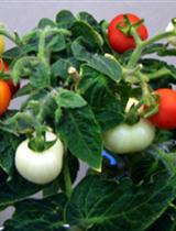
Hydroponic Culture of ‘Micro-Tom’ Tomato

水培法培养‘Micro-Tom’ 番茄

Reiko Motohashi Reiko Motohashi
HE Harumi Enoki
CF Chikako Fukazawa
YK Yoshikazu Kiriiwa
14500 Views
Oct 5, 2015
We use ‘Micro-Tom’ to study tomato fruit ripening and development mechanisms. ‘Micro-Tom’ is suitable for cultivation and experiments due to its small size of 10 to 20 cm in height and short life cycle of 3 months. There is also an abundance of publically available information on ‘Micro-Tom’ including EST, full-length cDNA clones and transcriptome data. ‘Micro-Tom’ plants are grown in hydroponic culture under fluorescents using Arabidopsis cultural shelves in greenhouses or plant rooms to get data with reproducibility for transcriptome and proteome analyses.
通过植株表型和抗氧化实验在烟草中进行盆位干旱胁迫耐受性分析

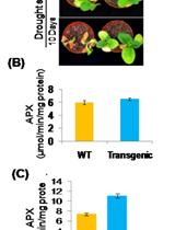
Pot Level Drought Stress Tolerance Assay in Tobacco through Plant Phenotyping and Antioxidant Assay

通过植株表型和抗氧化实验在烟草中进行盆位干旱胁迫耐受性分析

Priyanka Das Priyanka Das
NL Nita Lakra
KN Kamlesh Kant Nutan
Sneh L Singla-Pareek Sneh L Singla-Pareek
Ashwani Pareek Ashwani Pareek
12554 Views
Oct 5, 2015
Drought is an important abiotic factor which has a huge detrimental impact on crop productivity. Study of plant responses towards drought stress and investigating the mechanism of drought tolerance is crucial for achieving the target of developing drought-tolerant plants. Phenotyping is a cost effective approach which can be adopted to evaluate the severity of drought stress in a plant. Next to phenotyping parameters, biochemical parameters such as the study of antioxidant enzyme activity play significant roles in assessing the extent of drought stress caused injury in a plant. Among the antioxidant enzymes, ascorbate peroxidase is an enzyme which plays a crucial role in drought tolerance in plants. It has been well established that the activity of this enzyme increases under drought stress. Here, we present a simple and reproducible protocol to investigate the response of tobacco plants towards drought stress through measurement of phenotypic parameters and antioxidant enzyme activity. Though, these experiments have been conducted with tobacco plants, this protocol could be adopted for other crop species.
对水稻叶片中硅质小体进行组织化学染色

Histochemical Staining of Silica Body in Rice Leaf Blades

对水稻叶片中硅质小体进行组织化学染色

RY Ryusuke Yokoyama
NK Natsumi Kido
TY Tsuyoshi Yamamoto
JF Jun Furukawa
HI Hiroaki Iwai
SS Shinobu Satoh
KN Kazuhiko Nishitani
10740 Views
Oct 5, 2015
Silicon (Si) is a biologically important element for plants in the order Poales (Yamamoto et al., 2011; Kido et al., 2015). In rice, Si is mainly deposited in the motor cells and the cell walls of the leaf epidermis. However, the molecular basis of this overall process has not been elucidated. Thus, we propose a protocol for the histochemical staining of the silica body based on specific hydrogen bonding between silanol group and the carboxylate group of crystal violet lactone (Ichimura et al., 2008), as described by Isa et al. (2010), but with minor modifications. This modified protocol can be used for observing Si accumulation during rice development.
采用原子力显微镜对果胶纳米结构进行可视化

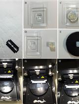
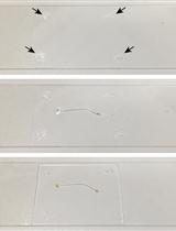
Pectin Nanostructure Visualization by Atomic Force Microscopy

采用原子力显微镜对果胶纳米结构进行可视化

SP Sara Posé
CP Candelas Paniagua
AK Andrew R Kirby
AG A. Patrick Gunning
VM Victor J Morris
MQ Miguel A Quesada
JM Jose A Mercado
9215 Views
Sep 20, 2015
Pectins, complex polysaccharides rich in galacturonic acid, are a major component of primary plant cell walls. These macromolecules regulate cell wall porosity and intercellular adhesion, being important in the control of cell expansion and differentiation through their effect on the rheological properties of the cell wall. In fruits, pectin disassembly during ripening is one the main event leading to textural changes and softening. Changes in pectic polymer size, composition and structure have been studied by conventional techniques, most of them relying on bulk analysis of a population of polysaccharides but studies of detailed structure of isolated polymer chains are scarce (Paniagua et al., 2014). Atomic force microscopy (AFM) is a versatile and powerful technique able to analyze force measurements, as well as to visualize roughness of biological samples at nanoscale (Morris et al., 2010). Using this technique, recent research has found a close relationship between pectin nanostructural complexity and texture and postharvest behavior in several fruits (Liu and Cheng, 2011; Cybulska et al., 2014; Posé et al., 2015). Here, we describe an AFM procedure to topographically visualize pectic polymers from fruit cell wall extracts that has successfully been used in the study of strawberry ripening (Posé et al., 2012; Posé et al., 2015). Thus, from AFM images the 3D structural analysis of isolated chains (length, height, and branch pattern) can be resolved at high magnification and with minimal sample preparation. A full description of AFM fundamentals and the different sampling modes are described in Morris et al. (2010).
使用转盘共聚焦显微镜分析拟南芥细胞中的纤维素生物合成

Analysis of in vivo Cellulose Biosynthesis in Arabidopsis Cells by Spinning Disk Confocal Microscopy

使用转盘共聚焦显微镜分析拟南芥细胞中的纤维素生物合成

TV Tamara Vellosillo
TY Trevor Yeats
NS Nadav Sorek
8897 Views
Oct 5, 2015
Cellulose is a main component of plant cell walls. Tools to analyze cellulose mainly rely on analytical chemistry, which yields information about cellulose amounts and structure, but cannot be applied to intact tissues. Moreover, these methods measure total cellulose and cannot be used to assay cellulose synthesis per se. Live cell imaging of the catalytic subunits of the cellulose synthesis complex (CSC) conjugated to fluorescent proteins is an important tool to understand the dynamics of the cellulose biosynthesis process (Paredez et al., 2006). This method can be used in various genetic backgrounds (Sorek et al., 2014) or with different chemical inhibitors (Brabham and Debolt, 2012). Here we describe in detail the procedure to visualize the movement of CSCs at the plasma membrane. As the movement of CSCs is likely caused by glucan synthesis and extrusion into the cell wall, live cell analysis of CSC velocity provides a method to directly measure cellulose synthesis in vivo.