往期刊物2022

卷册: 12, 期号: 18

left

Dec

20

Dec

5

Nov

20

Nov

5

Oct

20

Oct

5

Sep

20

Sep

5

Aug

20

Aug

5

Jul

20

Jul

5

Jun

20

Jun

5

May

20

May

5

Apr

20

Apr

5

Mar

20

Mar

5

Feb

20

Feb

5

Jan

20

Jan

5

right

生物化学

 蛋白酪氨酸磷酸酶生化抑制测定

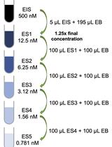
Protein Tyrosine Phosphatase Biochemical Inhibition Assays

蛋白酪氨酸磷酸酶生化抑制测定

MB Marek R. Baranowski
JW Jiaqian Wu
YH Ye Na Han
LL Lester J. Lambert
NC Nicholas D. P. Cosford
LT Lutz Tautz
2086 Views
Sep 20, 2022
Disturbance of the dynamic balance between protein tyrosine phosphorylation and dephosphorylation, modulated by protein tyrosine kinases (PTKs) and protein tyrosine phosphatases (PTPs), is known to be crucial for the development of many human diseases. The discovery of agents that restore this balance has been the subject of many drug research efforts, most of which have focused on tyrosine kinase inhibitors (TKIs), resulting in the development of more than 50 FDA-approved TKIs during the past two decades. More recently, accumulating evidence has suggested that members of the PTP superfamily are also promising drug targets, and efforts to discover tyrosine phosphatase inhibitors (TPIs) have increased dramatically. Here, we provide protocols for determining the potency of TPIs in vitro. We focus on the use of fluorescence-based substrates, which exhibit a dramatic increase in fluorescence emission when dephosphorylated by the PTP, and thus allow setting up highly sensitive and miniaturized phosphatase activity assays using 384-well or 1536-well microplates and a continuous (kinetic) assay format. The protocols cover PTP specific activity assays, Michaelis–Menten kinetics, dose-response inhibition assays, and dose-response data analysis for determining IC50 values. Potential pitfalls are also discussed. While advanced instrumentation is utilized for compound spotting and liquid dispensing, all the assays can be adapted to existing equipment in most laboratories. Assays are described for selected PTP drug targets, including SHP2 (PTPN11), PTP1B (PTPN1), STEP (PTPN5), and VHR (DUSP3). However, all protocols are applicable to members of the PTP enzyme family in general. Graphical abstract:
基于体外荧光成像的肌动蛋白捆绑测定

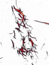
In vitro Fluorescence Imaging–based Actin Bundling Assay

基于体外荧光成像的肌动蛋白捆绑测定

AG Anjelika Gasilina
PR Paul A. Randazzo
1781 Views
Sep 20, 2022
Understanding the molecular and structural mechanisms that govern the assembly and organization of higher-order actin architecture requires the use of in vitro actin binding and bundling assays. Crosslinking of actin filaments into bundles can be monitored in vitro via several techniques, including negative staining/electron microscopy, low-speed co-sedimentation assay/SDS-PAGE, and fluorescence staining/confocal microscopy. We and others have previously characterized the N-BAR domain of ASAP1, an ADP-ribosylation factor GTPase-activating protein, as an actin-bundling module; we further identified key lysine residues responsible for actin cross-linking. Here, we use the ASAP1 BAR domain as an example and describe a detailed procedure for observing the actin bundle formation by confocal microscopy. This protocol requires small reaction volumes and takes advantage of bright commercially available fluorescent phalloidins, making it an ideal choice for medium-throughput screening of mutants or domain truncations in their ability to bundle actin.Graphical abstract:

生物信息学与计算生物学

使用 MapR对果蝇中的 R 环进行组织特异性、全基因组映射

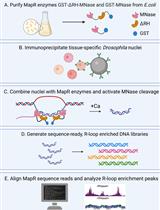
Tissue-Specific, Genome-wide Mapping of R-loops in Drosophila Using MapR

使用 MapR对果蝇中的 R 环进行组织特异性、全基因组映射

JJ Juan Jauregui-Lozano
KC Kendall Cottingham
HH Hana Hall
1512 Views
Sep 20, 2022
R-loops, or RNA:DNA hybrids, are structures that arise co-transcriptionally when a nascent RNA hybridizes back with the template ssDNA, leading to a displaced ssDNA. Because accumulation of R-loops can lead to genomic instability and loss of cellular homeostasis, it is important to determine the genome-wide distribution of R-loops in different physiological conditions. Current R-loop mapping strategies are based on R-loop enrichment—mediated by the S9.6 antibody, such as DRIP-seq, or by the exonuclease RNase H1, such as MapR—or the latest R-loop CUT&Tag, based on an artificial R-loop sensor derived from an RNase H1 sub-domain. Because some of these techniques often require high input material or expensive reagents, we sought to apply MapR, which does not require expensive reagents and has been shown to be compatible with low input samples. Importantly, we demonstrate that incorporation of improved CUT&RUN steps into the MapR protocol yields R-loop-enriched DNA when using low input Drosophila nuclei. Graphical abstract: Workflow for mapping tissue-specific, genome-wide R-loops in Drosophila. Purify GST-tagged and catalytically inactive RNase H1 tethered MapR enzymes, GST-ΔRH-MNase, and GST-MNase, from transformed E. coli. Perform tissue-specific nuclei immuno-enrichment from UAS-EGFP.KASH-Msp300 Drosophila using magnetic bead–bound green fluorescent protein (GFP) antibody. Incubate isolated nuclei with MapR enzymes and activate MNase DNA cleavage with low salt/high calcium buffers. Purify released, R-loopenriched DNA fragments and generate sequencing-ready libraries. Align MapR data to reference genome and compare R-loop enrichment peaks in genome browser.

生物物理学

一种用于肾活检标本现场评估的新型成像技术

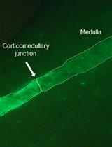
A Novel Imaging Technique for The On-site Assessment of Renal Biopsy Specimens

一种用于肾活检标本现场评估的新型成像技术

TT Tomoaki Takata
HI Hajime Isomoto
TI Takuji Iyama
KY Kentaro Yamada
1093 Views
Sep 20, 2022
When performing renal biopsy, it is necessary to identify the cortex, where glomeruli are exclusively distributed, to ensure the quality of the specimen for histological diagnosis. However, conventional methods using a stereomicroscope or magnifying lens often fail to clarify the quality of the specimen. We have established a fluorescent-based imaging technique for the on-site assessment of renal biopsy specimens. The fluorescent images can be easily obtained by adding an optical filter to the microscope and with a short incubation of an activatable fluorescent probe. This novel imaging technique can be applied to renal biopsy specimens for distinguishing the renal cortex.
人谷氨酰胺酶 C 与变构抑制剂复合物的系列室温晶体加载、数据收集和数据处理

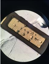
Serial Room Temperature Crystal Loading, Data Collection, and Data Processing of Human Glutaminase C in Complex with Allosteric Inhibitors

人谷氨酰胺酶 C 与变构抑制剂复合物的系列室温晶体加载、数据收集和数据处理

SM Shawn K. Milano
DS D. Marian Szebenyi
RC Richard A. Cerione
920 Views
Sep 20, 2022
Cancer cells often overexpress glutaminase enzymes, in particular glutaminase C (GAC). GAC resides in the mitochondria and catalyzes the hydrolysis of glutamine to glutamate. High levels of GAC have been observed in aggressive cancers and the inhibition of its enzymatic activity has been shown to reduce their growth and survival. Numerous GAC inhibitors have been reported, the most heavily investigated being a class of compounds derived from the small molecule BPTES (bis-2-(5-phenylacetamido-1,3,4-thiadiazol-2-yl)ethyl sulfide). X-ray structure determination under cryo-cooled conditions showed that the binding contacts for the different inhibitors were largely conserved despite their varying potencies. However, using the emerging technique serial room temperature crystallography, we were able to observe clear differences between the binding conformations of inhibitors. Here, we describe a step-by-step protocol for crystal handling, data collection, and data processing of GAC in complex with allosteric inhibitors using serial room temperature crystallography.Graphical abstract: Workflow for serial room temperature crystallography. Diagram showing the processing and scaling routine for crystals analyzed using serial room temperature crystallography.

细胞生物学

人体皮肤外植体制备和培养

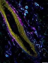
Human Skin Explant Preparation and Culture

人体皮肤外植体制备和培养

JS Jessica L. Shannon
SK Stephen J. Kirchner
JZ Jennifer Y. Zhang
2718 Views
Sep 20, 2022
The ex vivo experimentation with surgically discarded human skin represents a unique methodology amenable for mechanism and pharmacologic agent studies without the involvement of human subjects. Here, we describe a protocol that includes preparation, culture, and stimulation of human skin explants, and subsequent analyses by quantitative reverse transcription PCR and immunostaining. This protocol may also be applied for ex vivo studies of murine skin, reducing animal numbers and potentially harmful treatments. In our hands, this protocol has been used for wound healing, viral infection, and hair growth–related studies.Graphical abstract: Cartoon of explant skin culture. Skin explant sits on top of a gelatin surgical sponge saturated with culture medium at an air–liquid interface.

微生物学

裂殖酵母体内合成的脂联寡糖分析

Analysis of Lipid-linked Oligosaccharides Synthesized in vivo in Schizosaccharomyces pombe

裂殖酵母体内合成的脂联寡糖分析

AV Ayelen Valko
GG Giovanna L. Gallo
AW Ariel D. Weisz
AP Armando J. Parodi
CD Cecilia D’Alessio
1243 Views
Sep 20, 2022
Dolichol diphosphate-linked oligosaccharides (LLO) are the sugar donors in N-glycosylation, a fundamental protein post-translational modification of the eukaryotic secretory pathway. Defects in LLO biosynthesis produce human Congenital Disorders of Glycosylation Type I. The synthesis of LLOs and the transfer reactions to their protein acceptors is highly conserved among animal, plant, and fungi kingdoms, making the fission yeast Schizosaccharomyces pombe a suitable model to study these processes. Here, we present a protocol to determine the LLO patterns produced in vivo by S. pombe cells that may be easily adapted to other cell types. First, exponentially growing cultures are labeled with a pulse of [14C]-glucose. LLOs are then purified by successive extractions with organic solvents, and glycans are separated from the lipid moieties in mild acid hydrolysis and a new solvent extraction. The purified glycans are then run on paper chromatography. We use a deconvolution process to adjust the profile obtained to the minimal number of Gaussian functions needed to fit the data and determine the proportion of each species with respect to total glycan species present in the cell. The method we provide here might be used without any expensive or specialized equipment. The deconvolution process described here might also be useful to analyze species in non-completely resolved chromatograms. Graphical abstract: Workflow for the labeling, extraction, separation, and identification of LLO species in S. pombe. (A) Radioactive pulse of S. pombe cells with [14C]-glucose for 15 min at 28 °C. (B) Organic extraction of LLOs from labeled yeasts sequentially using methanol, chloroform, H2O, chloroform:methanol:H2O (1:1:0.3), 0.02 M HCl (to separate glycans from dolichol), and chloroform:methanol:H2O (1:16:16). (C) Preparation of the sample for chromatography on paper: drying by airflow and radioactivity check. (D) Loading of samples in chromatographic paper and descendent chromatography in a glass chamber. The obtained plots (CPM versus running distance) need to be analyzed to identify single glycan species.

神经科学

通过大鼠的味觉反应性测试来描述与内痛和恶心配对的味道的快乐反应

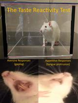
Characterizing Hedonic Responses to Flavors Paired with Internal Pain and Nausea through the Taste Reactivity Test in Rats

通过大鼠的味觉反应性测试来描述与内痛和恶心配对的味道的快乐反应

ML Matías López
DD Dominic M. Dwyer
PG Patricia Gasalla
CJ Claudia Jove
AB Azucena Begega
1551 Views
Sep 20, 2022
Feeding behavior is a complex experience that involves not only sensory (i.e., visual, odor, taste, or texture) but also affective or emotional aspects (i.e., pleasure, palatability, or hedonic value) of foods. As such, behavioral tests that assess the hedonic impact of foods are necessary to fully understand the factors involved in ingestive behavior. In this protocol, we use the taste reactivity (TR) test to characterize the hedonic responses of rats to flavors paired with either lithium chloride–induced nausea or internal pain produced by hypertonic NaCl, two treatments that reduce voluntary consumption. This application of the TR test demonstrates how emetic and non-emetic (somatic pain in particular) treatments produce dissociable patterns of hedonic reactions to fluids: only emetic treatments result in the production of aversive orofacial responses, reflecting conditioned nausea, whereas somatic pain produces immobility, reflecting conditioned fear. Other methods, such as the microstructural analysis of licking behavior, do not reliably distinguish conditioned nausea and fear, a key advantage of the more selective TR procedure. This protocol also contains guidance for adaptation to other species and designs.
用重组无磷酸酶进行原位去磷酸化测定

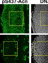
In situ Dephosphorylation Assay with Recombinant Nil Phosphatase

用重组无磷酸酶进行原位去磷酸化测定

NN Nilay Nandi
CT Charles Tracy
HK Helmut Krämer
1473 Views
Sep 20, 2022
The activity of numerous autophagy-related proteins depends on their phosphorylation status, which places importance on understanding the responsible kinases and phosphatases. Great progress has been made in identifying kinases regulating autophagy, but much less is known about the phosphatases counteracting their function. Genetic screens and modern proteomic approaches provide powerful tools to identify candidate phosphatases, but further experiments are required to assign direct roles for candidates. We have devised a novel protocol to test the role of purified phosphatases in dephosphorylating specific targets in situ. This approach has the potential to visualize context-specific differences in target dephosphorylation that are not easily detected by lysate-based approaches such as Western blots.Graphical abstract:

植物科学

拟南芥叶肉原生质体中的14C-百草枯外排试验

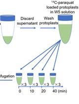
14C-paraquat Efflux Assay in Arabidopsis Mesophyll Protoplasts

拟南芥叶肉原生质体中的14C-百草枯外排试验

JX Jin-Qiu Xia
QL Qian-Qian Liu
CX Cheng-Bin Xiang
1063 Views
Sep 20, 2022
Weeds compete with crops for growth resources, causing tremendous yield losses. Paraquat is one of the three most common non-selective herbicides. To study the mechanisms of paraquat resistance, we need to trace the movement of paraquat in plants and within the cell. 14C is a radioactive carbon isotope widely used to trace substances of interest in various biological studies, especially in transport analyses. Here, we describe a detailed protocol using 14C-paraquat to demonstrate paraquat efflux in Arabidopsis protoplasts.