往期刊物2022

卷册: 12, 期号: 14

left

Dec

20

Dec

5

Nov

20

Nov

5

Oct

20

Oct

5

Sep

20

Sep

5

Aug

20

Aug

5

Jul

20

Jul

5

Jun

20

Jun

5

May

20

May

5

Apr

20

Apr

5

Mar

20

Mar

5

Feb

20

Feb

5

Jan

20

Jan

5

right

细胞生物学

小鼠室管膜纤毛干的纯化和免疫染色

Purification and Immunostaining of Mouse Ependymal Ciliary Shafts

小鼠室管膜纤毛干的纯化和免疫染色

KH Kai Hao
XZ Xueliang Zhu
Xiumin  Yan Xiumin Yan
2496 Views
Jul 20, 2022
Cilia and flagella are microtubule-based hair-like organelles protruding from the surface of most eukaryotic cells, and play essential roles in cell locomotion, left-right asymmetry, embryo development, and tissue homeostasis. With isolated cilia and flagella, great progress has been made in understanding the composition, structure, and function of cilia. However, the current cilia/flagella isolation methods are deficient in the integrity or productivity of purified cilia when applied to mammalian motile cilia. Here, we describe a new protocol that isolates cilia shafts from mouse ependymal cells, by horizontal shear force and mild detergent. This method enables the production of virtually integral cilia with high yields and less cell body contamination. It is suitable for immunostaining, puromycin labeling assay, and proximity ligation assay of mammalian motile cilia.Graphical abstract:

发育生物学

具有亚细胞分辨率的 3D 细胞模型的荧光成像

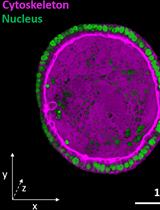
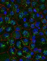
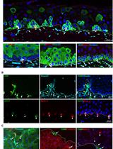
Fluorescence Imaging of 3D Cell Models with Subcellular Resolution

具有亚细胞分辨率的 3D 细胞模型的荧光成像

IV Indra Van Zundert
NM Nina Maenhoudt
SD Silke De Vriendt
HV Hugo Vankelecom
BF Beatrice Fortuni
SR Susana Rocha
4312 Views
Jul 20, 2022
Over the past years, research has made impressive breakthroughs towards the development and implementation of 3D cell models for a wide range of applications, such as drug development and testing, organogenesis, cancer biology, and personalized medicine. Opposed to 2D cell monolayer culture systems, advanced 3D cell models better represent the in vivo physiology. However, for these models to deliver scientific insights, appropriate investigation techniques are required. Despite the potential of fluorescence microscopy to visualize these models with high spatial resolution, sample preparation and imaging assays are not straightforward. Here, we provide different protocols of sample preparation for fluorescence imaging, for both matrix-embedded and matrix-free models (e.g., organoids and spheroids, respectively). Additionally, we provide detailed guidelines for imaging 3D cell models via confocal multi-photon fluorescence microscopy. We show that using these protocols, images of 3D cell culture systems can be obtained with sub-cellular resolution.Graphical abstract:

免疫学

B6/lpr小鼠肝脏、肺、胰腺和结肠中免疫细胞和与体内施用的抗体结合的细胞的免疫组织化学

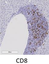
Immunohistochemistry of Immune Cells and Cells Bound to in vivo Administered Antibodies in Liver, Lung, Pancreas, and Colon of B6/lpr Mice

B6/lpr小鼠肝脏、肺、胰腺和结肠中免疫细胞和与体内施用的抗体结合的细胞的免疫组织化学

KA Kieran Adam
AM Adam Mor
2956 Views
Jul 20, 2022
Employing a novel mouse model of immune related adverse events (irAEs) induced by combination of anti-PD1 and anti-CTLA-4 antibodies, we visualized immune infiltration into the liver, lung, pancreas, and colon. Here, we describe the avidin-biotin conjugate (ABC) method used to stain T cells (CD4 and CD8), B cells (CD19), macrophages (F4/80), and cells bound by the in vivo administered rat anti-mouse antibodies for chromogenic immunohistochemistry (IHC). Using a biotinylated goat anti-rat antibody, we detected the localization of cells bound to the in vivo antibodies for PD-1 and CTLA-4. IHC has advantages over other techniques, namely antibody availability, resistance to photobleaching, and greater sensitivity. Additionally, detection and localization of in vivo antibodies can be used in mice models to infer their therapeutic efficacy, stability, and function.Graphical abstract:

微生物学

细菌素的基因组辅助鉴定、纯化和表征

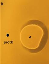
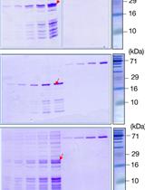
Genome-assisted Identification, Purification, and Characterization of Bacteriocins

细菌素的基因组辅助鉴定、纯化和表征

KO Kirill V. Ovchinnikov
TO Thomas F. Oftedal
SR Sebastian J. Reich
NB Nadav S. Bar
HH Helge Holo
MS Morten Skaugen
CR Christian U. Riedel
DD Dzung B. Diep
3008 Views
Jul 20, 2022
Bacteriocins are antimicrobial peptides with activity against antibiotic resistant bacterial pathogens. Here, we describe a set of methods aimed at purifying, identifying, and characterizing new bacteriocins. The purification consists of ammonium sulphate precipitation, cation-exchange chromatography, and reversed-phase chromatography. The yield of the bacteriocin is quantified by bacteriocin antimicrobial activity in a microtiter plate assay after each purification step. The mass of the purified bacteriocin is assessed by MALDI TOF MS analysis of the active fractions after reversed-phase chromatography. The mass is compared with the theoretical mass based on genetic information from the whole genome sequencing of the bacteriocin producer strain. Physicochemical characterization is performed by assessing antimicrobial activity following heat and protease treatments. Fluorescent techniques are used to examine the capacity of the bacteriocin to disrupt membrane integrity. Herein a set of protocols for purification and characterization of the bacteriocin nisin Z is used as a typical example in this paper.
使用微尺度热泳 (MST) 对噬菌体 Mu DNA 修饰蛋白Mom的结合亲和力定量

Binding Affinity Quantifications of the Bacteriophage Mu DNA Modification Protein Mom Using Microscale Thermophoresis (MST)

使用微尺度热泳 (MST) 对噬菌体 Mu DNA 修饰蛋白Mom的结合亲和力定量

SU Shubha Udupa
VN Valakunja Nagaraja
SK Shweta Karambelkar
2866 Views
Jul 20, 2022
Epigenetic modifications play diverse roles in biological systems. Nucleic acid modifications control gene expression, protein synthesis, and sensitivity to nucleic acid-cleaving enzymes. However, the mechanisms underlying the biosynthesis of nucleic acid modifications can be challenging to identify. Studying protein-ligand interactions helps decipher biosynthetic and regulatory pathways underlying biological reactions. Here, we describe a fluorescence labeling-based quantitative method for unraveling the biomolecular interactions of bacteriophage Mu DNA modification protein Mom with its ligands, using microscale thermophoresis (MST). Compared to traditional methods for studying protein-biomolecular interactions, MST requires significantly lower sample amounts, volumes, and analysis time, thus allowing screening of a large number of candidates for interaction with a protein of interest. Another distinguishing feature of the method is that it obviates the need for protein purification, often a time- and resource-consuming step, and works well with whole or partially purified cell extracts. Importantly, the method is sensitive over a broad range of molecular affinities while offering great specificity and can be used to interrogate ligands ranging from metal ions to macromolecules. Although we established this method for a DNA modification protein, it can easily be adapted to study a variety of molecular interactions engaged by proteins.
一个在不受干扰的营养素耗尽期间跟踪单个细胞动力学的微流体平台

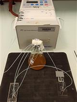
A Microfluidic Platform for Tracking Individual Cell Dynamics during an Unperturbed Nutrients Exhaustion

一个在不受干扰的营养素耗尽期间跟踪单个细胞动力学的微流体平台

TA Théo Aspert
BJ Basile Jacquel
gc Gilles Charvin
2272 Views
Jul 20, 2022
Microorganisms have evolved adaptive strategies to respond to the autonomous degradation of their environment. Indeed, a growing culture progressively exhausts nutrients from its media and modifies its composition. Yet, how single cells react to these modifications remains difficult to study since it requires population-scale growth experiments to allow cell proliferation to have a collective impact on the environment, while monitoring the same individuals exposed to this environment for days. For this purpose, we have previously described an integrated microfluidic pipeline, based on continuous separation of the cells from the media and subsequent perfusion of the filtered media in an observation chamber containing isolated single cells. Here, we provide a detailed protocol to implement this methodology, including the setting up of the microfluidic system and the processing of timelapse images.

神经科学

秀丽隐杆线虫化学感觉神经元衰老相关神经退行性变分析

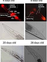
Analysis of Caenorhabditis elegans Aging-related Neurodegeneration in Chemosensory Neurons

秀丽隐杆线虫化学感觉神经元衰老相关神经退行性变分析

CC Cira Crespo
RG Roberto Grau
1990 Views
Jul 20, 2022
Aging and neuronal deterioration constitute important risk factors for the development of neuronal-related diseases, such as different dementia. The nematode Caenorhabditis elegans has emerged as a popular model system for studying neurodegeneration diseases, due to its complete neuronal connectivity map. DiI is a red fluorescent dye that can fill the worm amphid neurons and enables the visualization of their neurodegeneration over time. This protocol provides an efficient, fast, and safe method to stain worm amphid neurons to highlight the chemosensory structures of live nematodes.
最大化从牛眼中提取的视网膜中的杆外段产量

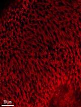
Maximizing the Rod Outer Segment Yield in Retinas Extracted from Cattle Eyes

最大化从牛眼中提取的视网膜中的杆外段产量

IP Isabella Panfoli
DC Daniela Calzia
SR Silvia Ravera
PB Paolo Bianchini
AD Alberto Diaspro
1614 Views
Jul 20, 2022
The retina is a thin neuronal multilayer responsible for the detection of visual information. The first step in visual transduction occurs in the photoreceptor outer segment. The studies on photoreception and visual biochemistry have often utilized rod outer segments (OS) or OS disks purified from mammalian eyes. Literature reports several OS and disk purification procedures that rarely specify the procedure utilized to collect the retina from the eye. Some reports suggest the use of scissors, while others do not mention the issue as they declare to utilize frozen retinas. Because the OS are deeply embedded in the retinal pigmented epithelium (RPE), the detachment of the retina by a harsh pull-out can cause the fracture of the photoreceptor cilium. Here, we present a protocol maximizing OS yield. Eye semi-cups, obtained by hemisecting the eyeball and discarding the anterior chamber structures and the vitreous, are filled with Mammalian Ringer. After 10–15 min of incubation, the retinas spontaneously detach with their wealth of OS almost intact. The impressive ability of the present protocol to minimize the number of OS stuck inside the RPE, and therefore lost, compared with the classic procedure, is shown by confocal laser scanning microscopy analysis of samples stained ex vivo with a dye (MitoTracker deep red) that stains both retinal mitochondria and OS. Total protein assay of OS disks purified by either procedure also shows a 300% total protein yield improvement. The advantage of the protocol presented is its higher yield of photoreceptor OS for subsequent purification procedures, while maintaining the physiological features of the retina.

干细胞

使用多孔 SYBR Green 定量 PCR 阵列在干细胞衍生的皮质神经元培养物中进行基因表达分析

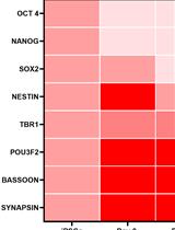
Gene Expression Analysis in Stem Cell-derived Cortical Neuronal Cultures Using Multi-well SYBR Green Quantitative PCR Arrays

使用多孔 SYBR Green 定量 PCR 阵列在干细胞衍生的皮质神经元培养物中进行基因表达分析

VS Vasavi Nallur Srinivasaraghavan
FZ Faria Zafar
BS Birgitt Schüle
2584 Views
Jul 20, 2022
To optimize differentiation protocols for stem cell-based in vitro modeling applications, it is essential to assess the change in gene expression during the differentiation process. This allows controlling its differentiation efficiency into the target cell types. While RNA transcriptomics provides detail at a larger scale, timing and cost are prohibitive to include such analyses in the optimization process. In contrast, expression analysis of individual genes is cumbersome and lengthy.Here, we developed a versatile and cost-efficient SYBR Green array of 27 markers along with two housekeeping genes to quickly screen for differentiation efficiency of human induced pluripotent stem cells (iPSCs) into excitatory cortical neurons. We first identified relevant pluripotency, neuroprogenitor, and neuronal markers for the array by literature search, and designed primers with a product size of 80-120 bp length, an annealing temperature of 60°C, and minimal predicted secondary structures. We spotted combined forward and reverse primers on 96-well plates and dried them out overnight. These plates can be prepared in advance in batches and stored at room temperature until use. Next, we added the SYBR Green master mix and complementary DNA (cDNA) to the plate in triplicates, ran quantitative PCR (qPCR) on a Quantstudio 6 Flex, and analyzed results with QuantStudio software.We compared the expression of genes for pluripotency, neuroprogenitor cells, cortical neurons, and synaptic markers in a 96-well format at four different time points during the cortical differentiation. We found a sharp reduction of pluripotency genes within the first three days of pre-differentiation and a steady increase of neuronal markers and synaptic markers over time. In summary, we built a gene expression array that is customizable, fast, medium-throughput, and cost-efficient, ideally suited for optimization of differentiation protocols for stem cell-based in vitro modeling.
边缘间充质基质细胞的分离和离体扩增

Isolation and ex vivo Expansion of Limbal Mesenchymal Stromal Cells

边缘间充质基质细胞的分离和离体扩增

NP Naresh Polisetti
LS Lyne Sharaf
TR Thomas Reinhard
GS Günther Schlunck
2202 Views
Jul 20, 2022
Limbal mesenchymal stromal cells (LMSC), a cellular component of the limbal stem cell niche, have the capability of determining the fate of limbal epithelial progenitor cells (LEPC), which are responsible for the homeostasis of corneal epithelium. However, the isolation of these LMSC has proven to be difficult due to the small fraction of LMSC in the total limbal population, and primary cultures are always hampered by contamination with other cell types. We recently published the efficient isolation and functional characterization of LMSC from the human corneal limbus using CD90 as a selective marker. We observed that flow sorting yielded a pure population of LMSC with superior self-renewal capacity and transdifferentiation potential, and supported the maintenance of the LEPC phenotype. Here, we describe an optimized protocol for the isolation of LMSC from cadaveric corneal limbal tissue by combined collagenase digestion and flow sorting with expansion of LMSC on plastic.Graphical abstract: