往期刊物2015

卷册: 5, 期号: 18

left

Dec

20

Dec

5

Nov

20

Nov

5

Oct

20

Oct

5

Sep

20

Sep

5

Aug

20

Aug

5

Jul

20

Jul

5

Jun

20

Jun

5

May

20

May

5

Apr

20

Apr

5

Mar

20

Mar

5

Feb

20

Feb

5

Jan

20

Jan

5

right

生物化学

吡啶血色原法测定纯化蛋白溶液中血红素的浓度

Pyridine Hemochromagen Assay for Determining the Concentration of Heme in Purified Protein Solutions

吡啶血色原法测定纯化蛋白溶液中血红素的浓度

Ian Barr Ian Barr
Feng Guo Feng Guo
32329 Views
Sep 20, 2015
Heme is a common cofactor in proteins, found in hemoglobin, myoglobin, cytochrome P450, DGCR8, and nitric oxide synthase, among others. This protocol describes a method for quantifying heme that works best in purified protein samples. This protocol might be used to, for example, determine whether a given heme-binding protein is fully occupied by heme, thus allowing correlation of heme content with activity. This requires the absolute heme concentration and an accurate protein concentration. Another use is to determine the extinction coefficients of a heme-bound protein. This assay is fast, easy, and reproducible if done correctly.

细胞生物学

采用Oxymax 开放环路间接能量测定仪测量小鼠中的静息能量新陈代谢

Measurement of Resting Energy Metabolism in Mice Using Oxymax Open Circuit Indirect Calorimeter

采用Oxymax 开放环路间接能量测定仪测量小鼠中的静息能量新陈代谢

Yaohui Nie Yaohui Nie
TG Timothy P. Gavin
SK Shihuan Kuang
10885 Views
Sep 20, 2015
Indirect calorimeter is a powerful tool to monitor resting energy metabolism through the measurement of oxygen (O2) consumption and carbon dioxide (CO2) production. From the measurement of VO2 and VCO2, the respiratory exchange ratio (RER) can be calculated to assess energy fuel utilization and energy expenditure (Evan et al., 2012). Previously, indirect calorimeter has been widely used in metabolic disease research in mice to reveal the potential roles of specific genes or treatments in regulating energy metabolism (for example: Bi et al., 2014; Feng et al., 2014). Here, we described a protocol to evaluate the resting energy metabolism of C57BL/6 mice during dark and light cycles using the Oxymax Open Circuit indirect calorimeter.
串行块面扫描电子显微成像检测细胞核和线粒体的形态特征

Morphological Quantification of Nuclei and Mitochondria in Serial Block-face Scanning Electron Microscopy Images

串行块面扫描电子显微成像检测细胞核和线粒体的形态特征

HL Haiyan Lu
NO Nobuhiko Ohno
RR Richard M. Ransohoff
8967 Views
Sep 20, 2015
Serial Block-face Scanning Electron Microscopy (SBF-SEM or 3D-EM) is a powerful tool to study biological structure in ultrastructural level. Quantification of cellular ultrastructure is useful to providing biological information. This technique requires not only high quality of tissue fixation and ideal sample embedding to preserve structures, but also delicate 3D image scanning and post-processing of images. We have adapted previous method to optimize the EM technique to detect and study cellular ultrastructure. Here we present the method to embed samples for 3D-EM technique and to quantify the morphological parameters of nucleus and mitochondria. Part I. Tissue embedding for 3D-EM images

微生物学

从绿脓杆菌生物膜和浮游种群中分离持留细胞

Isolation of Persister Cells from Biofilm and Planktonic Populations of Pseudomonas aeruginosa

从绿脓杆菌生物膜和浮游种群中分离持留细胞

Cláudia N. H. Marques Cláudia N. H. Marques
12436 Views
Sep 20, 2015
Persister cells are a stochastically produced sub-population of non-growing bacterial cells. Recently these cells have been more widely studied due to the recognition that they are tolerant to antimicrobials and thus, play a major role in the resilience of bacterial populations to antimicrobials, particularly in chronic biofilm infections. The following protocol describes the isolation/selection of persister cell sub-populations of Pseudomonas aeruginosa present in biofilms (sessile) and planktonic populations (free-living).
拟南芥根部的胞囊线虫感染实验

Infection Assay of Cyst Nematodes on Arabidopsis Roots

拟南芥根部的胞囊线虫感染实验

HB Holger Bohlmann
Krzysztof Wieczorek Krzysztof Wieczorek
11640 Views
Sep 20, 2015
Plant parasitic nematodes are devastating pests on many crops. Juveniles (J2) of cyst nematodes invade the roots to induce a syncytium. This feeding site is their only source of nutrients. Male nematodes leave the roots after the fourth molt to mate with females. The females stay attached to their syncytia throughout their life and produce hundreds of eggs, which are contained in their bodies. When the females die their bodies form the cysts, which protect the eggs. Cysts can survive for many years in the soil until favorable conditions induce hatching of the juveniles. The beet cyst nematode Heterodera schachtii is a pathogen of sugar beet (Beta vulgaris) but can also complete its life cycle on Arabidopsis roots growing on agar plates under sterile conditions. We present here protocols for a stock culture of H. schachtii and an infection assay on agar plates.
生物膜呼吸活性的量化

Quantification of Respiratory Activity in Biofilms

生物膜呼吸活性的量化

Cláudia N. H. Marques Cláudia N. H. Marques
SC Scott A. Craver
10248 Views
Sep 20, 2015
Bacteria live mostly as biofilms, not as planktonic cell populations. Bacterial cells living as biofilms are known to be in different physiological status. Persister cells are one of such physiological conditions and they are recognized as to be a stochastically produced sub-population of non-growing bacterial cells. The following protocol describes a method to determine the respiratory activity of cells within biofilms.
采用原位RT-PCR法检测和显示线虫感染的拟南芥根部组织中特定基因的转录水平

Detection and Visualization of Specific Gene Transcripts by in situ RT-PCR in Nematode-Infected Arabidopsis Root Tissue

采用原位RT-PCR法检测和显示线虫感染的拟南芥根部组织中特定基因的转录水平

Krzysztof Wieczorek Krzysztof Wieczorek
9717 Views
Sep 20, 2015
This protocol describes an effective method of in situ RT-PCR that was developed to localize specific gene expression directly in thin cross-sections of nematode feeding sites induced by the cyst nematode Heterodera schachtii (H. schachtii) or the root-knot nematode Meloidogyne incognita (M. incognita) in Arabidopsis roots using DIG (Digoxigenin-11dUTP) labeling coupled with AP (alkaline phosphatase) and nitro-blue tetrazolium/5-bromo-4-chloro-3'-indolylphosphate-based detection. This method is applicable to any other Arabidopsis root tissue.

神经科学

成年小鼠背根神经节神经元的分离和生长

Isolation and Growth of Adult Mouse Dorsal Root Ganglia Neurons

成年小鼠背根神经节神经元的分离和生长

Seong-il Lee Seong-il Lee
JL Joel Levine
26788 Views
Sep 20, 2015
Adult dorsal root ganglia neurons are among the few adult neuronal cell types that can be purified and grown relatively easily in dissociated cell culture. Here we describe a procedure for the isolation and growth of dissociated adult mouse DRG neurons using Percoll gradients and a chemically defined medium. These cultures can be used for morphological, biochemical and electrophysiological studies of neuronal growth and function.
线虫趋化性和跳跃运动试验

Chemotaxis and Jumping Assays in Nematodes

线虫趋化性和跳跃运动试验

TB Tiffany Baiocchi
Adler R. Dillman Adler R. Dillman
11175 Views
Sep 20, 2015
Nematodes have sensitive olfactory perception, which is used to detect and differentiate many volatile odorants. Some odorants are attractive, others repulsive, and yet others evoke no particular response. Chemotaxis assays can be used to determine the role of certain odors in many different behaviors including foraging, predator avoidance, and mate attraction. In addition to chemotaxis, some species of nematodes in the entomopathogenic genus Steinernema can jump, which is thought to play an important role in host-seeking and dispersal (Dillman and Sternberg, 2012). Jumping and chemotaxis assays have been successfully used to identify odorants that stimulate these behaviors in a variety of nematodes (Bargmann et al., 1993; Campbell and Kaya, 1999; Hallem et al., 2011; Dillman et al., 2012; Castelletto et al., 2014). Here a detailed protocol for chemotaxis and jumping assays is provided based on the growing body of literature.

植物科学

树脂包埋制备拟南芥横切片

Transverse Sectioning of Arabidopsis thaliana Leaves Using Resin Embedding

树脂包埋制备拟南芥横切片

Shweta Kalve Shweta Kalve
KS Kumud Saini
KV Kris Vissenberg
TB Tom Beeckman
GB Gerrit T. S. Beemster
13432 Views
Sep 20, 2015
The leaf is the major functional part of the shoot performing the bulk of photosynthetic activity. Its development is relatively plastic allowing the plant to adapt to environmental changes by modifying leaf size and anatomy. Moreover, a leaf is made up of various distinct cell layers, each having specialized functions. To understand functional adaptation and the development of the leaf it is essential to obtain cross sections throughout leaf development and at maturity (Kalve et al., 2014). Here, we describe a protocol for transverse sectioning of Arabidopsis thaliana leaves using resin embedding. This protocol provides a reliable platform to yield high quality images of cross sections allowing study of development of various tissue layers across the transversal axis of the leaf. As this method is an adaptation of the protocol developed for the Arabidopsis root tip by Beeckman and Viane (1999) and De Smet et al. (2004), it can easily be modified to accommodate other organs and species.
通过添加内标的超高效液相色谱法对植物组织中的脱落酸水平进行组织化学定量测定

Physicochemical Quantification of Abscisic Acid Levels in Plant Tissues with an Added Internal Standard by Ultra-Performance Liquid Chromatography

通过添加内标的超高效液相色谱法对植物组织中的脱落酸水平进行组织化学定量测定

Scott A M McAdam Scott A M McAdam
12751 Views
Sep 20, 2015
The phytohormone abscisic acid (ABA) is critical for a range of plant responses to the environment, most importantly in closing the stomata of seed plants during drought (Mittelheuser and Van Steveninck 1969; Brodribb et al. 2014). The high precision quantification of this hormone by physicochemical methods is a relatively simple process, essential for studies that aim to investigate the role or action of this hormone in plants. Outlined here is a method for the extraction, purification and quantification of ABA levels in plant tissues. This method involves methanolic extraction of ABA from homogenised tissue. Purification of ABA is then undertaken by a simple etheric partitioning method. A focus is placed on determining ABA levels in leaves; however this method is suitable for all tissue types, including plant solutes such as xylem sap, seeds (both dry and green) and large samples of tissue such as root systems.
对转基因植株样本中的农杆菌上T-DNA基因的cDNA 5'末端进行快速扩增(5'RACE)

5’ Rapid Amplification of cDNA Ends (5’ RACE) of Agrobacterial T-DNA Genes within Transformed Plant Sample

对转基因植株样本中的农杆菌上T-DNA基因的cDNA 5'末端进行快速扩增(5'RACE)

YZ Yi Zhang
12178 Views
Sep 20, 2015
The T-DNA (transferred-DNA) region of virulent Agrobacterium tumefaciens (A. tumefaciens) strain is transferred and integrated into the plant genome, and thereby the T-DNA genes are expressed in transformed plant cells. This protocol was used to analyze the transcription start sites (TSSs) of agrobacterial T-DNA genes within plant crown gall tumor. Firstly, the stems of Arabidopsis thaliana were inoculated by A. tumefaciens strain C58 and developed crown gall tumor. Subsequently, the mRNA was extracted from the crown gall tumor and then used for amplification of 5’ cDNA ends by 5’ Rapid Amplification of cDNA Ends (5’ RACE) assay. The full-length cDNAs were generated in reverse transcription reactions and used to analyze TSSs. Here, TSSs of three oncogenes, IaaH, IaaM and Ipt were analyzed as examples. This protocol also allows for identification of TSSs of the other agrobacterial T-DNA genes that expressed in plant cells.
外源化学物质处理后拟南芥原生质体中促分裂原活化蛋白激酶(MAPK)的检测

Detection of Mitogen-activated Protein Kinase (MAPK) Activation upon Exogenous Chemical Application in Arabidopsis Protoplasts

外源化学物质处理后拟南芥原生质体中促分裂原活化蛋白激酶(MAPK)的检测

Arsheed Hussain Sheikh Arsheed Hussain Sheikh
Alok Krishna Sinha Alok Krishna Sinha
11696 Views
Sep 20, 2015
The mitogen activated protein kinase cascade is a highly conserved signal pathway in plants. The exogenous chemicals, like hormones, can trigger a series of signalling cascades, including MAPK pathway, to modulate the plant physiology. Upon activation, some MAPKs are phosphorylated. It is important to develop methods that can detect changes in the phosphorylation status of MAPKs in plants when they come in contact with external chemicals. This method describes the exogenous treatment of Arabidopsis protoplasts with Kinetin and subsequent detection of the activated MAPKs. This method is useful for studying the effect of exogenously applied chemical compounds on the MAPK signaling cascade in Arabidopsis.
采用 EdU测定叶片细胞增殖区

Detection of the Cell Proliferation Zone in Leaves by Using EdU

采用 EdU测定叶片细胞增殖区

Hokuto Nakayama Hokuto Nakayama
KK Kensuke Kawade
Hirokazu Tsukaya Hirokazu Tsukaya
Seisuke Kimura Seisuke Kimura
8821 Views
Sep 20, 2015
Visualization of nuclei in S-phase cells in tissues is important for not only cell cycle research but also developmental research because morphogenesis is usually achieved by a combination of cell proliferation and cell expansion. Recently, DNA labeling with 5-ethynyl-2′-deoxyuridine (EdU), which is an analog of thymidine, has been used to visualize nuclei in S-phase cells to assess the activity of cell proliferation during development of plants. EdU is efficiently incorporated into newly synthesized DNA, and detection of EdU is based on the covalent reaction between EdU and Alexa Fluor® dye, which is one of useful fluorescent dyes; this allows us to use mild conditions for the assay without any DNA denaturation. This method could be easily applicable, and, indeed, has been used for various model and non-model plant species. Here, we have described a protocol developed for the detection of nuclei in S-phase cells in leaves.
通过农杆菌或粒子轰击法截断端粒介导染色体在植物中产生工程微型染色体

Telomere-mediated Chromosomal Truncation via Agrobacterium tumefaciens or Particle Bombardment to Produce Engineered Minichromosomes in Plants

通过农杆菌或粒子轰击法截断端粒介导染色体在植物中产生工程微型染色体

NG Nathaniel D. Graham
NS Nathan C. Swyers
RG Robert T. Gaeta
CZ Changzeng Zhao
JC Jon P. Cody
JB James A. Birchler
8612 Views
Sep 20, 2015
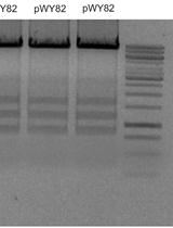
Minichromosomes are small, autonomously functioning chromosomes that exist separately from the normal chromosomal set. Creation of minichromosomes in plants relies on telomere truncation to remove the chromosome arms and the native telomere sequence and replace them with a transgene together with a new telomere sequence to generate a modifiable small chromosome. Telomere truncation has been accomplished utilizing both Agrobacterium tumefaciens, in which a telomere repeat sequence is cloned into the transformation vector near the right border, and particle bombardment, in which the genes of interest and telomere sequence are co-introduced into the plant. In this protocol we will describe the methods for introducing telomere sequences to both Agrobacterium and gold particles, as well as the methods required to verify that these sequences are intact. [Introduction] Engineered minichromosomes are autonomously functioning chromosomes that contain all of the necessary components required for maintenance through the cell cycle. The production of engineered minichromosomes has several potential applications for the next generation of genetic engineering (Gaeta et al., 2012). The construction of such chromosomes by assembling a centromere, origin of replication, and selectable marker all capped by telomere sequences, as originally performed in yeast, is not feasible in plants because of the epigenetic nature of centromere sequences (Birchler and Han, 2009; Birchler et al., 2011; Liu et al., 2015). In other words, functional centromeres in plants are determined by chromatin features independent of the underlying DNA and therefore the cloning and re-introduction of centromere sequences will not produce a minichromosome. In contrast to the centromere, the telomere is reliant on sequence, with most plant telomeres containing the same TTTAGGG repeat (Adams et al., 2001). As a result, introduction of telomere sequences during transformation has the potential to confer telomere function. Because of the epigenetic state of the centromere, engineered minichromosomes in plants need to be produced by cleaving away the chromosome arms from an endogenous centromere that never leaves a cell, a procedure known as the top-down method. This was accomplished with the finding that introduction of the chromosome end sequences, the telomere, would cleave chromosomes at the site of integration (Yu et al., 2006). By including genes of interest in addition to the telomere sequences, the foundation to build engineered minichromosomes to specification was established. This protocol describes the procedure to generate these initial truncated minichromosomes. The process of creating minichromosomes utilizes standard plant transformation protocols. The only modification is the addition of telomere sequences to the transformation construction so that both a transgene and telomere sequence are introduced into a double stranded break during the transformation process. In some cases, the introduced telomere sequence is recognized by telomere elongation machinery and converted into a functioning telomere. As a result, the acentric fragment distal to the insertion point will be lost, and a minichromosome will be created. Telomere truncation works well with both Agrobacterium tumefaciens and particle bombardment transformation techniques. Using Agrobacterium, successful minichromosome creation relies on inclusion of telomere sequences in the transformation construct. With particle bombardment, telomere sequences are simply added to the DNA mixture that is adhered to the gold beads before transformation is performed. While the length of telomere sequence required for telomere truncation is not known, a study in Arabidopsis thaliana successfully created truncated chromosomes with telomere repeats as short as 100 bp (Nelson et al., 2011). The study also found, however, that longer telomere sequences were more likely to induce truncation events. As a result, it is suggested that the largest amount of telomere that can be reasonably obtained be used during transformation. While the concept of including telomere sequences in transformation is relatively simple, working with telomere sequences using current molecular cloning techniques is challenging. The repeated nature of the sequences and the high GC content are inhibitive to polymerase function. As a result, protocols reliant on polymerase function, such as PCR or Sanger sequencing, are not efficient. Additionally, oligonucleotide synthesis technologies are limited for producing telomere repeat sequences at the time of this writing. Traditional cloning utilizing restriction enzymes has been most successful in our work. Isolated telomere sequences, when subjected to agarose gels, do not migrate at the expected sizes, but instead are found as discreet bands or smears throughout the gel, probably because they adopt various secondary structures. Additionally, purification of telomere sequences with gel or column purification is usually inefficient unless the DNA is present in large amounts, making traditional cloning difficult. Adding to these challenges is the observation that long telomere sequences are unstable in microbial cells, and have a tendency to be deleted and shortened over time. As a result, Stbl cells (Invitrogen), which possess the recA1 genotype and are specifically designed to prevent repeated sequences from recombination and thus rearrangement, must be used to maintain the repeat, and multiple clones should be isolated and screened to ensure the full size is present. Additionally, when a clone has been isolated, which contains the desired telomere insert, it is often useful to make a large plasmid extraction that is stored in addition to bacterial stocks. In order to generate engineered minichromosomes, the protocols presented below were developed. For cloning purposes, the telomere sequence is excised from a gel and ligated to the target plasmid within the agarose mixture. The source of the telomere sequence is plasmid pWY82 (Yu et al., 2006), which contains 2.6 kb of the telomere repeat (TTTAGGG). For particle bombardment, primers are used with a modified PCR program to amplify the telomere repeats, which are gel purified and added to gold particles together with the construct of interest. Whether truncation will be performed with Agrobacterium or particle bombardment, the standard transformation protocol for the species of interest can be followed. Fluorescence in-situ hybridization is then performed to determine if a minichromosome has successfully been produced (Yu et al., 2007).
采用14C 葡萄糖细胞壁掺入实验估测纤维素的生物合成

[14C] Glucose Cell Wall Incorporation Assay for the Estimation of Cellulose Biosynthesis

采用14C 葡萄糖细胞壁掺入实验估测纤维素的生物合成

CB Chad Brabham
JS Jozsef Stork
SD Seth Debolt
6919 Views
Sep 20, 2015
Cellulose is synthesized by Cellulose Synthase A proteins at the plasma membrane using the substrate UDP glucose. Herein, we provide a detailed method for measuring the incorporation of radiolabeled glucose into the cellulose fraction of the cell wall. In this method Arabidopsis seedlings are treated for 2 h with a cellulose biosynthesis inhibitor in the presence of radiolabeled glucose, and are subsequently boiled in acetic-nitric acid to solubilize non-cellulosic material. The radiolabeled glucose detected in the insoluble fraction indicates the amount of cellulose synthesized during the experimental timeframe. The short-term nature of this method is a useful tool in determining if inhibition of cellulose biosynthesis is the herbicides primary mode of action.