往期刊物2022

卷册: 12, 期号: 6

left

Dec

20

Dec

5

Nov

20

Nov

5

Oct

20

Oct

5

Sep

20

Sep

5

Aug

20

Aug

5

Jul

20

Jul

5

Jun

20

Jun

5

May

20

May

5

Apr

20

Apr

5

Mar

20

Mar

5

Feb

20

Feb

5

Jan

20

Jan

5

right

生物化学

将人类细胞中的天然表达与基于内含肽的亲和纯化和自切割标签相结合的新型蛋白质生产方法

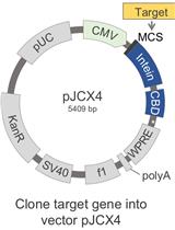
Novel Protein Production Method Combining Native Expression in Human Cells with an Intein-based Affinity Purification and Self-cleavable Tag

将人类细胞中的天然表达与基于内含肽的亲和纯化和自切割标签相结合的新型蛋白质生产方法

PC Peter J. Carman
RD Roberto Dominguez
1959 Views
Mar 20, 2022
The human proteins used in most biochemical studies are commonly obtained using bacterial expression. Owing to its relative simplicity and low cost, this approach has been extremely successful, but is inadequate for many proteins that require the mammalian folding machinery and posttranslational modifications (PTMs) for function. Moreover, the expressed proteins are typically purified using N- and/or C-terminal affinity tags, which are often left on proteins or leave non-native extra amino acids when removed proteolytically. Many proteins cannot tolerate such extra amino acids for function. Here we describe a protein production method that resolves both these issues. Our method combines expression in human Expi293F cells, which grow in suspension to high density and can process native PTMs, with a chitin-binding domain (CBD)-intein affinity purification and self-cleavable tag, which can be precisely removed after purification. In this protocol, we describe how to clone a target gene into our specifically designed human cell expression vector (pJCX4), and how to efficiently transfect the Expi293F cells and purify the expressed proteins using a chitin affinity resin.Graphic abstract:
使用薄层色谱法分析蓝藻中的异囊体和动体特异性糖脂

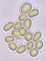
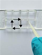
Analysis of Heterocyst and Akinete Specific Glycolipids in Cyanobacteria Using Thin-layer Chromatography

使用薄层色谱法分析蓝藻中的异囊体和动体特异性糖脂

RG Ritu Garg
RP Rebeca Perez
IM Iris Maldener
1640 Views
Mar 20, 2022
Several filamentous cyanobacteria like Nostoc differentiate specialized cells in response to changes in environmental factors, such as low light or nutrient starvation. These specialized cells are termed heterocysts and akinetes. Under conditions of nitrogen limitation, nitrogen-fixing heterocysts form in a semi-regular pattern and provide the filament with organic nitrogen compounds. Akinetes are spore-like dormant cells, which allow survival during adverse unfavorable conditions. Both cell types possess multilayered thick envelopes mainly composed of an outermost polysaccharide layer and inner layers of glycolipids, that are important for stress adaptation. To study these envelope glycolipids, a method for the isolation, separation and analysis of lipids from heterocysts and akinetes is essential. The present protocol describes a method involving the extraction of lipids from cyanobacteria using solvents and their separation and visualization on silica plates, to render analysis simple and easy. This protocol is relevant for studying mutants that are defective in glycolipid layer formation and for the comparison of glycolipid composition of heterocysts and akinetes under different environmental stresses.

生物工程

荧光法测定巨大单层囊泡中磷脂爬行酶活性的方法

A Fluorescence-based Assay for Measuring Phospholipid Scramblase Activity in Giant Unilamellar Vesicles

荧光法测定巨大单层囊泡中磷脂爬行酶活性的方法

PM Patricia P. M. Mathiassen
TP Thomas Günther Pomorski
2208 Views
Mar 20, 2022
Transbilayer movement of phospholipids in biological membranes is mediated by a diverse set of lipid transporters. Among them are scramblases that facilitate rapid bi-directional movement of lipids without metabolic energy input. In this protocol, we describe the incorporation of phospholipid scramblases into giant unilamellar vesicles (GUVs) formed from scramblase-containing large unilamellar vesicles by electroformation. We also describe how to analyze their activity using membrane-impermeant sodium dithionite, to bleach symmetrically incorporated fluorescent ATTO488-conjugated phospholipids. The fluorescence-based readout allows single vesicle tracking for a large number of settled/immobilized GUVs, and provides a well-defined experimental setup to directly characterize these lipid transporters at the molecular level.Graphic abstract: Giant unilamellar vesicles (GUVs) are formed by electroformation from large unilamellar vesicles (LUVs) containing phospholipid scramblases (purple) and trace amounts of a fluorescent lipid reporter (green). The scramblase activity is analyzed by a fluorescence-based assay of single GUVs, using the membrane-impermeant quencher dithionite. Sizes not to scale. Modified from Mathiassen et al. (2021).

生物物理学

离体血管的电生理测量

Electrophysiological Measurements of Isolated Blood Vessels

离体血管的电生理测量

SM Samuel A Molina
DM Daniela Maier-Begandt
BI Brant E Isakson
MK Michael Koval
1883 Views
Mar 20, 2022
The lumen of blood vessels is covered by endothelial cells, which regulate their permeability to ions and solutes. Endothelial permeability depends on the vascular bed and cell phenotype, and is influenced by different disease states. Most characterization of endothelial permeability has been carried out using isolated cells in culture. While analysis of cultured cells is a valuable approach, it does not account for factors of the native cell environment. Building on Ussing chamber studies of intact tissue specimens, here we describe a method to measure the electrophysiological properties of intact arteriole and venule endothelia, including transendothelial electrical resistance (TEER) and ion permselectivity. As an example, vessels isolated from the mesentery were treated ex vivo, then mounted in a custom-made tissue cassette that enable their analysis by classical approaches with an Ussing chamber. This method enables a detailed analysis of electrophysiological vessel responses to stresses such as proinflammatory cytokines, in the context of an intact vessel.Graphic abstract:

癌症生物学

基于线性复制和分裂的全基因组扩增法(LCS-WGA)分析单细胞损伤组

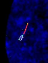
Single-cell Damagenome Profiling by Linear Copying and Splitting based Whole Genome Amplification (LCS-WGA)

基于线性复制和分裂的全基因组扩增法(LCS-WGA)分析单细胞损伤组

YN Yichi Niu
QZ Qiangyuan Zhu
CZ Chenghang Zong
2284 Views
Mar 20, 2022
Spontaneous DNA damage frequently occurs on the human genome, and it could alter gene expression by inducing mutagenesis or epigenetic changes. Therefore, it is highly desired to profile DNA damage distribution on the human genome and identify the genes that are prone to DNA damage. Here, we present a novel single-cell whole-genome amplification method which employs linear-copying followed by a split-amplification scheme, to efficiently remove amplification errors and achieve accurate detection of DNA damage in individual cells. In comparison to previous methods that measure DNA damage, our method uses a next-generation sequencing platform to detect misincorporated bases derived from spontaneous DNA damage with single-cell resolution.
使用共培养和多重 FACS 分析对原发性人类急性髓细胞白血病进行小分子筛选

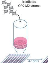
Small Molecule Screening of Primary Human Acute Myeloid Leukemia Using Co-culture and Multiplexed FACS Analysis

使用共培养和多重 FACS 分析对原发性人类急性髓细胞白血病进行小分子筛选

AB Aurelie Baudet
SH Simon Hultmark
FE Fredrik Ek
MM Mattias Magnusson
2057 Views
Mar 20, 2022
Ex vivo culture of primary acute myeloid leukemia (AML) cells is notoriously difficult due to spontaneous differentiation and cell death, which hinders mechanistic and translational studies. To overcome this bottleneck, we have implemented a co-culture system, where the OP9-M2 stromal cells support the growth, but most notably limit the differentiation of primary AML cells, thus allowing for mechanistic studies in vitro. Additionally, the co-culture on OP9-M2 stromal is superior in preserving surface marker expression of primary (adult and pediatric) AML cells in comparison to stroma-free culture. Thus, by combining the co-culture with multicolor, high-throughput FACS, we can evaluate the effect of hundreds of small molecules on multi-parametric processes including: cell survival, stemness (leukemic stem cells), and myeloid differentiation on the primary AML cells at a single-cell level. This method streamlines the identification of potential therapeutic agents, but also facilitates combinatorial screening aiming, for instance, at dissecting the regulatory pathways in a patient-specific manner.Graphic abstract: Schematic representation of the ex vivo small molecule screening of primary human acute myeloid leukemia. Irradiated, sub-confluent OP9-M2 stromal cells are plated in half-area 96 wells plates 4–16 h prior to adding primary AML cells. Compounds are added 36–48 h later and effects on cell number, leukemic stem cell population, and myeloid differentiation are quantifed by FACS after 4 days of treatment.

发育生物学

人类细胞系中心体的 3D 结构照明显微镜

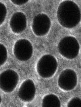
3D-Structured Illumination Microscopy of Centrosomes in Human Cell Lines

人类细胞系中心体的 3D 结构照明显微镜

KF Kari-Anne M. Frikstad
KS Kay O. Schink
SG Sania Gilani
LP Lotte B. Pedersen
SP Sebastian Patzke
2612 Views
Mar 20, 2022
The centrosome is the main microtubule-organizing center of animal cells, and is composed of two barrel-shaped microtubule-based centrioles embedded in protein dense pericentriolar material. Compositional and architectural re-organization of the centrosome drives its duplication, and enables its microtubule-organizing activity and capability to form the primary cilium, which extends from the mature (mother) centriole, as the cell exits the cell cycle. Centrosomes and primary cilia are essential to human health, signified by the causal role of centrosome- and cilia-aberrations in numerous congenic disorders, as well as in the etiology and progression of cancer. The list of disease-associated centrosomal proteins and their proximitomes is steadily expanding, emphasizing the need for high resolution mapping of such proteins to specific substructures of the organelle. Here, we provide a detailed 3D-structured illumination microscopy (3D-SIM) protocol for comparative localization analysis of fluorescently labeled proteins at the centrosome in fixed human cell lines, at approximately 120 nm lateral and 300 nm axial resolution. The procedure was optimized to work with primary antibodies previously known to depend on more disruptive fixation reagents, yet largely preserves centriole and centrosome architecture, as shown by transposing acquired images of landmark proteins on previously published transmission electron microscopy (TEM) images of centrosomes. Even more advantageously, it is compatible with fluorescent protein tags. Finally, we introduce an internal reference to ensure correct 3D channel alignment. This protocol hence enables flexible, swift, and information-rich localization and interdependence analyses of centrosomal proteins, as well as their disorder-associated mutations.
TUNEL 标记检测秀丽隐杆线虫性腺中的双链 DNA 断裂

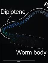
TUNEL Labeling to Detect Double-stranded DNA Breaks in Caenorhabditis elegans Gonads

TUNEL 标记检测秀丽隐杆线虫性腺中的双链 DNA 断裂

PK Peter A. Kropp
Kyle Rhodehouse Kyle Rhodehouse
AG Andy Golden
1920 Views
Mar 20, 2022
Analysis of DNA double strand breaks (DSBs) is important for understanding dyshomeostasis within the nucleus, impaired DNA repair mechanisms, and cell death. In the C. elegans germline, DSBs are important indicators of all three above-mentioned conditions. Although multiple methods exist to assess apoptosis in the germline of C. elegans, direct assessment of DSBs without the need for a reporter allele or protein-specific antibody is useful. As such, unbiased immunofluorescent approaches can be favorable. This protocol details a method for using terminal deoxynucleotidyl transferase dUTP nick end labeling (TUNEL) to assess DNA DSBs in dissected C. elegans germlines. Germlines are co-labeled with DAPI to allow for easy assessment of DNA DSBs. This approach allows for qualitative or quantitative measures of DNA DSBs.Graphic abstract: Schematic for TUNEL labeling of C. elegans germlines.
果蝇细胞化过程中使用双光子激光消融测量收缩环张力

Measurement of Contractile Ring Tension Using Two-photon Laser Ablation during Drosophila Cellularization

果蝇细胞化过程中使用双光子激光消融测量收缩环张力

SS Swati Sharma
RR Richa Rikhy
1564 Views
Mar 20, 2022
Cytokinesis occurs at the final step of cell division and leads to the separation of daughter cells. It requires assembly and constriction of the actomyosin contractile ring. The phases of assembly and constriction of the contractile ring show an increase in tension in the actomyosin complex. The measurement of tension in the contractile ring is of interest to probe the mechanics of contractile ring formation. Drosophila cellularization is a powerful genetic model system to study the mechanisms regulating actomyosin contractility during contractile ring constriction. Cellularization occurs in the interphase of syncytial cycle 14, where the plasma membrane extends around individual nuclei and forms complete cells with the help of a contractile ring at the bottom. The contractile ring forms at the furrow tip during the extension around individual nuclei and its assembly requires the coordinated action of cytoskeletal and plasma membrane-associated proteins. Laser ablation of the contractile ring enables the measurement of the contractility of the actomyosin network during cytokinesis. This protocol outlines the method used for estimating the contractility at the actomyosin ring during cellularization by laser ablation, in both control and mutant embryos for a Rho guanosine triphosphatase activating protein (RhoGAP) containing protein called GRAF (GTPase regulator associated with focal adhesion kinase-1). Physical cutting of the contractile ring by a two-photon laser at 800 nm leads to the displacement of the actomyosin ring edges, at a rate dependent upon the tension. This can be carried out at distinct steps of the contractile ring assembly during furrow extension in cellularization. Quantification of the extent of displacement and recoil velocity of movement of the edges at different stages of cellularization provides a quantitative measure of contractility in the system. This protocol describes the experimental procedure containing the preparation of live embryos, optimization of laser power, acquisition settings, and post-acquisition analysis of actomyosin contractility during Drosophila cellularization.

免疫学

人诱导多能干细胞向巨噬细胞的分化

Differentiation of Human Induced Pluripotent Stem Cell into Macrophages

人诱导多能干细胞向巨噬细胞的分化

HD Harriet Douthwaite
AA Aitor Bermejo Arteagabeitia
SM Subhankar Mukhopadhyay
3464 Views
Mar 20, 2022
As a model to interrogate human macrophage biology, macrophages differentiated from human induced pluripotent stem cells (hiPSCs) transcend other existing models by circumventing the variability seen in human monocyte-derived macrophages, whilst epitomizing macrophage phenotypic and functional characteristics over those offered by macrophage-like cell lines (Mukherjee et al., 2018). Furthermore, hiPSCs are amenable to genetic manipulation, unlike human monocyte-derived macrophages (MDMs) (van Wilgenburg et al., 2013; Lopez-Yrigoyen et al., 2020), proposing boundless opportunities for specific disease modelling.We outline an effective and efficient protocol that delivers a continual production of hiPSC-derived-macrophages (iMACs), exhibiting human macrophage surface and intracellular markers, together with functional activity.The protocol describes the resuscitation, culture, and differentiation of hiPSC into mature terminal macrophages, via the initial and intermediate steps of expansion of hiPSCs, formation into embryoid bodies (EBs), and generation of hematopoietic myeloid precursors.We offer a simplified, scalable, and adaptable technique that advances upon other protocols, utilizing feeder-free conditions and reduced growth factors, to produce high yields of consistent iMACs over a period of several months, economically.
CD8 T 细胞病毒抑制实验方法

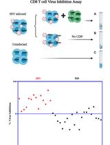
CD8 T Cell Virus Inhibition Assay Protocol

CD8 T 细胞病毒抑制实验方法

YX Yinyan Xu
AW Ann Marie Weideman
MA Maria Abad-Fernandez
KM Katie R. Mollan
SK Sallay Kallon
SS Shahryar Samir
JW Joanna A. Warren
GC Genevieve Clutton
NR Nadia Roan
AA Adaora A. Adimora
NA Nancie Archin
JK JoAnn Kuruc
CG Cindy Gay
MH Michael G. Hudgens
NG Nilu Goonetilleke
2473 Views
Mar 20, 2022
The human immunodeficiency virus (HIV)-1 viral inhibition assay (VIA) measures CD8+ T cell-mediated inhibition of HIV replication in CD4+ T cells and is increasingly used for clinical testing of HIV vaccines and immunotherapies. Different VIAs that differ in length of CD8:CD4 T cell culture periods (6–13 days), purity of CD4 cultures [isolated CD4+ T cells or CD8+ depleted peripheral blood mononuclear cells (PBMCs)], HIV strains (laboratory strains, isolates, reporter viruses) and read-outs of virus inhibition (p24 ELISA, intracellular measurement of p24, luciferase reporter expression, and viral gag RNA) have been reported.Here, we describe multiple modifications to a 7-day VIA protocol, the most impactful being the introduction of independent replicate cultures for both HIV infected-CD4 (HIV-CD4) and HIV-CD4:CD8 T cell cultures. Virus inhibition was quantified using a ratio of weighted averages of p24+ cells in replicate cultures and the corresponding 95% confidence intervals. We identify methodological and analysis changes that could be incorporated into other protocols to improve assay reproducibility. We found that in people living with HIV (PLWH) on antiretroviral therapy (ART), CD8 T cell virus inhibition was largely stable over time, supporting the use of this assay and/or analysis methods to examine therapeutic interventions.Graphic abstract:

微生物学

人肝类器官的产生、维持和HBV感染

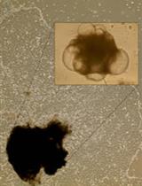
Generation, Maintenance and HBV Infection of Human Liver Organoids

人肝类器官的产生、维持和HBV感染

SR Shahla Romal
TH Tanvir Hossain
TM Tokameh Mahmoudi
3576 Views
Mar 20, 2022
Hepatitis B virus (HBV) infection represents a major public health problem infecting approximately 400 million people worldwide. Despite the availability of a preventive vaccine and anti-viral therapies, chronic HBV infection remains a major health issue because it increases the risk of developing liver cirrhosis and hepatocellular carcinoma (HCC). The lack of a relevant in vitro model for the study of the molecular mechanisms that drive HBV replication and latency, as well as HBV-related carcinogenesis, has been one of the major obstacles to the development of curative strategies. Here, we propose the use of human liver organoids as a platform for modeling HBV infection and related tumorigenesis. Human liver organoids can be seeded from both healthy and cirrhotic liver biopsies. They can be expanded in vitro when culturing in a medium containing a specific set of growth factors. When the culture medium is changed into a new medium containing growth factors that promote differentiation, organoids differentiate into functional hepatocytes, which makes them susceptible to infection with recombinant HBV. The novel in vitro primary model system described in this protocol can be utilized as a platform to study HBV pathogenesis and drug screening. Organoids generated from cirrhotic liver biopsies can be a potential tool for personalized medicine, and for modeling HCC and other liver diseases.Graphic abstract:
追踪细菌中的反向氧化三羧酸循环

Tracking the Reversed Oxidative Tricarboxylic Acid Cycle in Bacteria

追踪细菌中的反向氧化三羧酸循环

LS Lydia Steffens
EP Eugenio Pettinato
TS Thomas M. Steiner
WE Wolfgang Eisenreich
IB Ivan A. Berg
2819 Views
Mar 20, 2022
Different pathways for autotrophic CO2 fixation can be recognized by the presence of genes for their specific key enzymes. On this basis, (meta)genomic, (meta)transcriptomic, or (meta)proteomic analysis enables the identification of the role of an organism or a distinct pathway in primary production. However, the recently discovered variant of the reductive tricarboxylic acid (rTCA) cycle, the reverse oxidative tricarboxylic acid (roTCA) cycle, lacks unique enzymes, a feature that makes it cryptic for bioinformatics analysis. This pathway is a reversal of the widespread tricarboxylic acid (TCA) cycle. The functioning of the roTCA cycle requires unusually high activity of citrate synthase, the enzyme responsible for citrate cleavage, as well as elevated CO2 partial pressures. Here, we present a detailed description of the protocol we used for the identification of the roTCA cycle in members of Desulfurellaceae. First, we describe the anaerobic cultivation of Desulfurellaceae at different CO2 concentrations with a method that can be adapted to the cultivation of other anaerobes. Then, we explain how to measure activities of enzymes responsible for citrate cleavage, malate dehydrogenase reaction, and the crucial carboxylation step of the cycle catalyzed by pyruvate synthase in cell extracts. In conclusion, we describe stable isotope experiments that allow tracking of the roTCA cycle in vivo, through the position-specific incorporation of carbon-13 into amino acids. The label is provided to the organism as 13CO2 or [1-13C]glutamate. The same key methodology can be used for the reliable evaluation of the functioning of the roTCA cycle in any organism under study. This pathway is likely to participate, completely unseen, in the metabolism of various microorganisms.Graphic abstract:
基于CRISPR/Cas9系统的大豆疫霉转化

Phytophthora sojae Transformation Based on the CRISPR/Cas9 System

基于CRISPR/Cas9系统的大豆疫霉转化

JC Jingting Cao
MQ Min Qiu
WY Wenwu Ye
Yuanchao Wang Yuanchao Wang
2674 Views
Mar 20, 2022
Phytophthora sojae is a model species for the study of plant pathogenic oomycetes. The initial research on gene function using Phytophthora was mainly based on gene silencing technology. Recently, the CRISPR/Cas9-mediated genome editing technology was successfully established in P. sojae and widely used in oomycetes. In this protocol, we describe the operating procedures for the use of CRISPR/Cas9-based genome editing technology and PEG-mediated stable transformation of P. sojae protoplasts. Two plasmids were co-transformed into P. sojae: pYF515 expressing Cas9 and the single guide RNA, and the homologous replacement vector of the candidate gene. Finally, the ORF of candidate gene were replaced with the ORF of the entire hygromycin B phosphotransferase gene (HPH), to achieve precise knockout.
研究EB病毒感染的鼻咽三维模型

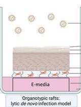
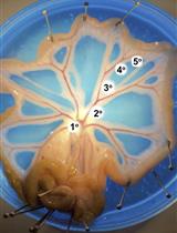
Three-dimensional Models of the Nasopharynx for the Study of Epstein-Barr Virus Infection

研究EB病毒感染的鼻咽三维模型

PZ Phillip Ziegler
AR Alex S. Reznik
SK Shweta P. Kitchloo
EW Eric Wang
SL Stella E. Lee
AG Anthony Green
MM Michael M. Myerburg
CS Clare E. Sample
KS Kathy Ho Yen Shair
2626 Views
Mar 20, 2022
The ubiquitous and cancer-associated Epstein-Barr virus (EBV) is associated with nearly all cases of nasopharyngeal carcinoma (NPC). Nasopharyngeal tissue is comprised of both pseudostratified and stratified epithelium, which are modeled in three-dimensional (3-D) cell culture. The cellular origin of EBV-associated NPC is as yet unknown, but both latent and lytic infections are likely important for preneoplastic mechanisms and replenishing the compartmentalized viral reservoir. Conventional 2-D cultures of nasopharyngeal epithelial cells (as primary cells or immortalized cell lines) are difficult to infect with EBV and cannot mimic the tissue-specific biology of the airway epithelium, which can only be captured in 3-D models. We have shown that EBV can infect the pseudostratified epithelium in air-liquid interface (ALI) culture using primary conditionally reprogrammed cells (CRCs) derived from the nasopharynx. In this protocol, we provide a step-by-step guide for the (i) conditional reprogramming of primary nasopharyngeal cells, (ii) differentiation of CRCs into pseudostratified epithelium in ALI culture (known as pseudo-ALI), and (iii) EBV infection of pseudo-ALI cultures. Additionally, we show that nasopharyngeal CRCs can be grown as organotypic rafts and subjected to EBV infection. These nasopharyngeal-derived 3-D cell cultures can be used to study EBV latent and lytic infection in relation to cell type and donor variation, by immunostaining and single-cell RNA-sequencing methods (Ziegler et al., 2021). These methods are useful for studies of EBV molecular pathogenesis, and can overcome many of the limitations associated with conventional 2-D cell cultures.Graphic abstract: Workflow of nasopharyngeal-derived conditionally reprogrammed cells grown into pseudostratified-ALI and organotypic rafts in 3-D cell culture. Created with Biorender.com.

植物科学

丝氨酸水解酶超家族酶的基于活性的蛋白质分析

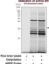
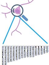
Activity-based Protein Profiling of Serine Hydrolase Superfamily Enzymes

丝氨酸水解酶超家族酶的基于活性的蛋白质分析

AD Achintya Kumar Dolui
ML Mahadev Latha
PV Panneerselvam Vijayaraj
2015 Views
Mar 20, 2022
Activity-based protein profiling (ABPP) is a chemoproteomics platform to assess the functional state of enzymes in complex biological systems. Over the two decades, ABPP has emerged from a gel-based to gel-free platform, for in-depth proteome analysis with enhanced resolution, sensitivity for target detection, and discovery of small molecule inhibitors. The gel-free format of ABPP coupled with advanced mass spectrometry is highly sensitive and provides more comprehensive knowledge for the targeted enzyme family than the gel-based method. ABPP strategy is applied across microbe, plant, and animal models. It can be performed both in vitro and in vivo studies, and there is no limitation on sample origin. Here, we report an ultrasensitive, gel-free format of ABPP called active site peptide profiling. This protocol describes the identification of authentic functional proteins, by tagging their active sites in a native biological system. It is high throughput in nature and helps enrich even low abundance functional proteins. Since protein identification is virtually based on a single peptide, the identified peptide should be a unique peptide to identify its parent protein. It can be performed in a facile manner and offers to consolidate identification of protein targets as well as the site of probe modification. We have validated this approach using a fluorophosphonate (FP) serine hydrolase probe in the native proteome of the cereal crop Oryza sativa.Graphic abstract: Serine hydrolase active site peptide profiling