往期刊物2022

卷册: 12, 期号: 4

left

Dec

20

Dec

5

Nov

20

Nov

5

Oct

20

Oct

5

Sep

20

Sep

5

Aug

20

Aug

5

Jul

20

Jul

5

Jun

20

Jun

5

May

20

May

5

Apr

20

Apr

5

Mar

20

Mar

5

Feb

20

Feb

5

Jan

20

Jan

5

right

生物信息学与计算生物学

使用 3DeeCellTracker 跟踪 3D 延时图像中的移动细胞

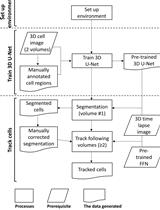
Tracking Moving Cells in 3D Time Lapse Images Using 3DeeCellTracker

使用 3DeeCellTracker 跟踪 3D 延时图像中的移动细胞

CW Chentao Wen
KK Koutarou D. Kimura
2922 Views
Feb 20, 2022
Recent advancements in 3D microscopy have enabled scientists to monitor signals of multiple cells in various animals/organs. However, segmenting and tracking the moving cells in three-dimensional time-lapse images (3D + T images), to extract their dynamic positions and activities, remains a considerable bottleneck in the field. We developed a deep learning-based software pipeline called 3DeeCellTracker, which precisely tracks cells with large movements in 3D + T images, obtained from different animals or organs, using highly divergent optical systems. In this protocol, we explain how to set up the computational environment, the required data, and the procedures to segment and track cells with 3DeeCellTracker. Our protocol will help scientists to analyze cell activities/movements in 3D + T image datasets that have been difficult to analyze.Graphic abstract: The flowchart illustrating how to use 3DeeCellTracker. See the Equipment and Procedure sections for detailed explanations.

生物工程

研究纳米粒子渗透的体外血脑屏障模型

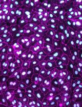
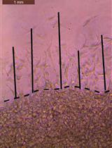
An in vitro Blood-brain Barrier Model to Study the Penetration of Nanoparticles

研究纳米粒子渗透的体外血脑屏障模型

SA Sezin Aday
WL Wen Li
JK Jeffrey M. Karp
NJ Nitin Joshi
10514 Views
Feb 20, 2022
The blood-brain barrier (BBB), a crucial protection mechanism in the central nervous system (CNS), is a selective barrier comprised of endothelial cells. It hampers the development of therapeutic and diagnostic tools for neurological diseases due to the poor penetration of most of these agents. Rationally engineered nanoparticles (NP) can facilitate the transport of therapeutic and diagnostic agents across the BBB. However, evaluating BBB penetration by NP majorly relies on the use of expensive and time-consuming animal experiments with low throughput. In vitro BBB models composed of brain endothelial cells can be a useful tool to rapidly screen multiple NP formulations to compare their BBB penetration ability and identify optimal formulations for in vivo validation. In this protocol, we present an in vitro model of BBB developed using murine cerebral cortex endothelial cells (bEnd.3). bEnd.3 is a commercially available, easy to manipulate cell line that forms tight junctions with potent paracellular barrier property. The protocol includes culturing of bEnd.3 cells, establishment of the in vitro model, and assessing NP permeability. We believe that, due to its simplicity and consistency, this step-by-step protocol can be easily used by researchers to screen NP-based drug delivery systems for BBB penetration.Graphic abstract:

生物物理学

将Escrt-Ⅲ蛋白注射到粘附的巨大单层囊泡中

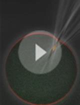
Femtoliter Injection of ESCRT-III Proteins into Adhered Giant Unilamellar Vesicles

将Escrt-Ⅲ蛋白注射到粘附的巨大单层囊泡中

VG Vasil N. Georgiev
YA Yunuen Avalos-Padilla
XF Xavier Fernàndez-Busquets
RD Rumiana Dimova
2791 Views
Feb 20, 2022
The endosomal sorting complex required for transport (ESCRT) machinery mediates membrane fission reactions that exhibit a different topology from that observed in clathrin-coated vesicles. In all of the ESCRT-mediated events, the nascent vesicle buds away from the cytosol. However, ESCRT proteins are able to act upon membranes with different geometries. For instance, the formation of multivesicular bodies (MVBs) and the biogenesis of extracellular vesicles both require the participation of the ESCRT-III sub-complex, and they differ in their initial membrane geometry before budding starts: the protein complex acts either from outside the membrane organelle (causing inward budding) or from within (causing outward budding). Several studies have reconstituted the action of the ESCRT-III subunits in supported bilayers and cell-sized vesicles mimicking the geometry occurring during MVBs formation (in-bud), but extracellular vesicle budding (out-bud) mechanisms remain less explored, because of the outstanding difficulties encountered in encapsulation of functional ESCRT-III in vesicles. Here, we provide a different approach that allows the recreation of the out-bud formation, by combining giant unilamellar vesicles as a membrane model and a microinjection system. The vesicles are immobilized prior to injection via weak adhesion to the chamber coverslip, which also ensures preserving the membrane excess area required for budding. After protein injection, vesicles exhibit outward budding. The approach presented in this work can be used in the future to disentangle the mechanisms underlying ESCRT-III-mediated fission, recreating the geometry of extracellular bud production, which remains a challenge. Moreover, the microinjection methodology can be also adapted to interrogate the action of other cytosolic components on the encapsulating membranous organelle.Graphic abstract: Out-bud formation after ESCRT-III protein injection into GUVs

癌症生物学

球形侵袭测定:一种测量癌细胞侵袭的新方法

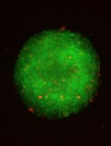
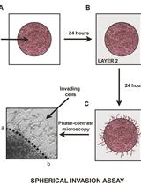
Spherical Invasion Assay: A Novel Method to Measure Invasion of Cancer Cells

球形侵袭测定:一种测量癌细胞侵袭的新方法

SR Stephen D. Richbart
JM Justin C. Merritt
EM Emily G. Moles
KB Kathleen C. Brown
AA Adeoluwa A. Adeluola
PF Paul T. Finch
JH Joshua A. Hess
MT Maria T. Tirona
SM Sarah L. Miles
MV Monica A. Valentovic
PD Piyali Dasgupta
4235 Views
Feb 20, 2022
The invasion of tumor cells into the neighboring blood vessels and lymph nodes is a vital step for distant metastasis. Traditionally, the invasive activity of growth factors (or the anti-invasive activity of drugs) is measured with the Boyden chamber assay. However, this assay has a few disadvantages like poor physiological relevance of transwell inserts and an inability to control chemokine gradients. The Boyden chamber assay is one of the most prevalent methods to measure the invasion of cancer cells. It would be advantageous to develop another assay that could validate the results of the Boyden chamber assay. With this in mind, our laboratory developed the spherical invasion assay (SIA) to measure the pro-invasive activity of human cancer cells. The SIA also circumvents some of the drawbacks of the Boyden chamber assay. The present manuscript measures the anti-invasive activity of the Src kinase inhibitor PP2 in A549 human non-small cell lung carcinoma (NSCLC) cells using the SIA. The SIA protocol is comprised of two steps. In the first step, A549 human NSCLC cells (treated or not with PP2) were mixed with Matrigel and seeded in the middle of an eight-well chamber slide. After 24 h, a second layer of Matrigel was overlaid over the first layer. Over the course of the next 24 h, the A549 cells invade from the primary to the secondary Matrigel layers. Subsequently, the cells are visualized by phase-contrast microscopy and the images obtained are quantified using ImageJ to calculate the anti-invasive activity of PP2 in A549 cells. The results of the SIA correlate well with Boyden chamber assays. The SIA may be adapted for multiple experimental designs, such as drug screening (to combat invasion and metastasis), measuring the pro-invasive activity of growth factors, and elucidating the signaling pathways underlying the pro-invasive/anti-invasive activity of biological modifiers.Graphic abstract:Diagrammatic illustration of the spherical invasion assay (Hurley et al., 2017). A. The first layer is comprised of human cancer cells mixed in a 1:1 suspension with Phenol Red containing Matrigel (represented as LAYER 1 in the figure). After 24 h, the cancer cells grow and extend up to the boundary of this first layer. B. A second layer of 1:1 solution Phenol Red-free Matrigel, in Phenol Red-free RPMI (represented as LAYER 2 in the figure) is added on top of the first Matrigel spot. The cells are incubated for 24 h at 37°C. C. Over these 24 h, the cancer cells invade from the primary layer into the secondary Matrigel layer. The chamber slides are observed by phase-contrast microscopy. D. A representative photograph of the images obtained by the SIA is shown. The black arrow indicates the cancer cells invading into the second layer of Matrigel. The dotted line represents the interface between the two layers. The distance to which the cells have traveled (into the secondary Matrigel layer) is measured at ten sites (for each photograph) in a randomized double-blind fashion by three independent observers, using NIH ImageJ Version 1.47. This process is repeated for three separate photographic fields per sample.
优化细胞外通量测定以测量不依赖锚定的肿瘤细胞球体的呼吸

Optimization of Extracellular Flux Assay to Measure Respiration of Anchorage-independent Tumor Cell Spheroids

优化细胞外通量测定以测量不依赖锚定的肿瘤细胞球体的呼吸

ZJ Zaineb Javed
BW Beth L. Worley
CS Coryn Stump
SS Sara S. Shimko
LC LaTaijah C. Crawford
KM Karthikeyan Mythreye
NH Nadine Hempel
3346 Views
Feb 20, 2022
Three-dimensional (3D) cell culture models are widely used in tumor studies to more accurately reflect cell-cell interactions and tumor growth conditions in vivo. 3D anchorage-independent spheroids derived by culturing cells in ultra-low attachment (ULA) conditions is particularly relevant to ovarian cancer, as such cell clusters are often observed in malignant ascites of late-stage ovarian cancer patients. We and others have found that cells derived from anchorage-independent spheroids vary widely in gene expression profiles, proliferative state, and metabolism compared to cells maintained under attached culture conditions. This includes changes in mitochondrial function, which is most commonly assessed in cultured live cells by measuring oxygen consumption in extracellular flux assays. To measure mitochondrial function in anchorage-independent multicellular aggregates, we have adapted the Agilent Seahorse extracellular flux assay to optimize measurements of oxygen consumption and extracellular acidification of ovarian cancer cell spheroids generated by culture in ULA plates. This protocol includes: (i) Methods for culturing tumor cells as uniform anchorage-independent spheroids; (ii) Optimization for the transfer of spheroids to the Agilent Seahorse cell culture plates; (iii) Adaptations of the mitochondrial and glycolysis stress tests for spheroid extracellular flux analysis; and (iv) Suggestions for optimization of cell numbers, spheroid size, and normalization of oxygen consumption rate (OCR) and extracellular acidification rate (ECAR) values. Using this method, we have found that ovarian cancer cells cultured as anchorage-independent spheroids display altered mitochondrial function compared to monolayer cultures attached to plastic dishes. This method allows for the assessment of mitochondrial function in a more relevant patho/physiological culture condition and can be adapted to evaluate mitochondrial function of various cell types that are able to aggregate into multicellular clusters in anchorage-independence. Graphic abstract:Workflow of the Extracellular Flux Assay to Measure Respiration of Anchorage-independent Tumor Cell Spheroids.
一种基于细胞的筛选方法,使用细胞内抗体发现靶向困难药物蛋白质的小分子

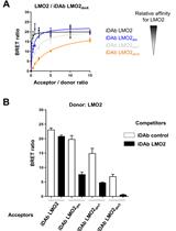
A Cell-based Screening Method Using an Intracellular Antibody for Discovering Small Molecules Targeting Hard-to-drug Proteins

一种基于细胞的筛选方法,使用细胞内抗体发现靶向困难药物蛋白质的小分子

NB Nicolas Bery
TR Terry H. Rabbitts
3214 Views
Feb 20, 2022
Targeting hard-to-drug proteins, such as proteins functioning by protein-protein interactions (PPIs) with small molecules, is difficult because of the lack of well-defined pockets. Fragment or computational-based methods are usually employed for the discovery of such compounds, but no generic method is available to quickly identify small molecules interfering with PPIs. Here, we provide a protocol describing a generic method to discover small molecules inhibiting the interaction between an intracellular antibody and its target, in particular for proteins that are hard to make in recombinant form. This protocol reports a versatile and generic method that can be applied to any target/intracellular antibody. Because it is a cell-based assay, it identifies chemical matters that are already displaying advantageous cell permeability properties.Graphic abstract:Cell-based intracellular antibody-guided small molecule screening.

细胞生物学

哺乳动物细胞系的 NBD 脂质摄取测定

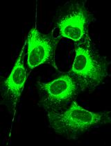
NBD-lipid Uptake Assay for Mammalian Cell Lines

哺乳动物细胞系的 NBD 脂质摄取测定

SH Sara Abad Herrera
MG Marta Grifell-Junyent
TP Thomas Günther Pomorski
3619 Views
Feb 20, 2022
All eukaryotic cells are equipped with transmembrane lipid transporters, which are key players in membrane lipid asymmetry, vesicular trafficking, and membrane fusion. The link between mutations in these transporters and disease in humans highlights their essential role in cell homeostasis. Yet, many key features of their activities, their substrate specificity, and their regulation remain to be elucidated. Here, we describe an optimized quantitative flow cytometry-based lipid uptake assay utilizing nitrobenzoxadiazolyl (NBD) fluorescent lipids to study lipid internalization in mammalian cell lines, which allows characterizing lipid transporter activities at the plasma membrane. This approach allows for a rapid analysis of large cell populations, thereby greatly reducing sampling variability. The protocol can be applied to study a wide range of mammalian cell lines, to test the impact of gene knockouts on lipid internalization at the plasma membrane, and to uncover the dynamics of lipid transport at the plasma membrane.Graphic abstract: Internalization of NBD-labeled lipids from the plasma membrane of CHO-K1 cells.

免疫学

再刺激诱导的细胞死亡 (RICD):通过流式细胞术分析在原代人类 T 细胞中建模、调查和量化 RICD 敏感性的方法

Restimulation-Induced Cell Death (RICD): Methods for Modeling, Investigating, and Quantifying RICD Sensitivity in Primary Human T Cells via Flow Cytometric Analysis

再刺激诱导的细胞死亡 (RICD):通过流式细胞术分析在原代人类 T 细胞中建模、调查和量化 RICD 敏感性的方法

KP Katherine Pohida
CL Camille M. Lake
DY Debra Yee
AS Andrew L. Snow
3220 Views
Feb 20, 2022
When the body mounts an immune response against a foreign pathogen, the adaptive arm of the immune system relies upon clonal expansion of antigen-specific T cell populations to exercise acquired effector and cytotoxic functions to clear it. However, T cell expansion must be modulated to effectively combat the perceived threat without inducing excessive collateral damage to host tissues. Restimulation-induced cell death (RICD) is an apoptotic program triggered in activated T cells when an abundance of antigen and IL-2 are present, imposing a negative feedback mechanism that constrains the growing T cell population. This autoregulatory process can be detected via increases in caspase activation, Annexin V binding, and loss of mitochondrial membrane potential. However, simple changes in T cell viability through flow cytometric analysis can reliably measure RICD sensitivity in response to T-cell receptor (TCR) restimulation. This protocol describes the in vitro polyclonal activation, expansion, and restimulation of human primary T cells isolated from donor peripheral blood mononuclear cells (PBMC). This simple procedure allows for accurate quantification of RICD via flow cytometry. We also describe strategies for interrogating the role of specific proteins and pathways that may alter RICD sensitivity. This straightforward protocol provides a quick and dependable tool to track RICD sensitivity in culture over time while probing critical factors that control the magnitude and longevity of an adaptive immune response.Graphic abstract:In-vitro simulation of restimulation-induced cell death in activated human T cells.

微生物学

 使用大肠杆菌无细胞表达系统对 mRNA 3' 端进行体外测定

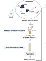
An in vitro Assay of mRNA 3’ end Using the E. coli Cell-free Expression System

使用大肠杆菌无细胞表达系统对 mRNA 3' 端进行体外测定

MN Monford Paul Abishek N
HL Heon M. Lim
2624 Views
Feb 20, 2022
At the end of about 80% of the operon in Escherichia coli, translation termination decouples transcription, leading to Rho-dependent transcription termination (RDT). However, no in vitro or in vivo assay system has proven to be good enough to see the 3’ end of the mRNA generated by RDT. Here, we present a cell-free assay system that could provide detailed information on the 3’ end of a transcript RNA generated by RDT. Our protocol shows how to extract transcript RNA generated by transcription reactions from a cell-free extract, followed by an RNA oligomer ligation to the 3’ end of a transcript RNA of interest. The 3' end of the RNA is amplified using RT-PCR. Its genetic location can be determined using a gene-specific primer extension reaction. The 3’ ends of mRNA can be visualized and quantified by polyacrylamide gel electrophoresis. One significant advantage of a cell-free assay system is that factors involved in the generation of the 3' end, such as proteins and sRNA, can be directly assayed by exogenously adding factor(s) to the reaction.Graphic abstract: An illustration of the experimental methodology.
基于活性的交联识别疟原虫中硫氧还蛋白结构域蛋白的底物

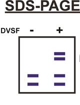
Activity-based Crosslinking to Identify Substrates of Thioredoxin-domain Proteins in Malaria Parasites

基于活性的交联识别疟原虫中硫氧还蛋白结构域蛋白的底物

DC David W. Cobb
GW Grace S. Woods
VM Vasant Muralidharan
2184 Views
Feb 20, 2022
Malaria remains a major public health issue, infecting nearly 220 million people every year. The spread of drug-resistant strains of Plasmodium falciparum around the world threatens the progress made against this disease. Therefore, identifying druggable and essential pathways in P. falciparum parasites remains a major area of research. One poorly understood area of parasite biology is the formation of disulfide bonds, which is an essential requirement for the folding of numerous proteins. Specialized chaperones with thioredoxin (Trx) domains catalyze the redox functions necessary for breaking incorrect and forming correct disulfide bonds in proteins. Defining the substrates of these redox chaperones is difficult and immunoprecipitation based assays cannot distinguish between substrates and interacting partners. Further, the substrate or client interactions with the redox chaperones are usually transient in nature. Activity based crosslinkers that rely on the nucleophilic cysteines on Trx domains and the disulfide bond forming cysteines on clients provide an easily scalable method to trap and identify the substrates of Trx-domain containing chaperones. The cell permeable crosslinker divinyl sulfone (DVSF) is active only in the presence of nucleophilic cysteines in proteins and, therefore, traps Trx domains with their substrates, as they form mixed disulfide bonds during the course of their catalytic activity. This allows the identification of substrates that rely on Trx activity for their folding, as well as discovering small molecules that interfere with Trx domain activity.Graphic abstract:Identification of thioredoxin domain substrates via divinylsulfone crosslinking and immunoprecipitation-mass spectrometry.

分子生物学

生物素化 RNA 和相关蛋白的下拉

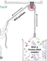
Pull-down of Biotinylated RNA and Associated Proteins

生物素化 RNA 和相关蛋白的下拉

JU Jagadeesh K. Uppala
CG Chandrima Ghosh
GS Grzegorz Sabat
MD Madhusudan Dey
6185 Views
Feb 20, 2022
Mapping networks of RNA-protein interactions in cells is essential for understanding the inner workings of many biological processes, including RNA processing, trafficking, and translation. Current in vivo methods for studying protein-RNA interactions rely mostly on purification of poly(A) transcripts, which represent only ~2–3% of total RNAs (Figure 1). Alternate robust methods for tagging RNA molecules with an RNA aptamer (e.g., MS2-, U1A- and biotin-RNA aptamer) and capturing the RNA-protein complex by the respective aptamer-specific partner are not extensively studied. Here, we describe a protocol (Figure 2) in which a biotin-RNA aptamer, referred to as the RNA mimic of biotin (RMB), was conjugated separately to two small RNA secondary structures that contribute to trafficking and translating HAC1 mRNA in the budding yeast Saccharomyces cerevisiae. The RMB-tagged RNA was expressed in yeast cells from a constitutive promoter. The biotinylated RNA bound to proteins was pulled down from the cell lysate by streptavidin agarose beads. RNA was detected by RT-PCR (Figure 3) and associated proteins by mass spectrometry (Figure 4). Our findings show that an RNA aptamer tag to RNA molecule is an effective method to explore the functional roles of RNA-protein networks in vivo.
标准化Ribo -Seq 用于量化翻译中的绝对全局和特定变化

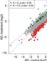
Normalized Ribo-Seq for Quantifying Absolute Global and Specific Changes in Translation

标准化Ribo -Seq 用于量化翻译中的绝对全局和特定变化

KH Katharina Hoerth
SR Sonja Reitter
JS Johanna Schott
3841 Views
Feb 20, 2022
Ribosome profiling (Ribo-Seq) is a highly sensitive method to quantify ribosome occupancies along individual mRNAs on a genome-wide scale. Hereby, ribosome-protected fragments (= footprints) are generated by nuclease digestion, isolated, and sequenced together with the corresponding randomly fragmented input samples, to determine ribosome densities (RD). For library preparation, equal amounts of total RNA are used. Subsequently, all transcript fragments are subjected to linker ligation, cDNA synthesis, and PCR amplification. Importantly, the number of reads obtained for every transcript in input and footprint samples during sequencing depends on sequencing depth and library size, as well as the relative abundance of the transcript in the sample. However, the information pertaining to the absolute amount of input and footprint sequences is lost during sample preparation, hence ruling out any conclusion whether translation is generally suppressed or activated in one condition over the other. Therefore, the RD fold-changes determined for individual genes do not reflect absolute regulation, but have to be interpreted as relative to bulk mRNA translation. Here, we modified the original ribosome profiling protocol that was first established by Ingolia et al. (2009), by adding small amounts of yeast lysate to the mammalian lysates of interest as a spike-in. This allows us to not only detect changes in the RD of specific transcripts relative to each other, but also to simultaneously measure global differences in RD (normalized ribosome density values) between samples. Graphic abstract:Global changes in translation efficiency can be detected with polysome profiling, where the proportion of polysomal ribosomes is interpreted as a proxy for ribosome density (RD) on bulk mRNA. Ribo-Seq measures changes in RD of specific mRNAs relative to bulk mRNA. The addition of a yeast-lysate, as a spike-in for normalization of read counts, allows for an absolute measurement of changes in RD.

神经科学

免疫沉淀法定量组织和细胞培养中与其相互作用蛋白相关的tRNAs的数量

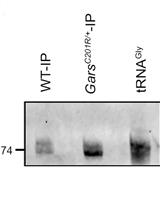
Immunoprecipation Assay to Quantify the Amount of tRNAs associated with Their Interacting Proteins in Tissue and Cell Culture

免疫沉淀法定量组织和细胞培养中与其相互作用蛋白相关的tRNAs的数量

SD Sarada Das
AZ Amila Zuko
RT Robin Thompson
ES Erik Storkebaum
ZI Zoya Ignatova
2855 Views
Feb 20, 2022
Transfer RNAs (tRNAs) are highly abundant species and, along their biosynthetic and functional path, they establish interactions with a plethora of proteins. The high number of nucleobase modifications in tRNAs renders conventional RNA quantification approaches unsuitable to study protein-tRNA interactions and their associated functional roles in the cell. We present an immunoprecipitation-based approach to quantify tRNA bound to its interacting protein partner(s). The tRNA-protein complexes are immunoprecipitated from cells or tissues and tRNAs are identified by northern blot and quantified by tRNA-specific fluorescent labeling. The tRNA interacting protein is quantified by an automated western blot and the tRNA amount is presented per unit of the interacting protein. We tested the approach to quantify tRNAGly associated with mutant glycyl-tRNA-synthetase implicated in Charcot-Marie-Tooth disease. This simple and versatile protocol can be easily adapted to any other tRNA binding proteins.Graphic abstract: Figure 1. Schematic of the tRNA-Immunoprecipitation approach.
调查斑马鱼判断偏差的行为分析

A Behavioural Assay to Investigate Judgment Bias in Zebrafish

调查斑马鱼判断偏差的行为分析

FE Felipe Espigares
RM Raquel R. Martins
RO Rui F. Oliveira
2321 Views
Feb 20, 2022
In this protocol, we describe for the first time a judgment bias paradigm to phenotype the way zebrafish assess ambiguous stimuli. We have developed and validated a protocol for a judgment bias test based on a Go/No-go task, and performed using a half radial maze. After a habituation phase, fish are trained to discriminate between two reference arms [positive (P) and negative (N)]. For this purpose, they experience a positive event (food reward in P), when presented with a specific location/color cue, and a negative event (chasing with net in N), when presented with a different location/color cue. Acquisition of the discrimination learning between P and N is revealed by the latencies to enter the experimental arms of the behavioral maze being significantly lower for the P arm than for the N arm. Once zebrafish are able to discriminate between P and N arms, their latency to enter other maze arms spatially located between P and N [(Near Positive (NP), Ambiguous (A) = half-way between P and N, and Near Negative (NN)] is analyzed. Latencies (L) to enter NP, A and NN maze arms are interpreted as indicating the individual expectancy to experience a reward/punishment on each of them. A judgment bias score (JBS) is calculated from the latencies to enter the P, N, and A arms for each fish [JBS = (LA–LP)*100/(LN–LP)], based on which fish can be classified into an optimistic/pessimistic axis. A JBS below 50 indicates that fish perceive the ambiguous stimulus as a positive one (optimistic bias), while JBS above 50 indicates that fish perceive the ambiguous stimulus as a negative one (pessimistic bias). However, for classification criteria, it could be advantageous to use the method of selecting extreme phenotypes (e.g., upper and lower quartiles of the JBS), since JBS in zebrafish falls into a bimodal distribution (unpublished data). Therefore, this protocol provides a unique, inexpensive, and effective alternative to other methods of measuring affective states in zebrafish that might be of great interest to a broad target audience and have a large number of applications.Graphic abstract: Flow chart of the judgment bias protocol in zebrafish.
触觉和食物奖励的预期性和完成性反应:一项人类研究方法

Anticipatory and Consummatory Responses to Touch and Food Rewards: A Protocol for Human Research

触觉和食物奖励的预期性和完成性反应:一项人类研究方法

EC Emilio Chiappini
GS Giorgia Silani
SK Sebastian Korb
1696 Views
Feb 20, 2022
Understanding the neural basis of reward processing is a major concern, as it holds the key to alleviating symptoms of addiction and poor mental health. However, this goal seems difficult to attain as long as research on reward processing cannot easily be compared across species and reward types, due to methodological differences and the presence of confounding factors. We recently developed an experimental paradigm that allows monitoring anticipatory and consummatory responses to matched social (touch) and nonsocial (food) rewards in adult humans. The following protocol describes in detail the materials and the paradigm, which measures reward wanting and liking with a real effort task and subjective ratings. It can also be used in combination with facial electromyography (EMG), brain imaging (e.g., fMRI), and pharmacological interventions. It is our firm belief that the field will profit greatly from more research being conducted on reward processing using this and similarly controlled paradigms, which allow for cross-species comparison.

植物科学

测量内源性 GA 和 IAA

Measuring Endogenous GA and IAA

测量内源性 GA 和 IAA

YC Yunping Chen
ZD Zhiwu Dan
Shaoqing  Li Shaoqing Li
2832 Views
Feb 20, 2022
Plant hormones regulate many physiological processes that largely influence growth, differentiation, and development. Contents of phytohormones were analyzed using a liquid chromatography-electrospray ionization-tandem mass spectrometry (LC-ESI-MS/MS) system. This protocol describes a detailed procedure to extract and quantify indole-3-acetic acid (IAA) and gibberellin acid (GA) in rice (Oryza sativa) tissues using high-performance liquid chromatography (HPLC)-based method.