往期刊物2022

卷册: 12, 期号: 3

left

Dec

20

Dec

5

Nov

20

Nov

5

Oct

20

Oct

5

Sep

20

Sep

5

Aug

20

Aug

5

Jul

20

Jul

5

Jun

20

Jun

5

May

20

May

5

Apr

20

Apr

5

Mar

20

Mar

5

Feb

20

Feb

5

Jan

20

Jan

5

right

癌症生物学

上皮性卵巢癌细胞临床相关获得性化疗耐药模型的建立

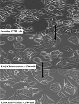
Developing Clinically Relevant Acquired Chemoresistance Models in Epithelial Ovarian Cancer Cell Lines

上皮性卵巢癌细胞临床相关获得性化疗耐药模型的建立

PS Priti S. Shenoy
SC Sourav Chakraborty
SG Snehal M. Gaikwad
AS Asmita Sakpal
PR Pritha Ray
3348 Views
Feb 5, 2022
Chemoresistance, the ability of cancer cells to overcome therapeutic interventions, is an area of active research. Studies on intrinsic and acquired chemoresistance have partly succeeded in elucidating some of the molecular mechanisms in this elusive phenomenon. Hence, drug-resistant cellular models are routinely developed and used to mimic the clinical scenario in-vitro. In an attempt to identify the underlying molecular mechanisms that allow ovarian cancer cells to gradually acquire chemoresistance, we have developed isogenic cellular models of cisplatin and paclitaxel resistance (singularly and in combination) over six months, using a clinically relevant modified pulse method. These models serve as important tools to investigate the underlying molecular players, modulation in genetics, epigenetics, and relevant signaling pathways, as well as to understand the role of drug detoxification and drug influx-efflux pathways in development of resistance. These models can also be used as screening tools for new therapeutic molecules. Additionally, repurposing therapeutic agents approved for diseases other than cancer have gained significant attention in improving cancer therapy. To investigate the effect of metformin on acquirement of chemoresistance, we have also developed a combinatorial model of metformin and platinum-taxol, using two different strategies. All these models were subsequently used to study modulation in receptor tyrosine kinase pathways, cancer stem cell functionalities, autophagy, metastasis, metabolic signatures, and various biological processes during development of chemoresistance. Herein, we outline the protocols used for developing these intricate resistant cellular models.Graphic abstract: Schematic of the step-wise development of cellular chemoresistant model. Schema illustrating the modified pulse method for the development of individual and combinatorial resistant models. Two different cell lines (A2780 and OAW42) were exposed to drug treatment (either alone or in various combinations) of one concentration for three consecutive cycles (3×). Furthermore, the surviving cells were sub-cultured subsequently with increasing drug concentrations. After every treatment cycle, 50% of the cells were cryo-preserved for further experiments. Additionally, to check for the development of chemoresistance and to assess the changes in cell cycle during resistance development, cell viability assays and flow cytometry were performed.
丰富酪氨酸磷酸化肽用于RTK信号动力学的定量质谱分析

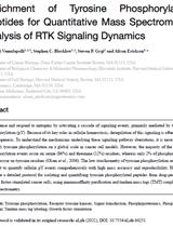
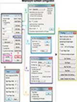
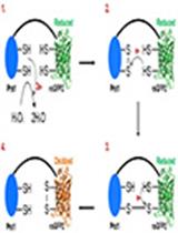
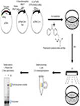
Enrichment of Tyrosine Phosphorylated Peptides for Quantitative Mass Spectrometry Analysis of RTK Signaling Dynamics

丰富酪氨酸磷酸化肽用于RTK信号动力学的定量质谱分析

VV Vidyasiri Vemulapalli
SB Stephen C. Blacklow
SG Steven P. Gygi
AE Alison Erickson
2775 Views
Feb 5, 2022
Cells sense and respond to mitogens by activating a cascade of signaling events, primarily mediated by tyrosine phosphorylation (pY). Because of its key roles in cellular homeostasis, deregulation of this signaling is often linked to oncogenesis. To understand the mechanisms underlying these signaling pathway aberrations, it is necessary to quantify tyrosine phosphorylation on a global scale in cancer cell models. However, the majority of the protein phosphorylation events occur on serine (86%) and threonine (12%) residues, whereas only 2% of phosphorylation events occur on tyrosine residues (Olsen et al., 2006). The low stoichiometry of tyrosine phosphorylation renders it difficult to quantify cellular pY events comprehensively with high mass accuracy and reproducibility. Here, we describe a detailed protocol for isolating and quantifying tyrosine phosphorylated peptides from drug-perturbed, growth factor-stimulated cancer cells, using immunoaffinity purification and tandem mass tags (TMT) coupled with mass spectrometry.
在Dunn Chamber中研究趋化迁移:应用于粘附癌细胞的案例

Studying Chemotactic Migration in Dunn Chamber: An Example Applied to Adherent Cancer Cells

在Dunn Chamber中研究趋化迁移:应用于粘附癌细胞的案例

KB Khedidja Benseddik
KZ Kossay Zaoui
2276 Views
Feb 5, 2022
Cell migration is a vital process in the development of multicellular organisms. When deregulated, it is involved in many diseases such as inflammation and cancer metastisation. Some cancer cells could be stimulated using chemoattractant molecules, such as growth factor Heregulin β1. They respond to the attractant or repellent gradients through a process known as chemotaxis. Indeed, chemotactic cell motility is crucial in tumour cell dissemination and invasion of distant organs. Due to the complexity of this phenomenon, the majority of available in vitro methods to study the chemotactic motility process have limitations and are mainly based on endpoint assays, such as the Boyden chamber assay. Nevertheless, in vitro time-lapse microscopy represents an interesting opportunity to study cell motility in a chemoattracting gradient, since it generates large volume image-based information, allowing the analysis of cancer cell behaviours. Here, we describe a detailed time-lapse imaging protocol, designed for tracking T47D human breast cancer cell line motility, toward a gradient of Heregulin β1 in a Dunn chemotaxis chamber assay. The protocol described here is readily adapted to study the motility of any adherent cell line, under various conditions of chemoattractant gradients and of pharmacological drug treatments. Moreover, this protocol could be suitable to study changes in cell morphology, and in cell polarity.

细胞生物学

超灵敏基因编码荧光指示剂 (roGFP2-Prx1) 在细胞微培养期间连续测量细胞内 H2O2

A Hypersensitive Genetically Encoded Fluorescent Indicator (roGFP2-Prx1) Enables Continuous Measurement of Intracellular H2O2 during Cell Micro-cultivation

超灵敏基因编码荧光指示剂 (roGFP2-Prx1) 在细胞微培养期间连续测量细胞内 H2O2

VG Veronica Gast
VS Verena Siewers
MM Mikael Molin
2506 Views
Feb 5, 2022
Hydrogen peroxide (H2O2) is a toxic oxidant produced as a byproduct of several biological processes. At too high levels of hydrogen peroxide cells will experience oxidative stress, leading to a cellular response to decrease its levels and to protect the cells. Previously, methods used to study and quantify intracellular H2O2 have been limited by both sensitivity and specificity. However, an increasing number of genetically encoded fluorescent indicators (GEFIs) are becoming available, which can specifically detect low levels of intracellular hydrogen peroxide. In this study, we use such a biosensor designed to monitor cytosolic H2O2 levels in the budding yeast Saccharomyces cerevisiae during continuous cultivation and in the absence of a fluorescence microscope. The fluorescent biosensor contains a peroxiredoxin protein fused to an engineered GFP molecule expressed from a commonly used yeast plasmid (pRS416-TEF1). The peroxiredoxin-based fluorescent indicator reduces H2O2, ultimately resulting in a GFP signal being emitted by the sensor. Here, we apply this biosensor to study cytosolic H2O2 levels in S. cerevisiae strains with and without recombinant protein production.Graphic abstract:Schematic overview of experimental steps.

药物发现

哺乳动物细胞中定位特异性病毒-宿主蛋白相互作用的琥珀抑制技术

Amber Suppression Technology for Mapping Site-specific Viral-host Protein Interactions in Mammalian Cells

哺乳动物细胞中定位特异性病毒-宿主蛋白相互作用的琥珀抑制技术

NI Nur Firdaus Isa
OB Olivier Bensaude
SM Shona Murphy
3290 Views
Feb 5, 2022
Probing the molecular interactions of viral-host protein complexes to understand pathogenicity is essential in modern virology to help the development of antiviral therapies. Common binding assays, such as co-immunoprecipitation or pull-downs, are helpful in investigating intricate viral-host proteins interactions. However, such assays may miss low-affinity and favour non-specific interactions. We have recently incorporated photoreactive amino acids at defined residues of a viral protein in vivo, by introducing amber stop codons (TAG) and using a suppressor tRNA. This is followed by UV-crosslinking, to identify interacting host proteins in live mammalian cells. The affinity-purified photo-crosslinked viral-host protein complexes are further characterized by mass spectrometry following extremely stringent washes. This combinatorial site-specific incorporation of a photoreactive amino acid and affinity purification-mass spectrometry strategy allows the definition of viral-host protein contacts at single residue resolution and greatly reduces non-specific interactors, to facilitate characterization of viral-host protein interactions.Graphic abstract:Schematic overview of the virus-host interaction assay based on an amber suppression approach. Mammalian cells grown in Bpa-supplemented medium are co-transfected with plasmids encoding viral sequences carrying a Flag tag, a (TAG) stop codon at the desired position, and an amber suppressor tRNA (tRNACUA)/aminoacyl tRNA synthetase (aaRS) orthogonal pair. Cells are then exposed to UV, to generate protein-protein crosslinks, followed by immunoprecipitation with anti-Flag magnetic beads. The affinity-purified crosslinks are probed by western blot using an anti-Flag antibody and the crosslinked host proteins are characterised by mass spectrometry.

免疫学

小鼠肺边缘单核细胞体外微囊泡摄取

Investigation of Microvesicle Uptake by Mouse Lung-marginated Monocytes in vitro

小鼠肺边缘单核细胞体外微囊泡摄取

YT Ying Ying Tan
KO Kieran P. O'Dea
BP Brijesh V. Patel
MT Masao Takata
3397 Views
Feb 5, 2022
Extracellular microvesicles (MVs) are released into the circulation in large numbers during acute systemic inflammation, yet little is known of their intravascular cell/tissue-specific interactions under these conditions. We recently described a dramatic increase in the uptake of intravenously injected MVs by monocytes marginated within the pulmonary vasculature, in a mouse model of low-dose lipopolysaccharide-induced systemic inflammation. To investigate the mechanisms of enhanced MV uptake by monocytes, we developed an in vitro model using in vivo derived monocytes. Although mouse blood is a convenient source, monocyte numbers are too low for in vitro experimentation. In contrast, differentiated bone marrow monocytes are abundant, but they are rapidly mobilized during systemic inflammation, and thus no longer available. Instead, we developed a protocol using marginated monocytes from the pulmonary vasculature as an anatomically relevant and abundant source. Mice are sacrificed by terminal anesthesia, the lungs inflated and perfused via the pulmonary artery. Perfusate cell populations are evaluated by flow cytometry, combined with in vitro generated fluorescently labelled MVs, and incubated in suspension for up to one hour. Washed cells are analyzed by flow cytometry to quantify MV uptake and confocal microscopy to localize MVs within cells (O'Dea et al., 2020). Using this perfusion-based method, substantial numbers of marginated pulmonary vascular monocytes are recovered, allowing multiple in vitro tests to be performed from a single mouse donor. As MV uptake profiles were comparable to those observed in vivo, this method is suitable for physiologically relevant high throughput mechanistic studies on mouse monocytes under in vitro conditions.Graphic abstract:Figure 1. Schematic of lung perfusate cell harvest and co-incubation with in vitro generated MVs. Created with BioRender.com.
HiSAT:一种快速诊断过敏的新方法

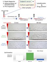
HiSAT: A Novel Method for the Rapid Diagnosis of Allergy

HiSAT:一种快速诊断过敏的新方法

HS Hirotomo Shibaguchi
YY Yuki Yasutaka
1782 Views
Feb 5, 2022
To identify causative substances for allergies to drugs or foods, the lymphocyte transformation test (LTT) is currently widely used as in vitro test, but its accuracy is not satisfactory. We have developed a novel method designated high-sensitivity allergy test (HiSAT) for determining allergy expression by measuring cell kinetics, using the chemotactic cells from non-allergic volunteers against a gradient field of cytokines released from immune cells when allergy develops. HiSAT requires a very small sample of 5 µL or less, and is applicable to three types of tests, depending on the situation in clinical practice: (i) diagnosis of the allergic expression, (ii) identification of the causative drug, and, in principle, (iii) pre-inspection.Graphic abstract:Schematic diagram of HiSAT. Serum from patients/subjects is used for rapid diagnosis in HiSAT. To identify the causative drug, the lymphocytes of interest are incubated with the candidate drug solution for 48 h to 72 h and then the culture supernatant is used in HiSAT. Before drug administration, it may possible to avoid the risk of allergies by performing pre-inspection, as well as the determination of the causative drug in HiSAT. A granulocyte-rich cell layer isolated from a non-allergic volunteer is used in HiSAT. Chemotactic cells migrate toward chemotactic factors in the test sample according to the concentration gradient. Cell kinetics (e.g., velocity or distance) are analyzed using sequential images of the test samples, and compared to the PHA-positive control.>

微生物学

细胞病变效应试验和空斑试验评估抗病毒化合物对人冠状病毒229E、OC43和NL63的体外活性

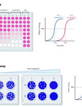
Cytopathic Effect Assay and Plaque Assay to Evaluate in vitro Activity of Antiviral Compounds Against Human Coronaviruses 229E, OC43, and NL63

细胞病变效应试验和空斑试验评估抗病毒化合物对人冠状病毒229E、OC43和NL63的体外活性

YH Yanmei Hu
CM Chunlong Ma
JW Jun Wang
5955 Views
Feb 5, 2022
Coronaviruses are important human pathogens, among which the severe acute respiratory syndrome coronavirus 2 (SARS-CoV-2) is the causative agent for the COVID-19 pandemic. To combat the SARS-CoV-2 pandemic, there is a pressing need for antivirals, especially broad-spectrum antivirals that are active against all seven human coronaviruses (HCoVs). For this reason, we are interested in developing antiviral assays to expedite the drug discovery process. Here, we provide the detailed protocol for the cytopathic effect (CPE) assay and the plaque assay for human coronaviruses 229E (HCoV-229E), HCoV-OC43, and HCoV-NL63, to identify novel antivirals against HCoVs. Neutral red was used in the CPE assay, as it is relatively inexpensive and more sensitive than other reagents. Multiple parameters including multiplicity of infection, incubation time and temperature, and staining conditions have been optimized for CPE and plaque assays for HCoV-229E in MRC-5, Huh-7, and RD cell lines; HCoV-OC43 in RD, MRC-5, and BSC-1 cell lines, and HCoV-NL63 in Vero E6, Huh-7, MRC-5, and RD cell lines. Both CPE and plaque assays have been calibrated with the positive control compounds remdesivir and GC-376. Both CPE and plaque assays have high sensitivity, excellent reproducibility, and are cost-effective. The protocols described herein can be used as surrogate assays in the biosafety level 2 facility to identify entry inhibitors and protease inhibitors for SARS-CoV-2, as HCoV-NL63 also uses ACE2 as the receptor for cell entry, and the main proteases of HCoV-OC43 and SARS-CoV-2 are highly conserved. In addition, these assays can also be used as secondary assays to profile the broad-spectrum antiviral activity of existing SARS-CoV-2 drug candidates.
用NanoLuc荧光素酶检测李斯特菌生物膜

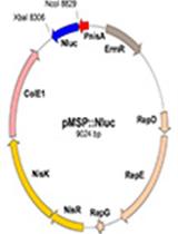
Listeria innocua Biofilm Assay Using NanoLuc Luciferase

用NanoLuc荧光素酶检测李斯特菌生物膜

AB Aleš Berlec
NJ Nika Janež
MS Meta Sterniša
AK Anja Klančnik
JS Jerica Sabotič
2390 Views
Feb 5, 2022
Biofilms serve as a bacterial survival strategy, allowing bacteria to persist under adverse environmental conditions. The non-pathogenic Listeria innocua is used as a surrogate organism for the foodborne pathogen Listeria monocytogenes, because they share genetic and physiological similarities and can be used in a Biosafety Level 1 laboratory. Several methods are used to evaluate biofilms, including different approaches to determine biofilm biomass or culturability, viability, metabolic activity, or other microbial community properties. Routinely used methods for biofilm assay include the classical culture-based plate counting method, biomass staining methods (e.g., crystal violet and safranin red), DNA staining methods (e.g., Syto 9), methods that use metabolic substrates to detect live bacteria (e.g., tetrazolium salts or resazurin), and PCR-based methods to quantify bacterial DNA. The NanoLuc (Nluc) luciferase biofilm assay is a viable alternative or complement to existing methods. Functional Nluc was expressed in L. innocua using the nisin-inducible expression system and bacterial detection was performed using furimazine as substrate. Concentration dependent bioluminescence signals were obtained over a concentration range greater than three log units. The Nluc bioluminescence method allows absolute quantification of bacterial cells, has high sensitivity, broad range, good day-to-day repeatability, and good precision with acceptable accuracy. The advantages of Nluc bioluminescence also include direct detection, absolute cell quantification, and rapid execution.Graphic abstract:Engineering Listeria innocua to express NanoLuc and its application in bioluminescence assay.

神经科学

经腔内缝合的短暂性大脑中动脉闭塞可重复诱导小鼠缺血性脑卒中

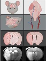
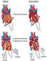
Transient Middle Cerebral Artery Occlusion with an Intraluminal Suture Enables Reproducible Induction of Ischemic Stroke in Mice

经腔内缝合的短暂性大脑中动脉闭塞可重复诱导小鼠缺血性脑卒中

LL Luke R. Lemmerman
HH Hallie N. Harris
MB Maria H.H. Balch
MR Maria A. Rincon-Benavides
NH Natalia Higuita-Castro
WA W. David Arnold
DG Daniel Gallego-Perez
6007 Views
Feb 5, 2022
Ischemic stroke is a leading cause of mortality and chronic disability worldwide, underscoring the need for reliable and accurate animal models to study this disease’s pathology, molecular mechanisms of injury, and treatment approaches. As most clinical strokes occur in regions supplied by the middle cerebral artery (MCA), several experimental models have been developed to simulate an MCA occlusion (MCAO), including transcranial MCAO, micro- or macro-sphere embolism, thromboembolisation, photothrombosis, Endothelin-1 injection, and – the most common method for ischemic stroke induction in murine models – intraluminal MCAO. In the intraluminal MCAO model, the external carotid artery (ECA) is permanently ligated, after which a partially-coated monofilament is inserted and advanced proximally to the common carotid artery (CCA) bifurcation, before being introduced into the internal carotid artery (ICA). The coated tip of the monofilament is then advanced to the origin of the MCA and secured for the duration of occlusion. With respect to other MCAO models, this model offers enhanced reproducibility regarding infarct volume and cognitive/functional deficits, and does not require a craniotomy. Here, we provide a detailed protocol for the surgical induction of unilateral transient ischemic stroke in mice, using the intraluminal MCAO model.Graphic abstract: Overview of the intraluminal monofilament method for transient middle cerebral artery occlusion (MCAO) in mouse.
对斑马鱼睡眠的大规模分析

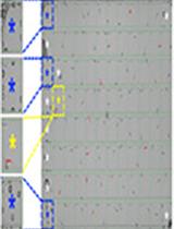
Large-scale Analysis of Sleep in Zebrafish

对斑马鱼睡眠的大规模分析

DL Daniel A. Lee
GO Grigorios Oikonomou
DP David A. Prober
3952 Views
Feb 5, 2022
Over the past decade, zebrafish have emerged as a powerful model for the study of vertebrate sleep and wake behaviors. Experimental evidence has demonstrated behavioral, anatomical, genetic, and pharmacological conservation of sleep between zebrafish and mammals, suggesting that discoveries in zebrafish can inform our understanding of mammalian sleep. Here, we describe a protocol for performing sleep behavioral experiments in larval zebrafish, using a high-throughput video tracking system. We explain how to set up a sleep behavioral experiment and provide guidelines on how to analyze the data. Using this protocol, a typical experiment can be completed in less than five days, and this method provides a scalable platform to perform genetic and pharmacological screens in a simple and cost-effective vertebrate model. By combining high-throughput behavioral assays with several advantageous features of zebrafish, this model system provides new opportunities to make discoveries that clarify the genetic and neurological mechanisms that regulate sleep.
大鼠持续性社交失败压力诱导

Induction of Repeated Social Defeat Stress in Rats

大鼠持续性社交失败压力诱导

SM Soumyabrata Munshi
AR Alexandra Ritger
JR J. Amiel Rosenkranz
2456 Views
Feb 5, 2022
Repeated social defeat stress (RSDS) is a model of chronic stress in rodents. There are several variants of social defeat procedures that exert robust effects in mice, but few published detailed protocols to produce a robust stress and altered immunological profile in rats. In this article, we describe the protocol for the induction of RSDS in adult male Sprague-Dawley rats. Using a resident-intruder paradigm, a physical component of stress is induced by direct attack from the resident aggressive retired breeder Long-Evans rats on the intruder experimental rats. A subsequent threat component is induced by the presence of the aggressor in the vicinity of the intruder, but with physical separation between them. The RSDS induced by this protocol produces robust immunological and behavioral changes in the experimental rats, as evidenced by development of anxiety-like behaviors in open field, social interaction, and elevated plus maze tests, as well as by changes in immune parameters (Munshi et al., 2020). This approach has been used as an ethologically relevant model of stressors that are potent enough to impact neural circuits that are similar to the neural circuits impacted in patients with depression and anxiety.

干细胞

将小鼠胚胎干细胞体外分化为巨噬细胞的有效方法

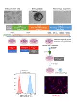
Efficient Method to Differentiate Mouse Embryonic Stem Cells into Macrophages in vitro

将小鼠胚胎干细胞体外分化为巨噬细胞的有效方法

QH Qimin Hai
JH Juying Han
SW Sophia Wells
JS Jonathan D. Smith
3113 Views
Feb 5, 2022
Macrophages are key cells in the innate immune system and play a role in a variety of diseases. However, macrophages are terminally differentiated and difficult to manipulate genetically via transfection or through CRISPR-Cas9 gene editing. To overcome this limitation, we provide a simplified protocol for the generation of mouse embryonic stem cells-derived macrophages (ESDM). Thus, genetic manipulation can be performed using embryonic stem cells, selecting for the desired changes, and finally producing macrophages to study the effects of the previous genetic manipulation. These studies can contribute to many areas of research, including atherosclerosis and inflammation. Production of ESDM has been previously achieved using embryoid body (EB) intermediates. Here, we optimized the EB method using a simplified medium, reducing the number of recombinant proteins and medium recipes required. Our EB-based differentiation protocol consists of three stages: 1) floating EB formation; 2) adherence of EBs and release of floating macrophage progenitors; and, 3) terminal differentiation of harvested macrophage progenitors. The advantages of this protocol include achieving independent floating EBs in stage 1 by using a rocker within the tissue culture incubator, as well as the exclusion of small EBs and cell clusters when harvesting macrophage progenitors via cell filtration.

更正

更正:使用磁性纳米颗粒进行单细胞测序从胚胎日 E13.5 小鼠胎肝中分离健康的 F4/80+ 巨噬细胞

Correction Notice: Isolation of Healthy F4/80+ Macrophages from Embryonic day E13.5 Mouse Fetal Liver Using Magnetic Nanoparticles for Single Cell Sequencing

更正:使用磁性纳米颗粒进行单细胞测序从胚胎日 E13.5 小鼠胎肝中分离健康的 F4/80+ 巨噬细胞

Kaustav Mukherjee Kaustav Mukherjee
JB James J. Bieker
792 Views
Feb 5, 2022