往期刊物2022

卷册: 12, 期号: 2

left

Dec

20

Dec

5

Nov

20

Nov

5

Oct

20

Oct

5

Sep

20

Sep

5

Aug

20

Aug

5

Jul

20

Jul

5

Jun

20

Jun

5

May

20

May

5

Apr

20

Apr

5

Mar

20

Mar

5

Feb

20

Feb

5

Jan

20

Jan

5

right

生物工程

通过粘度测量细胞裂解液中寡核苷酸的水解

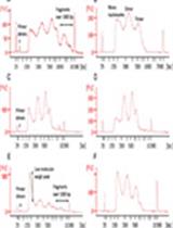
Measuring Oligonucleotide Hydrolysis in Cellular Lysates via Viscosity Measurements

通过粘度测量细胞裂解液中寡核苷酸的水解

RM Romel Menacho-Melgar
ML Michael D. Lynch
2190 Views
Jan 20, 2022
Cell lysis, a process that releases host oligonucleotides, is required in many biotechnological applications. However, intact oligonucleotides in crude cellular lysates increase the viscosity of lysates, which complicates downstream processes and routine laboratory workflows. To address this, nucleases that hydrolyze the intact oligonucleotides are commonly added, either as purified enzymes or co-expressed in genetically engineered bacterial strains. To measure oligonucleotide hydrolysis, common DNA quantification methods, such as qPCR or fluorescence-based, require expensive reagents and equipment, and cannot distinguish different-sized DNA fragments. Here, we outline a simple alternative method for measuring DNA/RNA hydrolysis in cellular lysates, by measuring their viscosity. This method only requires common laboratory supplies and a cell phone camera.

癌症生物学

小鼠结肠和小肠类器官的制备和培养,包括基因表达和类器官活力的分析

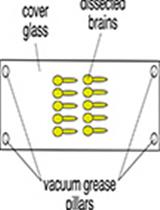
Preparation and Cultivation of Colonic and Small Intestinal Murine Organoids Including Analysis of Gene Expression and Organoid Viability

小鼠结肠和小肠类器官的制备和培养,包括基因表达和类器官活力的分析

LK Luisa Klemke
Joshua P. Blume Joshua P. Blume
TD Tiago De Oliveira
RS Ramona Schulz-Heddergott
6162 Views
Jan 20, 2022
Organoids are complex three-dimensional structures, which contain different cell types and help to overcome many limitations of conventional 2D cell culture techniques. Here, we present a protocol for the cultivation of murine matched-pairs of small intestinal and colonic epithelial organoids, and colonic tumor organoids derived from the chemical colorectal cancer (CRC) AOM/DSS mouse model. Therefore, intestinal crypts or tumor tissue containing stem cells are isolated from the same donor mouse and cultivated in Matrigel®. The culture medium is supplemented with different growth factors to model the intestinal stem cell niche, allowing their self-renewal and differentiation. Matched-pair organoids enable the analysis of pharmacological effects and the tumor selectivity of drugs.Graphic abstract: Schematic overview of colonic matched pair organoid preparation, generated from the chemical AOM/DSS colorectal cancer mouse model. Please note that normal colon-derived organoids (green) differ in their morphology from tumor-derived organoids (red). Normal colonic-derived organoids display a thicker and crypt-like epithelial layer, whereas tumor-derived organoids are round with a thin epithelial layer.
一种监测小鼠肺中单核细胞和肿瘤细胞与纳米颗粒实时相互作用的替代技术

An Alternative Technique for Monitoring the Live Interaction of Monocytes and Tumor Cells with Nanoparticles in the Mouse Lung

一种监测小鼠肺中单核细胞和肿瘤细胞与纳米颗粒实时相互作用的替代技术

FR Fernanda Ramos-Gomes
NF Nathalia Ferreira
FA Frauke Alves
MM M. Andrea Markus
2623 Views
Jan 20, 2022
Nanomaterials are increasingly used for the diagnosis and treatment of cancer, including lung cancer. For the clinical translation of nano-based theranostics, it is vital to detect and monitor their accumulation in the tumor, as well as their interaction with tumor, immune cells, and the tumor microenvironment (TME). While high resolution microscopy of fixed tumor specimens can provide some of this information from individual thin slices, it cannot capture cellular events over time and lacks 3D information of the tumor tissue. On the other hand, in vivo optical procedures either fall short of providing the necessary cellular resolution, as in the case of epifluorescence optical imaging, or are very demanding, as for instance intravital lung microscopy. We describe an alternative approach to investigate nanoparticle-cell interactions in entire mouse lung lobes, by longitudinal live cell confocal microscopy at nanometer resolution. By filling the lung ex vivo with 1% agarose, we were able to stabilize the lung lobes and visualize the interaction of fluorescent cells and nanoparticles for at least 4 hours post mortem. This high resolution ex vivo live cell imaging approach is an easy 4D tool for assessing several dynamic processes in tumor tissue, such as the traffic of cells, shedding of extracellular vesicles (EVs), and the accumulation of nanoparticles in tumor tissue.Graphic abstract: Schematic of the workflow for live cell imaging in the mouse lung.

发育生物学

结合免疫荧光和定量荧光原位杂交分析果蝇淋巴腺小细胞中的差异基因表达

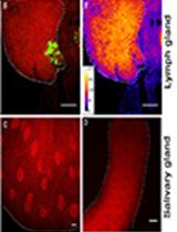
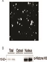
Combination of Immunofluorescence and Quantitative Fluorescence In-situ Hybridization for Analysing Differential Gene Expression in the Niche Cells of the Drosophila Lymph Gland

结合免疫荧光和定量荧光原位杂交分析果蝇淋巴腺小细胞中的差异基因表达

PR Parvathy Ramesh
SG Sushmit Ghosh
LM Lolitika Mandal
2867 Views
Jan 20, 2022
The Drosophila larval haematopoietic organ or lymph gland consists of multiple cell types arranged in zones. The smallest stem cell compartment consists of 40-45 cells that constitute the haematopoietic niche. In order to analyse the haematopoietic niche, it needs to be labelled with a specific antibody to differentiate it from the other cell types. To characterise a phenotype, it is often necessary to investigate the expression of a gene in a particular stem cell compartment within the lymph gland. In such a situation, in-situ hybridization is performed, as it indicates the localization of gene expression. Although chromogenic in-situ hybridization enables us to compare the signal and tissue morphology simultaneously, it fails to harness the information related to the degree of gene expression. Dual immunofluorescence and in-situ hybridization (IF-FISH) serves as the powerful technique that helps to visualize both protein and mRNA expression within the same cell type. This technique also provides reliable quantification regarding mRNA expression levels. When dealing with a few cells within the organ, like the niche of the larval lymph gland, fluorescently labelled riboprobes allows us to localize and assess the magnitude of gene expression within the niche cells, which are also immunolabelled with a niche-specific marker, to distinguish them from the adjoining cell types.

药物发现

使用生物发光病原菌的抗菌制剂的体内外快速评价

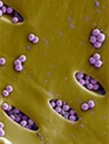
Rapid in vitro and in vivo Evaluation of Antimicrobial Formulations Using Bioluminescent Pathogenic Bacteria

使用生物发光病原菌的抗菌制剂的体内外快速评价

AS Artur Schmidtchen
MP Manoj Puthia
3202 Views
Jan 20, 2022
Basic and translational research needs rapid methods to test antimicrobial formulations. Bioluminescent bacteria and advanced imaging systems capable of acquiring bioluminescence enable us to quickly and longitudinally evaluate the efficacy of antimicrobials. Conventional approaches, such as radial diffusion and viable count assays, are time-consuming and do not allow for longitudinal analysis. Bioluminescence imaging is sensitive and gives vital spatial and temporal information on the infection status in the body. Here, using bioluminescent Pseudomonas aeruginosa, we describe an in vitro and an in vivo approach to rapidly evaluate the antimicrobial efficacy of the host-defense peptide TCP-25.Graphic abstract: Evaluation of antimicrobials using bioluminescent bacteria.

免疫学

一种高通量自动ELISA检测SARS-CoV-2刺突蛋白IgG抗体的方法

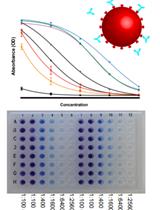
A High-throughput Automated ELISA Assay for Detection of IgG Antibodies to the SARS-CoV-2 Spike Protein

一种高通量自动ELISA检测SARS-CoV-2刺突蛋白IgG抗体的方法

JC Juliana Conkright-Fincham
CT Chieri Tomomori-Sato
RM Rich McGhee
EL Ella M. Leslie
CB Carolyn J. Beucher
LW Lauren E. Weems
SS Shigeo Sato
WR William B. Redwine
KW Kyle J. Weaver
BM Brandon D. Miller
KD Kym M. Delventhal
JK John J. Kary
AK Andrew B. Koebbe
AD Alexander Dean
JW Jessica L. Witt
LR Laura M. Remy
TP Tari J. Parmely
CZ Chongbei Zhao
YW Yan Wang
JC Joan W. Conaway
JU Jay R. Unruh
3812 Views
Jan 20, 2022
The SARS-CoV-2 pandemic and vaccination campaign has illustrated the need for high throughput serological assays to quantitatively measure antibody levels. Here, we present a protocol for a high-throughput colorimetric ELISA assay to detect IgG antibodies against the SARS-CoV-2 spike protein. The assay robustly distinguishes positive from negative samples, while controlling for potential non-specific binding from serum samples. To further eliminate background contributions, we demonstrate a computational pipeline for fitting ELISA titration curves, that produces an extremely sensitive antibody signal metric for quantitative comparisons across samples and time.

微生物学

秀丽隐杆线虫细菌负荷的定量研究

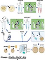
Quantification of Bacterial Loads in Caenorhabditis elegans

秀丽隐杆线虫细菌负荷的定量研究

AW Alyssa C. Walker
RB Rohan Bhargava
AV Alfonso S. Vaziriyan-Sani
AB Amanda S. Brust
DC Daniel M. Czyz
3394 Views
Jan 20, 2022
Caenorhabditis elegans is a ubiquitous free-living nematode that feeds on bacteria. The organism was introduced into a laboratory setting in the 1970s and has since gained popularity as a model to study host-bacteria interactions. One advantage of using C. elegans is that its intestine can be colonized by the bacteria on which it feeds. Quantifying the bacterial load within C. elegans is an important and easily obtainable metric when investigating host-bacteria interactions. Although quantification of bacteria harbored in C. elegans via whole-worm lysis is not a novel assay, there is great variation between existing methods. To lyse C. elegans, many protocols rely on the use of a hand-held homogenizer, which could introduce systematic error and subsequent variation between researchers performing the same experiment. Here, we describe a method of lysing the intestines of C. elegans to quantify the bacterial load within the intestine. Our method has been optimized for removing exogenous bacteria while maintaining worm paralysis, to ensure no bactericidal agents are swallowed, which could kill bacteria within the intestine and affect results. We utilize and compare the efficiency of two different homogenization tools: a battery-powered hand-held homogenizer, and a benchtop electric homogenizer, where the latter minimizes variability. Thus, our protocol has been optimized to reduce systematic error and decrease the potential for variability among experimenters.Graphic abstract: Simplified overview of the procedure used to quantify the bacterial load within C. elegans. The two different methods are herein described for worm lysis: “Option 1” is a hand-held homogenizer, and “Option 2” is a benchtop homogenizer.
利用两阶段自诱导细胞自溶和DNA/RNA自水解在大肠杆菌中表达和提取蛋白

Simple Scalable Protein Expression and Extraction Using Two-stage Autoinducible Cell Autolysis and DNA/RNA Autohydrolysis in Escherichia coli

利用两阶段自诱导细胞自溶和DNA/RNA自水解在大肠杆菌中表达和提取蛋白

RM Romel Menacho-Melgar
ML Michael D. Lynch
2977 Views
Jan 20, 2022
Recombinant protein expression is extensively used in biological research. Despite this, current protein expression and extraction methods are not readily scalable or amenable for high-throughput applications. Optimization of protein expression conditions using traditional methods, reliant on growth-associated induction, is non-trivial. Similarly, protein extraction methods are predominantly restricted to chemical methods, and mechanical methods reliant on expensive specialized equipment more tuned for large-scale applications. In this article, we outline detailed protocols for the use of an engineered autolysis/autohydrolysis E. coli strain, in two-stage fermentations in shake-flasks. This two-stage fermentation protocol does not require optimization of expression conditions and results in high protein titers. Cell lysis in an engineered strain is tightly controlled and only triggered post-culture by addition of a 0.1% detergent solution. Upon cell lysis, a nuclease digests contaminating host oligonucleotides, which facilitates sample handling. This method has been validated for use at different scales, from microtiter plates to instrumented bioreactors.Graphic abstract: Two-stage protein expression, cell autolysis and DNA/RNA autohydrolysis. Reprinted with permission from Menacho-Melgar et al. (2020a). Copyright 2020 John Wiley and Sons.
酵母暴露于可见光下的高通量生长测量

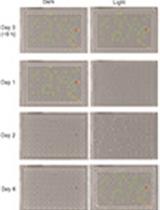
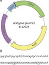
High-throughput Growth Measurements of Yeast Exposed to Visible Light

酵母暴露于可见光下的高通量生长测量

KL Katarina Logg
MA Mikael Andersson
AB Anders Blomberg
MM Mikael Molin
2396 Views
Jan 20, 2022
Light is a double-edged sword: it is essential for life on the planet but also causes cellular damage and death. Consequently, organisms have evolved systems not only for harvesting and converting light energy into chemical energy but also for countering its toxic effects. Despite the omnipresence and importance of such light-dependent effects, there are very few unbiased genetic screens, if any, investigating the mechanistic consequences that visible light has on cells. Baker’s yeast, Saccharomyces cerevisiae, is one of the best annotated organisms thanks to several easily available mutant collections and its amenability to high-throughput genetic screening. However, until recently this yeast was thought to lack receptors for visible light, therefore its response to visible light was poorly understood. Nevertheless, a couple of years ago it was discovered that yeast senses light via a novel and unconventional pathway involving a peroxisomal oxidase, hydrogen peroxide, and a particular type of antioxidant protein, called peroxiredoxin. Here, we describe in detail a protocol for scoring yeast genes involved in the resistance to visible light (400-700 nm) on a genome-wide scale. Because cells in dense cultures shield each other from light exposure, resulting in apparent light resistance, our method involves adaptations to reduce inoculum size under conditions amenable to high-throughput screens, to properly be able to identify light-sensitive mutants. We also describe how to measure growth in the presence of light, including two follow-up validation tests. In this way, this method makes it possible to score light-sensitivity on a genome-wide scale with high confidence.Graphic abstract: Overview of strategy for high-throughput determination of yeast growth upon visible light stress.

分子生物学

哺乳动物冷冻保存细胞的ATAC测序方法

ATAC Sequencing Protocol For Cryopreserved Mammalian Cells

哺乳动物冷冻保存细胞的ATAC测序方法

JC Juan Manuel Caravaca
MM Monika Mehta
SG Sujatha Gowda
BT Bao Tran
4367 Views
Jan 20, 2022
ATAC-seq (assay for transposase-accessible chromatin with high-throughput sequencing) is a powerful method to evaluate chromatin accessibility and nucleosome positioning at a genome-wide scale. This assay uses a hyperactive Tn5 transposase, to simultaneously cut open chromatin and insert adapter sequences. After sequencing, the reads generated through this technique are generally indicative of transcriptional regulatory elements that are located in accessible chromatin. This method was originally developed by Buenrostro et al. (2013), and since then it has been improved by the same authors several times, until their last update called OMNI ATAC-seq (Corces et al., 2017). Here, we describe an ATAC-seq protocol based on the OMNI-ATAC method, with a special focus on the initial steps of thawing cryopreserved cells, and the final steps of library purification using magnetic beads. This protocol can be of interest for laboratories working in a fast-paced environment.Graphic abstract: Flowchart of the protocol

神经科学

斑马鱼幼虫三氯乙酸固定及抗体染色

Trichloroacetic Acid Fixation and Antibody Staining of Zebrafish Larvae

斑马鱼幼虫三氯乙酸固定及抗体染色

EM E. Anne Martin
SI Sundas Ijaz
AP Alberto E. Pereda
AM Adam C. Miller
3195 Views
Jan 20, 2022
Larval zebrafish have been established as an excellent model for examining vertebrate biology, with many researchers using the system for neuroscience. Controlling a fast escape response of the fish, the Mauthner cells and their associated network are an attractive model, given their experimental accessibility and fast development, driving ethologically relevant behavior in the first five days of development. Here, we describe methods for immunostaining electrical and chemical synapse proteins at 3-7 days post fertilization (dpf) in zebrafish using tricholoracetic acid fixation. The methods presented are ideally suited to easily visualize neural circuits and synapses within the fish.
膜包裹的纳米颗粒上g蛋白信号通路的膜相关组分的重构

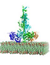
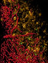
Reconstitution of Membrane-associated Components of a G-protein Signaling Pathway on Membrane-coated Nanoparticles (Lipobeads)

膜包裹的纳米颗粒上g蛋白信号通路的膜相关组分的重构

MI Michael J. Irwin
XW Xin Wang
RC Rick H. Cote
2264 Views
Jan 20, 2022
G-protein coupled signaling pathways are organized into multi-protein complexes called signalosomes that are located within and on cellular membranes. We describe the use of silica nanoparticles coated with a unilamellar phospholipid bilayer (lipobeads) to reconstitute the activated photoreceptor G-protein α-subunit (Gtα*) with its cognate effector (phosphodiesterase-6; PDE6) for biochemical and structural studies of the activation mechanism regulating this GPCR signaling pathway. Lipobeads are prepared by resuspending dried-down phospholipid mixtures with monodisperse 70 nm silica particles, followed by extrusion through a 100 nm membrane filter. This uniform and supported liposomal preparation is easily sedimented, permitting the separation of soluble from membrane-associated proteins. Upon loading lipobeads with Gtα* and PDE6, we find that activation of PDE6 catalysis by Gtα* occurs much more efficiently than in the absence of membranes. Chemical cross-linking of membrane-confined proteins allows detection of changes in protein-protein interactions, resulting from G-protein activation of PDE6. The advantages of using lipobeads over partially purified membrane preparations or traditional liposomal preparations are generally applicable to the study of other membrane-confined signal transduction pathways.

植物科学

拟南芥幼苗核蛋白的分离与提取

Fractionation and Extraction of Crude Nuclear Proteins From Arabidopsis Seedlings

拟南芥幼苗核蛋白的分离与提取

JZ Jiajia Zhao
JB Jinsong Bao
FX Feifei Xu
5326 Views
Jan 20, 2022
The plant nucleus is an important subcellular organelle that contains the genome, ribosomal RNA, and regulatory proteins, and performs a central role in the functioning and metabolism of the cell. Fractionation of intact nuclei is a crucial process to elucidate the function of nuclear proteins. Here, we present a simple method for the fractionation of crude nuclei and extraction of nuclear proteins, based on previously established methods. This protocol provides an easy and quick method to isolate crude nuclei and extract nuclear proteins from Arabidopsis seedlings, which is useful for the research on the nuclear proteins, without requirement for high-purity nuclei.Graphic abstract: Schematic procedure for the isolation of crude nuclei and extraction of nuclear proteins from Arabidopsis seedlings.
甜菜和拟南芥幼苗丝核枯菌侵染试验

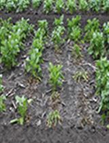
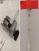
Rhizoctonia solani Infection Assay of Young Sugar Beet and Arabidopsis plantlets

甜菜和拟南芥幼苗丝核枯菌侵染试验

FD Fredrik Dölfors
LH Louise Holmquist
GT Georgios Tzelepis
CD Christina Dixelius
3242 Views
Jan 20, 2022
Rhizoctonia solani is a soil-borne fungus, which rarely produces any spores in culture. Hence, all inoculation procedures are based on mycelia, often as a coat on cereal kernels, placed in close vicinity to the plant to be infected. In this protocol, an inoculation method is described where the fungus is first allowed to infest a perlite-maize flour substrate for 10 days, followed by thorough soil mixing to generate uniform fungal distribution. Pre-grown seedlings are then replanted in the infested soil. Plant materials can be harvested, five (sugar beet) and ten days (Arabidopsis) post infection, followed by a rapid cleaning step ahead of any nucleic acid preparation. Commercial DNA or RNA extraction kits can be used or, if higher DNA yield is required, a CTAB extraction method. Our purpose was to develop a reliable and reproducible protocol to determine the infection levels in planta upon infection with R. solani. This protocol is less laborious compared to previous ones, improves the consistency of plant infection, reproducibility between experiments, and suits both a root crop and Arabidopsis.Graphic abstract: Overview of the R. solani infection procedure.

干细胞

从三维到二维:不同物种肠道类器官细胞培养插件的二维培养方法的协调

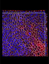
From 3D to 2D: Harmonization of Protocols for Two-dimensional Cultures on Cell Culture Inserts of Intestinal Organoids from Various Species

从三维到二维:不同物种肠道类器官细胞培养插件的二维培养方法的协调

DW David Warschkau
ED Estefanía Delgado-Betancourt
DH David Holthaus
AM Antonia Müller
GK Gudrun Kliem
SK Susanne M. Krug
JS Joerg-Dieter Schulzke
TA Toni Aebischer
CK Christian Klotz
FS Frank Seeber
4743 Views
Jan 20, 2022
In the expanding field of intestinal organoid research, various protocols for three- and two-dimensional organoid-derived cell cultures exist. Two-dimensional organoid-derived monolayers are used to overcome some limitations of three-dimensional organoid cultures. They are increasingly used also in infection research, to study physiological processes and tissue barrier functions, where easy experimental access of pathogens to the luminal and/or basolateral cell surface is required. This has resulted in an increasing number of publications reporting different protocols and media compositions for organoid manipulation, precluding direct comparisons of research outcomes in some cases. With this in mind, here we describe a protocol aimed at the harmonization of seeding conditions for three-dimensional intestinal organoids of four commonly used research species onto cell culture inserts, to create organoid-derived monolayers that form electrophysiologically tight epithelial barriers. We give an in-depth description of media compositions and culture conditions for creating these monolayers, enabling also the less experienced researchers to obtain reproducible results within a short period of time, and which should simplify the comparison of future studies between labs, but also encourage others to consider these systems as alternative cell culture models in their research.Graphic abstract: Schematic workflow of organoid-derived monolayer generation from intestinal spheroid cultures. ECM, extracellular matrix; ODM, organoid-derived monolayer.
利用DNA和线粒体染料流式细胞术分析涡虫干细胞

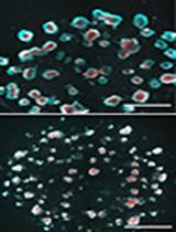
Flow Cytometry Analysis of Planarian Stem Cells Using DNA and Mitochondrial Dyes

利用DNA和线粒体染料流式细胞术分析涡虫干细胞

MM Mohamed Mohamed Haroon
PV Praveen Kumar Vemula
DP Dasaradhi Palakodeti
2744 Views
Jan 20, 2022
Planarians are free-living flatworms that emerged as a crucial model system to understand regeneration and stem cell biology. The ability to purify neoblasts, the adult stem cell population of planaria, through fluorescence-activated cell sorting (FACS) has tremendously increased our understanding of pluripotency, specialization, and heterogeneity. To date, the FACS-based purification methods for neoblasts relied on nuclear dyes that discriminate proliferating cells (>2N), as neoblasts are the only dividing somatic cells. However, this method does not distinguish the functional states within the neoblast population. Our work has shown that among the neoblasts, the pluripotent stem cells (PSCs) are associated with low mitochondrial content and this property could be leveraged for purification of the PSC-enriched population. Using the mitochondrial dye MitoTracker Green (MTG) and the nuclear dye SiR-DNA, we have described a method for isolation of PSCs that are viable and compatible with downstream experiments, such as transplantation and cell culture. In this protocol, we provide a detailed description for sample preparation and FACS gating for neoblast isolation in planaria.