往期刊物2021

卷册: 11, 期号: 21

left

Dec

20

Dec

5

Nov

20

Nov

5

Oct

20

Oct

5

Sep

20

Sep

5

Aug

20

Aug

5

Jul

20

Jul

5

Jun

20

Jun

5

May

20

May

5

Apr

20

Apr

5

Mar

20

Mar

5

Feb

20

Feb

5

Jan

20

Jan

5

right

生物化学

弓形虫热蛋白质组分析以识别顶复体寄生虫中的蛋白质-配体相互作用

Thermal Proteome Profiling to Identify Protein-ligand Interactions in the Apicomplexan Parasite Toxoplasma gondii

弓形虫热蛋白质组分析以识别顶复体寄生虫中的蛋白质-配体相互作用

AH Alice L. Herneisen
SL Sebastian Lourido
4909 Views
Nov 5, 2021
Toxoplasma gondii is a single-celled eukaryotic parasite that chronically infects a quarter of the global population. In recent years, phenotypic screens have identified compounds that block parasite replication. Unraveling the pathways and molecular mechanisms perturbed by such compounds requires target deconvolution. In parasites, such deconvolution has been achieved via chemogenomic approaches—for example, directed evolution followed by whole-genome sequencing or genome-wide knockout screens. As a proteomic alternative that directly probes the physical interaction between compound and protein, thermal proteome profiling (TPP), also known as the cellular thermal shift assay (CETSA), recently emerged as a method to identify small molecule–target interactions in living cells and cell extracts in a variety of organisms, including unicellular eukaryotic pathogens. Ligand binding induces a thermal stability shift—stabilizing or destabilizing proteins that change conformationally in response to the ligand—that can be measured by mass spectrometry (MS). Cells are incubated with different concentrations of ligand and heated, causing thermal denaturation of proteins. The soluble protein is extracted and quantified with multiplexed, quantitative MS, resulting in thousands of thermal denaturation profiles. Proteins engaging the ligand can be identified by their compound-dependent thermal shift. The protocol provided here can be used to identify ligand-target interactions and assess the impact of environmental or genetic perturbations on the thermal stability of the proteome in T. gondii and other eukaryotic pathogens.Graphic abstract: Thermal proteome profiling for target identification in the apicomplexan parasite T. gondii.

生物工程

开发具有 BMP-2 和 VEGF 时空定义模式的 3D 生物打印支架,用于大骨缺损的再生

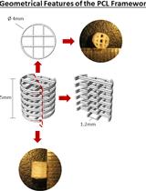
Development of a 3D Bioprinted Scaffold with Spatio-temporally Defined Patterns of BMP-2 and VEGF for the Regeneration of Large Bone Defects

开发具有 BMP-2 和 VEGF 时空定义模式的 3D 生物打印支架,用于大骨缺损的再生

FF Fiona E. Freeman
PP Pierluca Pitacco
Lv Lieke H. A. van Dommelen
JN Jessica Nulty
DB David C. Browe
JS Jung-Youn Shin
EA Eben Alsberg
DK Daniel J. Kelly
2860 Views
Nov 5, 2021
The local delivery of growth factors such as BMP-2 is a well-established strategy for the repair of bone defects. The limitations of such approaches clinically are well documented and can be linked to the need for supraphysiological doses and poor spatio-temporal control of growth factor release in vivo. Using bioprinting techniques, it is possible to generate implants that can deliver cytokines or growth factors with distinct spatiotemporal release profiles and patterns to enhance bone regeneration. Specifically, for bone healing, several growth factors, including vascular endothelial growth factor (VEGF) and bone morphogenic proteins (BMPs), have been shown to be expressed at different phases of the process. This protocol aims to outline how to use bioprinting strategies to deliver growth factors, both alone or in combination, to the site of injury at physiologically relevant dosages such that repair is induced without adverse effects. Here we describe: the printing parameters to generate the polymer mechanical backbone; instructions to generate the different bioinks and allow for the temporal control of both growth factors; and the printing process to develop implants with spatially defined patterns of growth factors for bone regeneration. The novelty of this protocol is the use of multiple-tool fabrication techniques to develop an implant with spatio-temporal control of growth factor delivery for bone regeneration. While the overall aim of this protocol was to develop an implant for bone regeneration, the technique can be modified and used for a variety of regenerative purposes.Graphic abstract: 3D Bioprinting Spatio-Temporally Defined Patterns of Growth Factors to Tightly Control Bone Tissue Regeneration.

癌症生物学

通过荧光测量实时 DNA/RNA 核酸酶活性

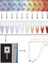
Measuring Real-time DNA/RNA Nuclease Activity through Fluorescence

通过荧光测量实时 DNA/RNA 核酸酶活性

PW Paulina Wyrzykowska
SR Sally Rogers
RC Richard Chahwan
3122 Views
Nov 5, 2021
DNA and RNA nucleases are wide-ranging enzymes, taking part in broad cellular processes from DNA repair to immune response control. Growing interest in the mechanisms and activities of newly discovered nucleases inspired us to share the detailed protocol of our nuclease assay (Sheppard et al., 2019). This easy and inexpensive method can provide data that enables understanding of the molecular mechanism for novel or tested nucleases, from substrate preference and cofactors involved to catalytic rate of reaction.

发育生物学

APEX 介导的细胞外囊泡靶向细胞中蛋白质的邻近标记

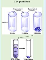
APEX-mediated Proximity Labeling of Proteins in Cells Targeted by Extracellular Vesicles

APEX 介导的细胞外囊泡靶向细胞中蛋白质的邻近标记

LS Lu Song
JC Jun Chen
AS Angela Sun
RS Randy Schekman
4098 Views
Nov 5, 2021
Extracellular vesicles (EVs) are thought to mediate intercellular communication through the delivery of cargo proteins and RNA to target cells. The uptake of EVs is often followed visually using lipophilic-dyes or fluorescently-tagged proteins to label membrane constituents that are then internalized into recipient cells (Christianson et al., 2013; De Jong et al., 2019). However, these methods do not probe the exposure of EV cargo to intracellular compartments, such as the cytoplasm and nucleus, where protein or RNA molecules could elicit functional changes in recipient cells. In this protocol, we employ an EV cargo protein-APEX fusion to detect proximity interactions with recipient cell cytoplasmic/nuclear targets. This approach results in the biotinylation of proteins in close contact with the reporter fusion and thus permits profiling of biotinylated proteins affinity purified on immobilized streptavidin beads.Graphic abstract: Schematic showing three steps of APEX-mediated proximity labeling of proteins in cells targeted by EVs.
黑腹果蝇淋巴腺的完整原位制备用于综合幼虫造血分析

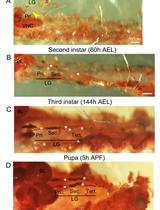
Intact in situ Preparation of the Drosophila melanogaster Lymph Gland for a Comprehensive Analysis of Larval Hematopoiesis

黑腹果蝇淋巴腺的完整原位制备用于综合幼虫造血分析

DR Diana Rodrigues
KV K. VijayRaghavan
LW Lucas Waltzer
MI Maneesha S. Inamdar
3372 Views
Nov 5, 2021
Blood cells have a limited lifespan and are replenished by a small number of hematopoietic stem and progenitor cells (HSPCs). Adult vertebrate hematopoiesis occurs in the bone marrow, liver, and spleen, rendering a comprehensive analysis of the entire HSPC pool nearly impossible. The Drosophila blood system is well studied and has developmental, molecular, and functional parallels with that of vertebrates. Unlike vertebrates, post-embryonic hematopoiesis in Drosophila is essentially restricted to the larval lymph gland (LG), a multi-lobed organ that flanks the dorsal vessel. Because the anterior-most or primary lobes of the LG are easy to dissect out, their cellular and molecular characteristics have been studied in considerable detail. The 2-3 pairs of posterior lobes are more delicate and fragile and have largely been ignored. However, posterior lobes harbor a significant blood progenitor pool, and several hematopoietic mutants show differences in phenotype between the anterior and posterior lobes. Hence, a comprehensive analysis of the LG is important for a thorough understanding of Drosophila hematopoiesis. Most studies focus on isolating the primary lobes by methods that generally dislodge and damage other lobes. To obtain preparations of the whole LG, including intact posterior lobes, here we provide a detailed protocol for larval fillet dissection. This allows accessing and analyzing complete LG lobes, along with dorsal vessel and pericardial cells. We demonstrate that tissue architecture and integrity is maintained and provide methods for quantitative analysis. This protocol can be used to quickly and effectively isolate complete LGs from first instar larval to pupal stages and can be implemented with ease.
靶向小鼠卫星细胞中长链非编码 RNA 的表达

Targeting the Expression of Long Noncoding RNAs in Murine Satellite Cells from Single Myofibers

靶向小鼠卫星细胞中长链非编码 RNA 的表达

MM Martina Macino
BB Beatrice Biferali
AC Andrea Cipriano
MB Monica Ballarino
CM Chiara Mozzetta
2659 Views
Nov 5, 2021
LncRNAs have been recently implicated in the epigenetic control of muscle differentiation and their functional characterization has traditionally relied upon in vitro models of myogenic differentiation. However, the use of experimental paradigms to specifically target lncRNAs expression in muscle stem cells (MuSCs), also known as satellite cells, represents an important requisite to interrogate their function in more physiological contexts. Since isolation and culture of single myofibers preserves satellite cells within their physiological niche underneath the surrounding basal lamina, this procedure represents the optimal approach to follow satellite cell dynamics ex-vivo, such as activation from quiescence, expansion of committed progenitors, differentiation, and self-renewal. Here, we detail an optimized protocol to isolate viable single myofibers from the extensor digitorum longus (EDL) skeletal muscle of adult mice and to manipulate the expression of lncRNAs by antisense LNA GapmeRs-mediated knock-down (KD). Furthermore, we describe a method of EdU incorporation that, coupled to lncRNA KD and subsequent immunofluorescence analysis of proliferating, differentiating, and satellite cell-specific markers, permits the inference of lncRNAs function on muscle stem cells dynamics.Graphic abstract: Graphical representation of the single myofiber isolation method. Experimental workflow showing the main steps of the protocol procedure: EDL muscle harvesting from the mouse hindlimb; EDL digestion into single myofibers; transfection with antisense oligos and culture for 96h; immunofluorescence protocol and image outcome.

免疫学

慢病毒介导的条件基因表达

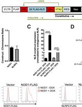
Lentivirus-mediated Conditional Gene Expression

慢病毒介导的条件基因表达

LR Leah Rommereim
AA Ajay Suresh Akhade
RG Ronald N. Germain
IF Iain D. C. Fraser
NS Naeha Subramanian
5350 Views
Nov 5, 2021
The ability to identify the role of a particular gene within a system is dependent on control of the expression of that gene. In this protocol, we describe a method for stable, conditional expression of Nod-Like receptors (NLRs) in THP-1 cells using a lentiviral expression system. This system combines all the necessary components for tetracycline-inducible gene expression in a single lentivector with constitutive co-expression of a selection marker, which is an efficient means for controlling gene expression using a single viral infection of cells. This is done in a third generation lentiviral expression platform that improves the safety of lentiviruses and allows for greater gene expression than previous lentiviral platforms. The lentiviral expression plasmid is first engineered to contain the gene of interest driven by a TRE (tetracycline response element) promoter in a simple gateway cloning step and is then co-transfected into HEK293T cells, along with packaging and envelope plasmids to generate the virus. The virus is used to infect a cell type of interest at a low MOI so that the majority of the transduced cells contain a single viral integration. Infected cells are grown under selection, and viral integration is validated by qPCR. Gene expression in stably transduced cells is induced with doxycycline and validated by qPCR, immunoblot, and flow cytometry. This flexible lentiviral expression platform may be used for stable and robust induction of a gene of interest in a range of cells for multiple applications.Graphic abstract: Schematic overview of lentiviral transduction of THP-1 cells.
调节性 T 细胞抑制人树突状细胞

Suppression of Human Dendritic Cells by Regulatory T Cells

调节性 T 细胞抑制人树突状细胞

QH Qing Huang
AL Avery J. Lam
DB Dominic A. Boardman
ND Nicholas A. J. Dawson
ML Megan K. Levings
3603 Views
Nov 5, 2021
Regulatory T cells (Tregs) suppress immune responses via a variety of mechanisms and can be used as a cellular therapy to induce tolerance. The function of Tregs is commonly assessed in vitro using assays that measure suppression of effector T cell proliferation and/or cytokine production. However, Tregs can also suppress the function of antigen presenting cells, creating a need for methodology to routinely measure this aspect of their function. This protocol describes a method to measure human Treg-mediated suppression of CD80 and CD86 expression on mature, monocyte-derived dendritic cells. Representative data show suppression mediated by polyclonal Tregs as well as antigen-specific Tregs generated using chimeric antigen receptor (CAR) technology. This method can be used in parallel to T cell suppression assays to measure the functional activity of human Tregs.
携带 SARS-CoV 或 SARS-CoV-2 附着尖峰糖蛋白的重组复制缺陷慢病毒的生产及其在受体向性和中和试验中的应用

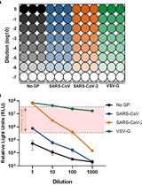
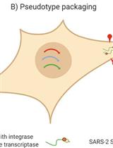
Production of Recombinant Replication-defective Lentiviruses Bearing the SARS-CoV or SARS-CoV-2 Attachment Spike Glycoprotein and Their Application in Receptor Tropism and Neutralisation Assays

携带 SARS-CoV 或 SARS-CoV-2 附着尖峰糖蛋白的重组复制缺陷慢病毒的生产及其在受体向性和中和试验中的应用

NT Nazia Thakur
GG Giulia Gallo
AE Ahmed M. E. Elreafey
DB Dalan Bailey
3521 Views
Nov 5, 2021
For enveloped viruses, such as SARS-CoV-2, transmission relies on the binding of viral glycoproteins to cellular receptors. Conventionally, this process is recapitulated in the lab by infection of cells with isolated live virus. However, such studies can be restricted due to the availability of high quantities of replication-competent virus, biosafety precautions and associated trained staff. Here, we present a protocol based on pseudotyping to produce recombinant replication-defective lentiviruses bearing the SARS-CoV or SARS-CoV-2 attachment Spike glycoprotein, allowing the investigation of viral entry in a lower-containment facility. Pseudoparticles are produced by cells transiently transfected with plasmids encoding retroviral RNA packaging signals and Gag-Pol proteins, for the reconstitution of lentiviral particles, and a plasmid coding for the viral attachment protein of interest. This approach allows the investigation of different aspects of viral entry, such as the identification of receptor tropism, the prediction of virus host range, and zoonotic transmission potential, as well as the characterisation of antibodies (sera or monoclonal antibodies) and pharmacological inhibitors that can block entry.Graphic abstract: SARS-CoV and SARS-CoV-2 pseudoparticle generation and applications.

微生物学

在细菌培养和培养基中定量度洛西汀以研究药物-肠道微生物组的相互作用

Quantification of Duloxetine in the Bacterial Culture and Medium to Study Drug-gut Microbiome Interactions

在细菌培养和培养基中定量度洛西汀以研究药物-肠道微生物组的相互作用

Prasad B. Phapale Prasad B. Phapale
SB Sonja Blasche
KP Kiran R. Patil
TA Theodore Alexandrov
2602 Views
Nov 5, 2021
Expanding our understanding of drug-gut bacteria interactions requires high-throughput drug measurements in complex bacterial cultures. Quantification of drugs in the cultures, media, and cell pellets is prone to strong matrix effects. We have developed a liquid chromatography–high resolution mass spectrometry (LC–HRMS) method for quantifying duloxetine from high-throughput gut-drug interaction experiments. The method is partially validated for its reproducibility, sensitivity, and accuracy, which makes it suitable for largescale drug screens. We extensively used this method to study biotransformation and bioaccumulation of duloxetine and other drugs in several species of gut bacteria.
真菌对重金属的耐受指数的测定和重金属抗性试验

Determination of Fungal Tolerance Index to Heavy Metals and Heavy Metal Resistance Tests

真菌对重金属的耐受指数的测定和重金属抗性试验

RC Rosa Paulina Calvillo-Medina
2216 Views
Nov 5, 2021
Fungal metallo-tolerance has been described in different species and plays an important role in bioremediation of contaminated environments. Metallo-tolerance is mainly documented by microdilution assays and agar well diffusion methods using equipment that can be expensive. The tolerance index can be calculated to determine the efficiency of a fungus to degrade and resist heavy metals. The present protocol is based on analyzing the tolerance index and minimum inhibitory concentration of the metallo-tolerance potential of culturable fungi on solid media. This can be calculated by daily measurements of colony size on agar supplemented with different concentrations of heavy metals. This method is an easy approach to determine fungal heavy metal resistance using simple laboratory equipment without spectroscopy.
测定 NAD(P)H 的分析:光滑念珠菌细胞提取物中的醌氧化还原酶活性

An Assay to Determine NAD(P)H: Quinone Oxidoreductase Activity in Cell Extracts from Candida glabrata

测定 NAD(P)H 的分析:光滑念珠菌细胞提取物中的醌氧化还原酶活性

AB Anamika Battu
RP Rajaram Purushotham
RK Rupinder Kaur
2208 Views
Nov 5, 2021
Flavodoxin-like proteins (Fld-LPs) are an important constituent of the oxidative stress defense system in several organisms and highly conserved from bacteria to humans. These proteins possess NAD(P)H:quinone oxidoreductase activity and convert quinones to hydroquinones through two-electron reduction, using NAD(P)H and quinone as electron donor and acceptor, respectively. Purified yeast and bacterial Fld-LPs exhibit NAD(P)H:quinone oxidoreductase activity in vitro. Here, we describe a protocol to measure oxidoreductase activity of Fld-LPs that are present in extracts of whole cells. We have recently shown that the assembly and activity of a Fld-LP, CgPst2, is regulated by an aspartyl protease-mediated cleavage of its C-terminus in the pathogenic yeast Candida glabrata. Mutant yeast where the CgPST2 gene was deleted lacked cellular NAD(P)H:quinone oxidoreductase activity and displayed elevated susceptibility to menadione stress. The protocol described herein is based on the measurement of NADH oxidation (conversion of NADH to NAD+) by endogenous Fld-LPs in the presence of quinone menadione. This assay can be performed with whole cell lysates prepared by the mechanical lysis of C. glabrata cells and does not require expression and purification of Fld-LPs from a heterogeneous system, thereby allowing researchers to study the effect of different posttranslational modifications and varied structural states of Fld-LPs on their enzymatic activities. Since many FLP-LPs are known to exist in dimeric and tetrameric states possessing differential activities, our efficient and easy-to-use assay can reliably detect and validate their quinone reductase activities. Although we have used menadione with CgPst2 enzyme in our study, the protocol can easily be modified to examine the presence of Fld-LPs with specificity for other quinones. As this assay does not require many expensive chemicals, it can readily be scaled up and adapted for other medically important fungi and potentially be a useful tool to characterize fungal oxidative stress response systems and screen inhibitors specific for fungal Fld-LPs, thereby contributing to our understanding of fungal pathogenesis mechanisms.

分子生物学

严重急性呼吸系统综合症冠状病毒 2 (SARS-CoV-2) 慢病毒假型的生产、滴定、中和、储存和冻干

Production, Titration, Neutralisation, Storage and Lyophilisation of Severe Acute Respiratory Syndrome Coronavirus 2 (SARS-CoV-2) Lentiviral Pseudotypes

严重急性呼吸系统综合症冠状病毒 2 (SARS-CoV-2) 慢病毒假型的生产、滴定、中和、储存和冻干

CD Cecilia Di Genova
AS Alex Sampson
SS Simon Scott
DC Diego Cantoni
MM Martin Mayora-Neto
EB Emma Bentley
GM Giada Mattiuzzo
EW Edward Wright
MD Mariliza Derveni
BA Bethany Auld
BF Bill T. Ferrara
DH Dale Harrison
MS Mohamed Said
AS Arwa Selim
ET Elinor Thompson
CT Craig Thompson
GC George Carnell
NT Nigel Temperton
4670 Views
Nov 5, 2021
This protocol details a rapid and reliable method for the production and titration of high-titre viral pseudotype particles with the SARS-CoV-2 spike protein (and D614G or other variants of concern, VOC) on a lentiviral vector core, and use for neutralisation assays in target cells expressing angiotensin-converting enzyme 2 (ACE2) and transmembrane serine protease 2 (TMPRSS2). It additionally provides detailed instructions on substituting in new spike variants via gene cloning, lyophilisation and storage/shipping considerations for wide deployment potential. Results obtained with this protocol show that SARS-CoV-2 pseudotypes can be produced at equivalent titres to SARS-CoV and Middle East respiratory syndrome coronavirus (MERS-CoV) pseudotypes, neutralised by human convalescent plasma and monoclonal antibodies, and stored at a range of laboratory temperatures and lyophilised for distribution and subsequent application.
基于核衣壳的 SARS-CoV-2 转互补细胞培养系统,重现完整的病毒生命周期

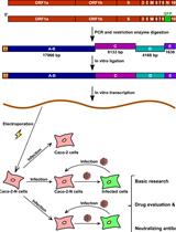
A Nucleocapsid-based Transcomplementation Cell Culture System of SARS-CoV-2 to Recapitulate the Complete Viral Life Cycle

基于核衣壳的 SARS-CoV-2 转互补细胞培养系统,重现完整的病毒生命周期

YY Yanying Yu
XJ Xiaohui Ju
QD Qiang Ding
3134 Views
Nov 5, 2021
The ongoing COVID-19 pandemic is caused by severe acute respiratory syndrome coronavirus 2 (SARS-CoV-2). As this virus is classified as a biosafety level-3 (BSL-3) agent, the development of countermeasures and basic research methods is logistically difficult. Recently, using reverse genetics, we developed a BSL-2 cell culture system for production of transcription- and replication-component virus-like-particles (trVLPs) by genetic transcomplementation. The system consists of two parts: SARS-CoV-2 GFP/ΔN genomic RNA, in which the nucleocapsid (N) gene, a critical gene for virion packaging, is replaced by a GFP reporter gene; and a packaging cell line for ectopic expression of N (Caco-2-N). The complete viral life cycle can be recapitulated and confined to Caco-2-N cells, with GFP positivity serving as a surrogate readout for viral infection. In addition, we utilized an intein-mediated protein splicing technique to split the N gene into two independent vectors and generated the Caco-2-Nintein cells as a packaging cell line to further enhance the security of this cell culture model. Altogether, this system provides for a safe and convenient method to produce trVLPs in BSL-2 laboratories. These trVLPs can be modified to incorporate desired mutations, permitting high-throughput screening of antiviral compounds and evaluation of neutralizing antibodies. This protocol describes the details of the trVLP cell culture model to make SARS-CoV-2 research more readily accessible.
从拟南芥幼苗中富集细胞质 RNA 颗粒

Enrichment of Cytoplasmic RNA Granules from Arabidopsis Seedlings

从拟南芥幼苗中富集细胞质 RNA 颗粒

ZL Zhen Lei
EK Eun Yu Kim
JC Jungnam Cho
2551 Views
Nov 5, 2021
RNA granules (RGs) are membraneless intracellular compartments that play important roles in the post-transcriptional control of gene expression. Stress granules (SGs) are a type of RGs that form under environmental challenges and/or internal cellular stresses. Stress treatments lead to strong mRNAs translational inhibition and storage in SGs until the normal growth conditions are restored. Intriguingly, we recently showed that plant stress granules are associated with siRNA bodies, where the RDR6-mediated and transposon-derived siRNA biogenesis occurs (Kim et al., 2021). This protocol provides a technical workflow for the enrichment of cytoplasmic RGs from Arabidopsis seedlings. We used the DNA methylation-deficient ddm1 mutant in our study, but the method can be applied to any other plant samples with strong RG formation. The resulting RG fractions can be further tested for either RNAs or proteins using RNA-seq and mass spectrometry-based proteomics.

神经科学

小鼠膈肌神经肌肉接头的整体染色

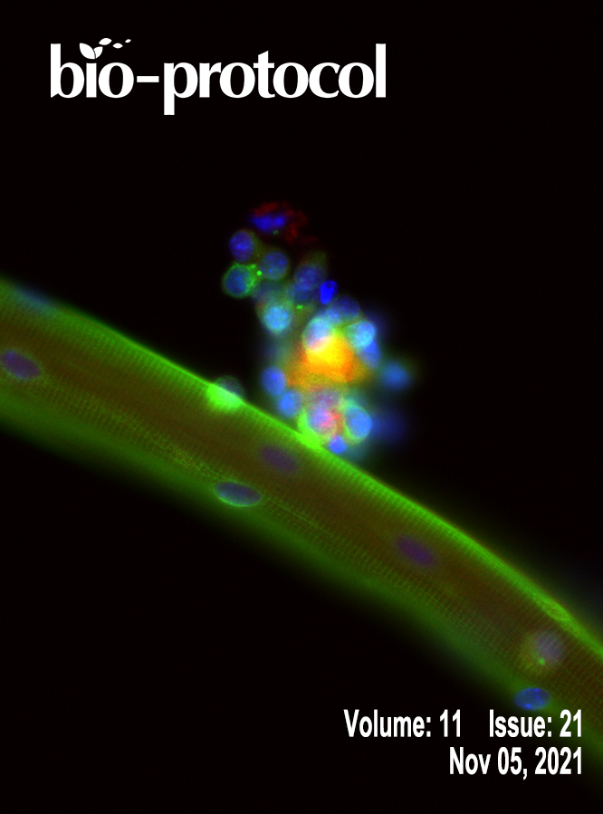
Whole-mount Staining of Mouse Diaphragm Neuromuscular Junctions

小鼠膈肌神经肌肉接头的整体染色

RS Rula Sha
ZW Zhenzhen Wang
XY Xue You
YL Yuguo Liu
ZX Zhiqin Xie
YF Ying Feng
3446 Views
Nov 5, 2021
The neuromuscular junction (NMJ) is a specialized synapse that connects the terminal end of a motor neuron and a skeletal muscle fiber. Defects in NMJ cause abnormalities of neuromuscular transmission, leading to NMJ disorders. The mammalian diaphragm muscle is essential for respiration and has been widely used to study NMJ formation. Here, we provide a simple and straightforward protocol for preparing diaphragms from embryonic, neonatal, and adult mice and for subsequent NMJ staining.

植物科学

小菜蛾幼虫对DMNT处理的偏好试验

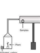
Preference Test of Plutella xylostella Larvae upon DMNT Treatment

小菜蛾幼虫对DMNT处理的偏好试验

CC Chen Chen
ZT Zhen Tao
CW Chuanhong Wang
TW Tengyue Wang
HC Hongyi Chen
TJ Taoshan Jiang
PL Peijin Li
1680 Views
Nov 5, 2021
We describe a method to test the preference of insects in response to (3E)-4,8-dimethyl-1,3,7-nonatriene (DMNT). We use a device that includes a horizontal glass tube, two grooves (with activated carbon), air flow, rubber stoppers/tubes, transparent glass containers (optional), and a holder for the glass tube (optional). Equal amounts of activated carbon in the groove (removable) are placed at both ends to avoid air contamination. The air flow is generated by an air pump. In the closed device, different samples are placed at each end of the glass tube. The air pump at the top of the glass tube forms an air flow that converges to the middle site of the glass tube. In each test, insect larvae are located in the middle of the glass test tube. If the test samples release DMNT that can be sensed by insects, the insects will selectively move to one specific end of the glass tube. The number of insects that move to each end will be recorded for further studies. This method can also be used to test the preference of insects in response to other volatile compounds.

干细胞

利用微细血管EZSPHERETM从人类iPS细胞中进行高通量三维球状细胞形成和有效的心肌细胞分化

High-throughput 3D Spheroid Formation and Effective Cardiomyocyte Differentiation from Human iPS Cells Using the Microfabric Vessels EZSPHERETM

利用微细血管EZSPHERETM从人类iPS细胞中进行高通量三维球状细胞形成和有效的心肌细胞分化

TM Tatsuaki Miwa
AI Alimjan Idiris
HK Hiromichi Kumagai
2682 Views
Nov 5, 2021
High-throughput 3D spheroid formation from human induced pluripotent stem cells (hiPSCs) can be easily performed using the unique microfabric vessels EZSPHERE, resulting in effective and large scale generation of differentiated cells such as cardiomyocytes or neurons. Such hiPSC-derived cardiomyocytes (hiPSC-CMs) or neurons are very useful in the fields of regenerative medicine or cell-based drug safety tests. Previous studies indicated that 3D spheroids arising from hiPSCs are effectively differentiated into high quality hiPSC-CMs by controlling Wnt signals through utilization of the microfabric vessels EZSPHERE. Here, we describe a simple and highly efficient protocol for generating a large number of uniformly sized hiPSC spheroids and inducing them for cardiac differentiation using the EZSPHERE. This method comprises the collection and dissociation of the spheroids from cardiac differentiation medium, in the middle stage of the whole cardiac differentiation process, and re-seeding the obtained single cells into the EZSPHERE to re-aggregate them into uniform hiPSC-CM spheroids with controlled size. This re-aggregation process promotes non-canonical Wnt signal-related cardiac development and improves the purity and maturity of the hiPSC-CMs generated.Graphic abstract: Overview of cardiac differentiation from iPSCs by spheroid formation and reaggregation using EZSPHERE.

系统生物学

拟南芥植株上的组蛋白修饰ChIP-seq

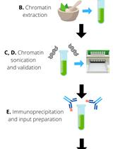
Histone modification ChIP-seq on Arabidopsis thaliana Plantlets

拟南芥植株上的组蛋白修饰ChIP-seq

JR Juan S. Ramirez-Prado
DL David Latrasse
MB Moussa Benhamed
4075 Views
Nov 5, 2021
Characterizing the molecular mechanisms regulating gene expression is crucial for understanding the regulatory processes underlying physiological responses to environmental and developmental signals in eukaryotes. The covalent modification of histones contributes to the compaction levels of chromatin, as well as the recruitment of the transcriptional machinery to specific loci, facilitating metastable changes in gene activity. ChIP-seq (Chromatin Immunoprecipitation followed by sequencing) has become the gold standard method for determining histone modification profiles among different organisms, tissues, and genotypes. In the current protocol, we describe a highly robust method for performing ChIP-seq of histone modifications in Arabidopsis thaliana plantlets. Besides its robustness, this method uses in-house-prepared buffers for chromatin extraction, immunoprecipitation, washing, and elusion, making it cost-effective in contrast to commercial kits.