往期刊物2021

卷册: 11, 期号: 19

left

Dec

20

Dec

5

Nov

20

Nov

5

Oct

20

Oct

5

Sep

20

Sep

5

Aug

20

Aug

5

Jul

20

Jul

5

Jun

20

Jun

5

May

20

May

5

Apr

20

Apr

5

Mar

20

Mar

5

Feb

20

Feb

5

Jan

20

Jan

5

right

生物化学

用于蛋白质组和相互作用组分析的斑马鱼中的邻近依赖性生物素标记

Proximity Dependent Biotin Labelling in Zebrafish for Proteome and Interactome Profiling

用于蛋白质组和相互作用组分析的斑马鱼中的邻近依赖性生物素标记

ZX Zherui Xiong
HL Harriet P. Lo
KM Kerrie-Ann McMahon
RP Robert G. Parton
TH Thomas E. Hall
5110 Views
Oct 5, 2021
Identification of protein interaction networks is key for understanding intricate biological processes, but mapping such networks is challenging with conventional biochemical methods, especially for weak or transient interactions. Proximity-dependent biotin labelling (BioID) using promiscuous biotin ligases and mass spectrometry (MS)-based proteomics has emerged in the past decade as a powerful method for probing local proteomes and protein interactors. Here, we describe the application of an engineered biotin ligase, TurboID, for proteomic mapping and interactor screening in vivo in zebrafish. We generated novel transgenic zebrafish lines that express TurboID fused to a conditionally stabilised GFP-binding nanobody, dGBP, which targets TurboID to the GFP-tagged proteins of interest. The TurboID-dGBP zebrafish lines enable proximity-dependent biotin labelling in live zebrafish simply through outcrossing with existing GFP-tagged lines. Here, we outline a detailed protocol of the BLITZ method (Biotin Labelling In Tagged Zebrafish) for utilising TurboID-dGBP fish lines to map local proteomes and screen novel interactors.Graphic abstract:Schematic overview of the BLITZ method. TurboID-dGBP fish are crossed with GFP-tagged lines to obtain embryos co-expressing TurboID-dGBP (indicated by mKate2) and the GFP-POI (protein of interest). Embryos expressing only TurboID are used as a negative control. Embryos (2 to 7 dpf) are incubated overnight with a 500 μM biotin-supplemented embryo medium. This biotin incubation step allows TurboID to catalyse proximity-dependent biotinylation in live zebrafish embryos. After biotin incubation, embryos are solubilised in lysis buffer, and free biotin is removed using a PD-10 desalting column. The biotinylated proteins are captured by streptavidin affinity purification, and captured proteins are analysed by MS sequencing.
通过静脉注射脂质纳米颗粒包裹的核苷修饰的 mRNA 蛋白质在小鼠肝脏中瞬时而稳定地表达

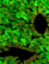
Transient yet Robust Expression of Proteins in the Mouse Liver via Intravenous Injection of Lipid Nanoparticle-encapsulated Nucleoside-modified mRNA

通过静脉注射脂质纳米颗粒包裹的核苷修饰的 mRNA 蛋白质在小鼠肝脏中瞬时而稳定地表达

EE Elissa Everton
FR Fatima Rizvi
AS Anna R. Smith
MB Mitchell Beattie
YT Ying Tam
NP Norbert Pardi
DW Drew Weissman
VG Valerie Gouon-Evans
4953 Views
Oct 5, 2021
With the recent availability of the SARS-CoV-2 mRNA-based vaccines, public attention has been drawn to this new technology and how it may be applied to other indications. Temporal activation of key hepatic regenerative pathways can induce liver regeneration, overcoming the lack of donor organs for liver transplantation and ineffectiveness of alternative treatments. Recombinant protein therapies and genetic therapies that target these pathways require frequent and repeated injections or, when integrated into the genome, may lead to deleterious effects. In contrast, nucleoside-modified mRNA encapsulated in lipid nanoparticles (mRNA-LNP) are non-integrative and induce transient yet robust expression of proteins that could serve as an ideal therapeutic tool to treat specific liver diseases. For instance, our recent publication in Nature Communications used mRNA-LNP to express hepatic mitogens, hepatocyte growth factor, and epidermal growth factor to induce liver regeneration following both acute and chronic liver injuries. Initial testing with firefly luciferase mRNA-LNP transfection and in vivo imaging confirmed specific hepatotropic delivery. In this protocol, we describe in detail the necessary steps to deliver mRNA-LNP to the murine liver and, following intravenous injection of eGFP mRNA-LNP, verify transfection efficiency using flow cytometry and liver cell specificity using immunofluorescence analyses. This procedure presents an unprecedented tool that can be customized with mRNA-LNP encoding any protein of interest to be expressed by virtually all hepatocytes, ~70% endothelial cells, and ~40% Kupffer cells for promoting liver function and/or regeneration.Graphic abstract:Experimental Design of mRNA-LNP IV Injection and Analysis of Liver Cell Specificity and Efficiency of Transfection (Created with BioRender.com)

生物工程

斑马鱼胚胎作为研究体内纳米粒子行为的预测动物模型

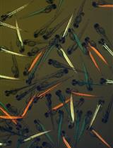
Zebrafish Embryos as a Predictive Animal Model to Study Nanoparticle Behavior in vivo

斑马鱼胚胎作为研究体内纳米粒子行为的预测动物模型

GA Gabriela Arias-Alpizar
JB Jeroen Bussmann
FC Frederick Campbell
3230 Views
Oct 5, 2021
A failure to fully understand the complex in vivo behavior of systemically administered nanomedicines has stymied clinical translation. To bridge this knowledge gap, new in vivo tools are needed to rapidly and accurately assess the nearly infinite array of possible nanoparticle designs. Zebrafish embryos are small, transparent, and easily manipulated animals that allow for whole organism visualization of fluorescently labeled nanoparticles in real time and at cellular resolution using standard microscope setups. Furthermore, key nano-bio interactions present in higher vertebrates are fully conserved in zebrafish embryos, making these animal models a highly predictive and instructive addition to the nanomedicine design pipeline. Here, we present a step-by-step protocol to intravenously administer, image, and analyze nanoparticle behavior in zebrafish embryos and highlight key nano-bio interactions within the embryonic zebrafish corresponding to those commonly found within the mammalian liver. In addition, we outline practical steps required to achieve light-triggered activation of nanoparticles within the transparent embryo. Graphic abstract:Zebrafish embryos to study nanoparticle behavior in vivo. Formulation, intravenous administration, imaging, and analysis of nanoparticles.

癌症生物学

小鼠白血病细胞代谢的稳定同位素示踪分析

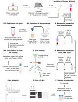
Analysis of Leukemia Cell Metabolism through Stable Isotope Tracing in Mice

小鼠白血病细胞代谢的稳定同位素示踪分析

Nv Nick van Gastel
JS Jessica B. Spinelli
MH Marcia C. Haigis
DS David T. Scadden
3904 Views
Oct 5, 2021
Once thought to be a mere consequence of the state of a cell, intermediary metabolism is now recognized as a key regulator of mammalian cell fate and function. In addition, cell metabolism is often disturbed in malignancies such as cancer, and targeting metabolic pathways can provide new therapeutic options. Cell metabolism is mostly studied in cell cultures in vitro, using techniques such as metabolomics, stable isotope tracing, and biochemical assays. Increasing evidence however shows that the metabolic profile of cells is highly dependent on the microenvironment, and metabolic vulnerabilities identified in vitro do not always translate to in vivo settings. Here, we provide a detailed protocol on how to perform in vivo stable isotope tracing in leukemia cells in mice, focusing on glutamine metabolism in acute myeloid leukemia (AML) cells. This method allows studying the metabolic profile of leukemia cells in their native bone marrow niche.

细胞生物学

间充质体的结构和功能图谱

Structural and Functional Mapping of Mesenchymal Bodies

间充质体的结构和功能图谱

SS Sébastien Sart
RT Raphaël F.-X. Tomasi
AB Antoine Barizien
GA Gabriel Amselem
AC Ana Cumano
CB Charles N. Baroud
3914 Views
Oct 5, 2021
The formation of spheroids with mesenchymal stem/stromal cells (MSCs), mesenchymal bodies (MBs), is usually performed using bioreactors or conventional well plates. While these methods promote the formation of a large number of spheroids, they provide limited control over their structure or over the regulation of their environment. It has therefore been hard to elucidate the mechanisms orchestrating the structural organization and the induction of the trophic functions of MBs until now. We have recently demonstrated an integrated droplet-based microfluidic platform for the high-density formation and culture of MBs, as well as for the quantitative characterization of the structural and functional organization of cells within them. The protocol starts with a suspension of a few hundred MSCs encapsulated within microfluidic droplets held in capillary traps. After droplet immobilization, MSCs start clustering and form densely packed spherical aggregates that display a tight size distribution. Quantitative imaging is used to provide a robust demonstration that human MSCs self-organize in a hierarchical manner, by taking advantage of the good fit between the microfluidic chip and conventional microscopy techniques. Moreover, the structural organization within the MBs is found to correlate with the induction of osteo-endocrine functions (i.e., COX-2 and VEGF-A expression). Therefore, the present platform provides a unique method to link the structural organization in MBs to their functional properties.Graphic abstract:Droplet microfluidic platform for integrated formation, culture, and characterization of mesenchymal bodies (MBs). The device is equipped with a droplet production area (flow focusing) and a culture chamber that enables the culture of 270 MBs in parallel. A layer-by-layer analysis revealed a hierarchical developmental organization within MBs.
人循环中性粒细胞微泡的分离

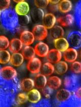
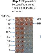
Isolation of Microvesicles from Human Circulating Neutrophils

人循环中性粒细胞微泡的分离

DZ Dong Zhan
EM Ellie McConachie
SE Steven W. Edwards
HW Helen L. Wright
RM Robert J. Moots
Sittisak Honsawek Sittisak Honsawek
3190 Views
Oct 5, 2021
Neutrophil-derived microvesicles (NDMVs) are liberated by neutrophils upon cell activation by molecules. Once activated, neutrophils are primarily involved in acute inflammation; however, the microvesicles they produce are largely anti-inflammatory. NDMVs have been shown to protect cartilage during inflammatory arthritis. They exert these effects by inhibiting or affecting the function of target cells, including macrophages. NDMVs have the potential to act as disease-modifying agents, especially for inflammatory diseases. This protocol describes a method using differential centrifugation to separate neutrophils from whole human blood. Subsequently, neutrophils are identified by cytospin and Wright’s staining, and then the NDMVs are isolated using differential centrifugation.
秀丽隐杆线虫内源性蛋白质的固定和免疫染色或翻译后修饰

Fixation and Immunostaining of Endogenous Proteins or Post-translational Modifications in Caenorhabditis elegans

秀丽隐杆线虫内源性蛋白质的固定和免疫染色或翻译后修饰

RO Robert O'Hagan
IT Irini Topalidou
2982 Views
Oct 5, 2021
Although the advent of genetically-encoded fluorescent markers, such as the green fluorescent protein (GFP; Chalfie et al., 1994), has enabled convenient visualization of gene expression in vivo, this method is generally not effective for detecting post-translational modifications because they are not translated from DNA sequences. Genetically-encoded, fluorescently-tagged transgene products can also be misleading for observing expression patterns because transgenes may lack endogenous regulatory DNA elements needed for precise regulation of expression that could result in over or under expression. Fluorescently-tagged proteins created by CRISPR genome editing are less prone to defective expression patterns because the loci retain endogenous DNA elements that regulate their transcription (Nance and Frøkjær-Jensen, 2019). However, even CRISPR alleles encoding heritable fluorescently-tagged protein markers can result in defects in function or localization of the gene product if the fluorescent tag obstructs or otherwise interferes with important protein interaction domains or affects the protein structure.Indirect immunofluorescence is a method for detecting endogenous gene expression or post-translational modifications without the need for transgenesis or genome editing. Here, we present a reliable protocol in which C. elegans nematodes are fixed, preserved, and permeabilized for staining with a primary antibody to bind proteins or post-translational modifications, which are then labeled with a secondary antibody conjugated to a fluorescent dye. Use of this method may be limited by the availability of (or ability to generate) a primary antibody that binds the epitope of interest in fixed animals. Thousands of animals are simultaneously subjected to a series of chemical treatments and washes in a single centrifuge tube, allowing large numbers of identically-treated stained animals to be examined. We have successfully used this protocol (O’Hagan et al., 2011 and 2017; Power et al., 2020) to preserve and detect post-translational modifications of tubulin in C. elegans ciliated sensory neurons and to detect non-modified endogenous protein (Topalidou and Chalfie, 2011).

发育生物学

秀丽隐杆线虫肌动蛋白丝的Phalloidin染色用于肌纤维可视化

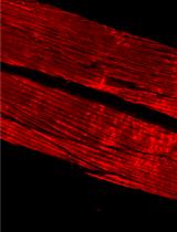
Phalloidin Staining of Actin Filaments for Visualization of Muscle Fibers in Caenorhabditis elegans

秀丽隐杆线虫肌动蛋白丝的Phalloidin染色用于肌纤维可视化

MR Mario Romani
JA Johan Auwerx
4042 Views
Oct 5, 2021
Advances in C. elegans research have allowed scientists to recapitulate different human disorders, from neurodegenerative diseases to muscle dysfunction, in these nematodes. Concomitantly, the interest in visualizing organs affected by these conditions has grown, leading to the establishment of different antibody- and dye-based staining protocols to verify tissue morphology. In particular, the quality of muscle tissue has been largely used in nematodes as a readout for fitness and healthspan. Phalloidin derivatives, which are commonly used to stain actin filaments in cells and tissues, have been implemented in the context of C. elegans research for visualization of muscle fibers. However, the majority of the phalloidin-based protocols depend on fixation steps using harmful compounds, preparation of specific buffers, and large amounts of worms. Herein, we implemented a safer and more flexible experimental procedure to stain actin filaments in C. elegans using phalloidin-based dyes. Lyophilization of the worms followed by their acetone permeabilization allows bypassing the fixation process while also providing the opportunity to suspend the experiment at different steps. Moreover, by using conventional buffers throughout our protocol, we avoid the additional preparation of solutions. Finally, our protocol requires a limited number of worms, making it suitable for slow-growing C. elegans strains. Overall, this protocol provides an efficient, fast, and safer method to stain actin filaments and visualize muscle fibers in C. elegans.Graphic abstract:Schematic overview of phalloidin staining in C. elegans for assessing muscle fiber morphology.
足月人胎盘原代细胞滋养层细胞的分离

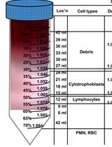
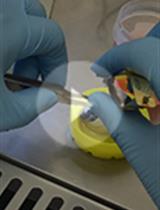
Isolation of Primary Cytotrophoblasts From Human Placenta at Term

足月人胎盘原代细胞滋养层细胞的分离

AC Arthur Colson
CD Christophe Louis Depoix
CH Corinne Hubinont
FD Frédéric Debiève
2794 Views
Oct 5, 2021
The placenta is a multifaceted organ, fulfilling critical functions for the fetus and the mother. Therefore, it is a critical regulator of the pregnancy, and its dysfunction leads to diseases, including fetal growth restriction and preeclampsia. Studying the placenta is a difficult task since its existence is transient, and its structure is specific to our species. In vitro differentiation of primary cytotrophoblast isolated from term human placenta has been widely used in the placental research field as it represents a reliable model to study cellular differentiation and function. Direct alternatives include trophoblastic cell lines, explants, and organoids, but this protocol, based on the separation of the cells on a Percoll gradient, presents the advantage of being relatively cheap and easy to perform in every research laboratory. Furthermore, the 2D culture is a flexible method that can be adapted to various experimental conditions (transfection, drug exposure, metabolic study, observations, etc.), allowing mechanistic explorations of cellular processes.

免疫学

T 淋巴细胞中的钙成像:与遗传编码或化学Ca2+指示一起使用的实验方案

Calcium Imaging in T Lymphocytes: a Protocol for Use with Genetically Encoded or Chemical Ca2+ Indicators

T 淋巴细胞中的钙成像:与遗传编码或化学Ca2+指示一起使用的实验方案

AJ Amit Jairaman
MC Michael D. Cahalan
3965 Views
Oct 5, 2021
Elevations in cytosolic calcium (Ca2+) drive a wide array of immune cell functions, including cytokine production, gene expression, and cell motility. Live-cell imaging of cells loaded with ratiometric chemical Ca2+ indicators remains the gold standard for visualization and quantification of intracellular Ca2+ signals; ratiometric imaging can be accomplished with dyes such as Fura-2, the combination of Fluo-4 and Fura-Red, or, alternatively, by expressing genetically-encoded Ca2+ indicators (GECI) such as GCaMPs. Here, we describe a detailed protocol for Ca2+ imaging of T cells in vitro using genetically encoded or chemical indicators that can also be applied to a wide variety of cell types. The protocol addresses the challenge of facilitating T cell attachment on various substrates prepared on glass-bottom dishes to enable T cell imaging on an inverted microscope. The protocol also emphasizes cell preparation steps that ensure optimal cell viability – an essential requirement for recording dynamic changes in cytosolic Ca2+ levels – and that ensure reproducibility between multiple samples. Finally, we describe a simple algorithm to analyze single-cell Ca2+ signals over time using Fiji (ImageJ) software.
测量小鼠补体通路的各个组成部分的总经典通路和活动

Measuring Total Classical Pathway and Activities of Individual Components of the Mouse Complement Pathway

测量小鼠补体通路的各个组成部分的总经典通路和活动

WZ Wioleta M. Zelek
2407 Views
Oct 5, 2021
The complement system is a central component of innate immunity, responsible for recognition and killing of bacteria by tagging invaders through opsonisation, thereby promoting phagocytosis, and by direct lysis. Complement activity is routinely measured using functional assays that utilise erythrocytes as targets. The classical pathway haemolytic assay (CH50) with antibody sensitised sheep erythrocytes as target is used worldwide in clinical and research laboratories to measure complement activity in human and rodent sera. While there are no particular limitations in the human assay, measuring complement in mouse serum is more difficult and usually requires large amounts of serum, which is challenging to collect in experiments. In particular, it is challenging to measure the activities of individual mouse complement proteins. To overcome this hurdle, we have developed protocols that employ human sera depleted of single complement proteins as the source of the other complement proteins and test mouse serum to restore the relevant component. This simple haemolytic assay is a useful tool for confirming natural or engineered complement deficiencies and complement dysregulation in mouse models.

微生物学

胶体几丁质、无细胞上清液的简易制备方法及昆布多糖酶的测定

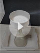
Simple Methods for the Preparation of Colloidal Chitin, Cell Free Supernatant and Estimation of Laminarinase

胶体几丁质、无细胞上清液的简易制备方法及昆布多糖酶的测定

AK Ananthamurthy Koteshwara
3930 Views
Oct 5, 2021
Colloidal chitin (CC) is a common substrate used in research work involving chitin-active enzymes (chitinases). Cell free supernatant (CFS) is prepared from fermented broth. Preparation of CC and CFS usually involve large amounts of liquid, which must be separated from the solids. This necessitates the use of a large volume centrifugation facility, which may not be accessible to everyone. Filtration is a viable alternative to centrifugation, and several filter elements are described in the literature. Each of those elements has its own set of disadvantages like non-availability, high cost, fragility, and non-reusability. Here we describe the use of lab coat clothing material (LCCM) for the preparation of CC and CFS. For filtration purposes, the LCCM was found to be functional, rugged, reusable, and cost-effective. Also described here is a new method for the estimation of laminarinase using a laminarin infused agarose gel plate. An easily available optical fabric brightener (OFB) was used as a stain for the agarose plate. The laminarin infused agarose plate assay is simple, inexpensive, and was found to be impervious to high amounts of ammonium sulfate (AS) in enzyme precipitates.

分子生物学

通过 AFM 成像确定 DNA 和核小体构象的高通量流程

A High-throughput Pipeline to Determine DNA and Nucleosome Conformations by AFM Imaging

通过 AFM 成像确定 DNA 和核小体构象的高通量流程

SK Sebastian F. Konrad
WV Willem Vanderlinden
JL Jan Lipfert
2433 Views
Oct 5, 2021
Atomic force microscopy (AFM) is a powerful tool to image macromolecular complexes with nanometer resolution and exquisite single-molecule sensitivity. While AFM imaging is well-established to investigate DNA and nucleoprotein complexes, AFM studies are often limited by small datasets and manual image analysis that is slow and prone to user bias. Recently, we have shown that a combination of large scale AFM imaging and automated image analysis of nucleosomes can overcome these previous limitations of AFM nucleoprotein studies. Using our high-throughput imaging and analysis pipeline, we have resolved nucleosome wrapping intermediates with five base pair resolution and revealed how distinct nucleosome variants and environmental conditions affect the unwrapping pathways of nucleosomal DNA. Here, we provide a detailed protocol of our workflow to analyze DNA and nucleosome conformations focusing on practical aspects and experimental parameters. We expect our protocol to drastically enhance AFM analyses of DNA and nucleosomes and to be readily adaptable to a wide variety of other protein and protein-nucleic acid complexes.

神经科学

人视网膜原代 Müller 细胞的分离、培养和鉴定

Isolation, Culture, and Identification of Primary Müller Cells from Human Retina

人视网膜原代 Müller 细胞的分离、培养和鉴定

YC Yingying Chen
TZ Ting Zhang
SZ Shaoxue Zeng
MY Michelle Yam
MG Mark C. Gillies
LZ Ling Zhu
3082 Views
Oct 5, 2021
Müller cells, the major glial cells of the retina, play vital roles in maintaining redox homeostasis and retinal metabolism. An immortalized human Müller cell line (MIO-M1) is widely used as an in vitro model to study Müller cells’ function, but they may not be exactly the same as primarily cultured human Müller cells. The use of human primary Müller cells (huPMCs) in culture has been limited by the requirement for complicated culture systems or particular age ranges of donors. We have successfully grown huPMCs using our established protocol. The cell type was pure, and cultured cells expressed Müller cell-specific markers strongly. The cultured huPMCs were used for morphologic, metabolic, transcriptomic, and functional studies.Graphic abstract:Timeline for human primary Müller cell (huPMC) culture
诱导、预防和治疗小鼠创伤后应激障碍样记忆的方案:光遗传学和行为方法

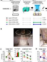
Protocols to Induce, Prevent, and Treat Post-traumatic Stress Disorder-like Memory in Mice: Optogenetics and Behavioral Approaches

诱导、预防和治疗小鼠创伤后应激障碍样记忆的方案:光遗传学和行为方法

AA Aline S. Al Abed
AS Azza Sellami
ED Eva-Gunnel Ducourneau
CB Chloé Bouarab
AM Aline Marighetto
AD Aline Desmedt
2810 Views
Oct 5, 2021
One of the cardinal features of post-traumatic stress disorder (PTSD) is a paradoxical memory alteration including both emotional hypermnesia for salient trauma-related cues and amnesia for the surrounding traumatic context. Interestingly, some clinical studies have suggested that contextual amnesia would causally contribute to the PTSD-related hypermnesia insofar as decontextualized, traumatic memory is prone to be reactivated in contexts that can be very different from the original traumatic context. However, most current animal models of PTSD-related memory focus exclusively on the emotional hypermnesia, i.e., the persistence of a strong fear memory, and do not distinguish normal (adaptive) from pathological (PTSD-like) fear memory, leaving unexplored the hypothetical critical role of contextual amnesia in PTSD-related memory formation, and thus challenging the development of innovative treatments. Having developed the first animal model that precisely recapitulates the two memory components of PTSD in mice (emotional hypermnesia and contextual amnesia), we recently demonstrated that contextual amnesia, induced by optogenetic inhibition of the hippocampus (dorsal CA1), is a causal cognitive process of PTSD-like hypermnesia formation. Moreover, the hippocampus-dependent contextualization of traumatic memory, by optogenetic activation of dCA1 in traumatic condition, prevents PTSD-like hypermnesia formation. Finally, once PTSD-like memory has been formed, the re-contextualization of traumatic memory by its reactivation in the original traumatic context normalizes this pathological fear memory. Revealing the key role of contextual amnesia in PTSD-like memory, this procedure opens a therapeutic perspective based on trauma contextualization and the underlying hippocampal mechanisms.

植物科学

拟南芥芽的暗呼吸测量

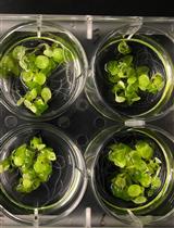
Dark Respiration Measurement from Arabidopsis Shoots

拟南芥芽的暗呼吸测量

JF Jose P. Fonseca
MG Marcus Griffiths
LY Larry M. York
KM Kirankumar S. Mysore
3086 Views
Oct 5, 2021
Dark respiration refers to experimental measures of leaf respiration in the absence of light, done to distinguish it from the photorespiration that occurs during photosynthesis. Dark aerobic respiration reactions occur solely in the mitochondria and convert glucose molecules from cytoplasmatic glycolysis and oxygen into carbon dioxide and water, with the generation of ATP molecules. Previous methods typically use oxygen sensors to measure oxygen depletion or complicated and expensive photosynthesis instruments to measure CO2 accumulation. Here, we provide a detailed, step-by-step approach to measure dark respiration in plants by recording CO2 fluxes of Arabidopsis shoot and root tissues. Briefly, plants are dark acclimated for 1 hour, leaves and roots are excised and placed separately in airtight chambers, and CO2 accumulation is measured over time with standard infrared gas analyzers. The time-series data is processed with R scripts to produce dark respiration rates, which can be standardized by fresh or dry tissue mass. The current method requires inexpensive infrared gas analyzers, off-the-shelf parts for chambers, and publicly available data analysis scripts.